

Introduction
The two-spotted spider mite (TSSM) Tetranychus urticae Koch (Acari: Tetranychidae) is a highly polyphagous mite and a pest of many cultivated plants (Jeppson et al., Reference Jeppson, Keifer and Baker1975; Smith Meyer, Reference Smith Meyer1996). It has been reported on more than 200 host plants (Bolland et al., Reference Bolland, Gutierrez and Flechtmann1998). In semi-arid areas of Kenya, T. urticae is a serious pest of beans, tomatoes and several other vegetables (Knapp et al., Reference Knapp, Wagener and Navajas2003; Varela et al., Reference Varela, Seif and Löhr2003). The control of this species mainly relies on the use of synthetic chemical acaricides. However, one of the problems associated with this strategy is the ability of this pest to rapidly develop resistance after only a few applications of the same product. Therefore, there has been a shift of emphasis towards biological control (Gerson et al., Reference Gerson, Smiley and Ochoa2003; Zhang, Reference Zhang2003; Oliveira et al., Reference Oliveira, Janssen, Pallini, Venzon, Fadini and Duarte2007), including the use of entomopathogenic fungi (EPF) (Tamai et al., Reference Tamai, Alves, de Almeida and Faion2002; Chandler et al., Reference Chandler, Davidson and Jacobson2005; Maniania et al., Reference Maniania, Bugeme, Wekesa, Delalibera and Knapp2008). The mitosporic EPF Beauveria bassiana (Balsamo-Crivelli) Vuillemin and Metarhizium anisopliae (Metchnikoff) Sorokin (Hypocreales: Clavicipitaceae) have been reported to be pathogenic to various arthropods including mites, and are being developed as components of integrated pest management (IPM) programmes (Chandler et al., Reference Chandler, Davidson, Pell, Ball, Shaw and Sunderland2000, Reference Chandler, Davidson and Jacobson2005; Tamai et al., Reference Tamai, Alves, de Almeida and Faion2002; Wekesa et al., Reference Wekesa, Maniania, Knapp and Boga2005; Maniania et al., Reference Maniania, Bugeme, Wekesa, Delalibera and Knapp2008).
The susceptibility of arthropods to infection by EPF is influenced by a number of factors related to the pathogen, the host and the environment (Benz, Reference Benz, Fuxa and Tanada1987; Inglis et al., Reference Inglis, Goettel, Butt, Strasser, Butt, Jackson and Magan2001). Among the host factors, the developmental stage has mostly been reported to influence the susceptibility of arthropods to EPF (Feng et al., Reference Feng, Carruthers, Roberts and Robson1985; Ekesi and Maniania, Reference Ekesi and Maniania2000; Samish et al., Reference Samish, Gindin, Alekseev and Glazer2001; Wekesa et al., Reference Wekesa, Knapp, Maniania and Boga2006). Oduor (Reference Oduor1995) found that immature stages of the cassava green mite Mononychellus tanajoa (Bondar) (Acari: Tetranychidae) were less susceptible to fungal infection than their mature stages. Similar results have been reported for T. urticae (Irigaray et al., Reference Irigaray, Marco-Mancebón and Pérez-Moreno2003) and the tomato red spider mite Tetranychus evansi Baker & Pritchard (Wekesa et al., Reference Wekesa, Knapp, Maniania and Boga2006). Therefore, it is imperative to assess the biological control potential of an EPF, by testing it against more than one developmental stage of the host. Thus, the aim of the study was to evaluate the susceptibility of the developmental stages of the TSSM to infection by the isolates of B. bassiana and M. anisopliae.
Materials and methods
Mite cultures
The different developmental stages (eggs, larvae, protonymphs, deutonymphs and adults) of T. urticae were obtained from its stock culture that was established in the laboratory at icipe's Headquarters in Nairobi, Kenya. The TSSM was reared on the common bean, Phaseolus vulgaris L. (Fabaceae), variety GLP-2 at 25 ± 2 °C, 60–70% relative humidity (RH) and a 12 h light–12 h dark photoperiod. The initial culture originated from mites collected in 2004 from rose plants obtained from Homegrown (K) Ltd located in Naivasha, Kenya. To obtain eggs, approximately 15 adult females were collected from the colony of mite culture and placed on each of three bean leaf discs positioned on wet cotton wool in 10 Petri dishes (diameter 90 mm) to allow them to lay eggs for 24 h. For the motile stages (larvae, protonymphs, deutonymphs and adults), eggs were used to rear the required developmental stages. Emerging adults were transferred to new bean leaf discs that were replaced after every 5 days.
Fungal isolates and preparation of conidial suspensions
One isolate of B. bassiana (ICIPE279) and three isolates of M. anisopliae (ICIPE7, ICIPE78 and ICIPE84) were used. They were obtained from the icipe's Arthropod Germplasm Centre and were selected for their virulence against adult T. urticae at a broad temperature range (Bugeme et al., Reference Bugeme, Knapp, Boga, Wanjoya and Maniania2009). They were isolated from different sources: soil (B. bassiana isolate ICIPE279); Amblyomma variegatum Fabricius (M. anisopliae isolate ICIPE7); Temnoschoita nigroplagiata Marshall (M. anisopliae isolate ICIPE78); Ornithacris turbida cavroisi (Finot 1907) (M. anisopliae isolate ICIPE84). Conidia were harvested by scrapping the surface of 3-week-old sporulating cultures grown on Sabouraud dextrose agar (SDA) in Petri dishes at 26 ± 2 °C. Conidia were suspended in 20 ml sterile distilled water containing 0.05% Triton X-100. The conidial suspension was vortexed for 5 min to produce a homogeneous conidial suspension. The viability of conidia was determined by spread-plating 0.1 ml of the suspension (titrated to 3.0 × 10⁶ conidia/ml) on the SDA plates. A sterile microscope cover slip was placed on each plate. Plates were incubated at 26 ± 2 °C and examined after 24 h. The percentage of germination was determined by counting 100 spores for each plate, and over 85% of the spores germinated.
Inoculation of eggs
A total of 20 freshly laid TSSM eggs (less than 24 h-old) were transferred to bean leaf discs (diameter 25 mm) in four Petri dishes (diameter 60 mm) lined with wet cotton wool and were sprayed with 10 ml of conidial suspensions using Burgerjon's spray tower (INRA, Dijon, France) (Burgerjon, Reference Burgerjon1956). The concentrations of 3 × 10⁵, 1 × 10⁶, 3 × 10⁶ and 1 × 10⁷ conidia/ml were used. Control lots were treated with sterile 0.05% Triton X-100. Petri dishes containing eggs were kept in an incubator at 26 ± 2 °C and 60–70% RH. Egg viability was observed daily for 7 days, and the experiment was repeated four times.
Inoculation of larvae, protonymphs, deutonymphs and adults
Both sides of the fresh bean leaf discs (diameter 25 mm) were sprayed with 10 ml of conidial suspensions using Burgerjon's spray tower, as described above, and then placed in Petri dishes lined with wet cotton wool. The 20 TSSM eggs were transferred to the treated bean leaf discs using a fine camel hairbrush. The concentrations of 3 × 10⁵, 1 × 10⁶, 3 × 10⁶ and 1 × 10⁷ conidia/ml were used for each developmental stage. Control lots were treated with sterile distilled water containing 0.05% Triton X-100. Mites were exposed to the treated bean leaf discs for 5 days after which they were removed and replaced with the fresh and untreated ones. They were maintained in an incubator at 26 ± 2 °C and 80–95% RH. Mortality was recorded daily for 10 days, and the treatments were repeated four times.
Statistical analysis
The percentage of egg viability and the percentage of mortality in the motile stages were corrected for natural mortality (Abbott, Reference Abbott1925) and arcsine-transformed to normalize the data before analysis of variance (SAS Institute, 1999–2001). Differences between means were compared by the Student–Newman–Keuls test, and P= 0.05 was considered as significant (SAS Institute, 1999–2001). Lethal concentration to 50% mortality (LC50) and lethal concentration to 90% mortality (LC90) were estimated with repeated-measures logistic regression using generalized estimating equations (Stokes et al., Reference Stokes, Davis and Koch2000). All analyses were carried out using the GENMOD procedure of SAS (SAS Institute, 1999–2001).
Results
Effect of fungal infection on egg viability
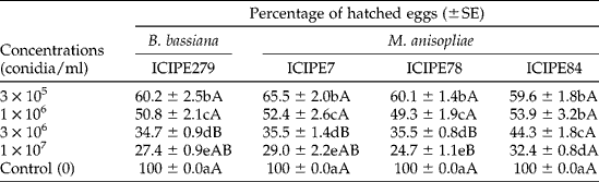
In the controls, 100% of the eggs hatched 7 days after the treatment. However, there were significant differences between the different concentrations of fungal isolates used in relation to egg viability: B. bassiana ICIPE279 (F= 343.42; degree of freedom (df) = 4, 15; P< 0.0001); M. anisopliae ICIPE78 (F= 590.96; df = 4, 15; P< 0.0001); M. anisopliae ICIPE84 (F= 186.33; df = 4, 15; P< 0.0001); M. anisopliae ICIPE7 (F= 220.86; df = 4, 15; P< 0.0001) (Table 1). Egg viability was dose-dependent, with the lowest viability occurring at the highest concentration of 1 × 10⁷ conidia/ml. Significant differences were also observed between the fungal isolates at the concentrations of 3 × 10⁶ conidia/ml (F= 12.19; df = 3, 12; P= 0.0006) and 1 × 10⁷ conidia/ml (F= 5.60; df = 3, 12; P= 0.0123). However, there was no significant difference observed between the fungal isolates at the concentrations of 3 × 10⁵ conidia/ml (F= 3.32; df = 3, 12; P= 0.0567) and 1 × 10⁶ conidia/ml (F= 0.61; df = 3, 12; P= 0.6214) (Table 1). Eggs that did not hatch developed mycosis.
Effect of Beauveria bassiana and Metarhizium anisopliae infection on the egg viability of Tetranychus urticae 7 days after treatment at 26±2 °C

Means ( ± SE) within a column followed by the different lower-case letters and within a row followed by the different upper-case letters are significantly different (P= 0.05; Student–Newman–Keuls test).
Effect of fungal infection on larvae, protonymphs, deutonymphs and adults
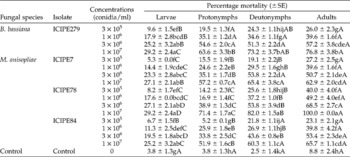
Mortality in the controls was 3.8, 3.8, 2.5 and 8.8% for larvae, protonymphs, deutonymphs and adults, respectively, 10 days post-infection. In all the fungal isolates tested, the highest conidial concentration was found to induce the highest mortality: larvae (F= 16.71; df = 16, 51; P< 0.0001); protonymphs (F= 93.73; df = 16, 51; P< 0.0001); deutonymphs (F= 79.94; df = 16, 51; P< 0.0001); adults (F= 83.00; df = 16, 51; P< 0.0001) (Table 2).
Effect of Metarhizium anisopliae and Beauveria bassiana infection on the motile developmental stages of Tetranychus urticae +

Means ( ± SE) within a column followed by the different lower-case letters and within a row followed by the different upper-case letters are significantly different (P= 0.05; Student–Newman–Keuls test).
+ Mortality was recorded at the different concentrations for 10 days post-infection at 26 ± 2 °C.
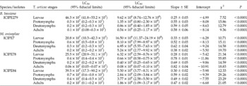
The LC50 values varied between 20.8 and 46.3 × 10⁷ conidia/ml for larvae, 0.3 and 0.7 × 10⁷ conidia/ml for protonymphs, 0.2 and 0.4 × 10⁷ conidia/ml for deutonymphs, and 0.06 and 0.2 × 10⁷ conidia/ml for adults (Table 3). The LC90 values varied between 5.50 and 16.50 × 10⁸ conidia/ml for larvae, 0.66 and 8.10 × 10⁸ conidia/ml for protonymphs, 0.40 and 6.95 × 10⁸ conidia/ml for deutonymphs, and 0.08 and 5.24 × 10⁸ conidia/ml for adults (Table 3).
Effect of Metarhizium anisopliae and Beauveria bassiana infection on the motile developmental stages of Tetranychus urticae

LC, lethal concentration.
Discussion
Egg viability varied with conidial concentrations and fungal isolates: the higher the concentration, the higher the egg mortality. B. bassiana and M. anisopliae have been reported to infect and kill mite eggs (Irigaray et al., Reference Irigaray, Marco-Mancebón and Pérez-Moreno2003; Shi and Feng, Reference Shi and Feng2004; Wekesa et al., Reference Wekesa, Knapp, Maniania and Boga2006). Shi and Feng (Reference Shi and Feng2004) observed that mortality varied significantly among fungal isolates and conidial concentrations. As observed in our study, high conidial concentrations generally led to higher mortalities. Similar observations were made by Wekesa et al. (Reference Wekesa, Knapp, Maniania and Boga2006) who found that, at a high concentration of 1 × 10⁸ conidia/ml, the isolates of B. bassiana and M. anisopliae were able to kill about 80% of T. evansi eggs.
The susceptibility of the different motile developmental stages of T. urticae also varied with fungal isolate, fungal species and mite stage. Mature stages (deutonymphs and adults) were generally more susceptible than immature ones (larvae and protonymphs), which corroborates the findings of Irigaray et al. (Reference Irigaray, Marco-Mancebón and Pérez-Moreno2003) and Wekesa et al. (Reference Wekesa, Knapp, Maniania and Boga2006). The motile-stage mortality of T. urticae was also dose-dependent, with the highest mortality occurring at the highest conidial concentration. Similar results have been reported for the cassava green mite M. tanajoa (Bondar) (Barreto et al., Reference Barreto, Marques, Gondim and de Oliveira2004), the citrus rust mite Phyllocoptruta oleivora (Ashmead) (Alves et al., Reference Alves, Tamai, Rossi and Castiglioni2005), the TSSM (Irigaray et al., Reference Irigaray, Marco-Mancebón and Pérez-Moreno2003) and T. evansi (Wekesa et al., Reference Wekesa, Knapp, Maniania and Boga2006). Oduor (Reference Oduor1995) also found that deutonymphs and adults of the cassava green mite were more susceptible to infection by Neozygites floridana Weiser & Muma than its larvae and protonymphs. The higher susceptibility of deutonymphs and adults to fungal infection observed in the laboratory may explain the higher prevalence of infection by Entomophthora sp. in field-collected deutonymphs and adult TSSM compared with its larvae and protonymphs (Carner and Canerday, Reference Carner and Canerday1970). The high susceptibility of adult arthropods to fungal infection compared with immature stages has also been reported for other groups of arthropods such as the legume flower thrips Megalurothrips sjostedti (Trybom) (Ekesi and Maniania, Reference Ekesi and Maniania2000) and the western flower thrips Frankliniella occidentalis (Pergande) (Maniania et al., Reference Maniania, Ekesi, Löhr and Mwangi2001). The differences observed in the susceptibility of the different developmental stages of arthropods to fungal infection have been attributed to the characteristics of their cuticle: the interaction between the integument of arthropods being penetrated by the fungus and moulting. Moulting plays a significant role in the resistance of arthropods to fungal infection, especially when the time interval between consecutive moulting stages is short (Vey and Fargues, Reference Vey and Fargues1977), which is the case with larvae and protonymphs of the TSSM. Oduor (Reference Oduor1995) speculated that the differences in the susceptibility of different developmental stages of mites to fungal infection may also be attributed to the smaller size and limited movement of the younger stages of mites, which reduces the encounters with the conidia. This may explain why mature stages are more susceptible to fungal infection than younger (immature) ones.
Although the egg viability and motile-stage mortality of T. urticae varied with conidial concentrations, the highest concentration (1 × 10⁷ conidia/ml) of both the isolates of B. bassiana and M. anisopliae was more efficient than the other tested concentrations (3 × 10⁵, 1 × 10⁶ and 3 × 10⁶ conidia/ml) in reducing egg viability and inducing mortality in motile stages. Infection at immature stages required a higher fungal inoculum than that at mature stages. While testing the virulence of B. bassiana against T. urticae, Irigaray et al. (Reference Irigaray, Marco-Mancebón and Pérez-Moreno2003) found that a concentration of 3184.4 conidia/ml was required to kill 50% of T. urticae larvae, but only a concentration of 1949.4 conidia/ml was required to kill 50% of adults. Since crops are often infested with different developmental stages of the pest, understanding which stage is most susceptible and most refractory to infection is important for the development of IPM strategies (Ferron, Reference Ferron, Kerkut and Gilbert1985). Therefore, the fungal concentration of 1 × 10⁷ conidia/ml (approximately 1012 conidia/ha) should be considered for the effective control of T. urticae.
Acknowledgements
We thank Mr R. Rotich, Mr B. Muia and Ms E. Ouna for their technical assistance. We are grateful to Mr A. Wanjoya for providing assistance on the statistical analysis. We are also grateful to the SII-Dutch government fund through the African Regional Postgraduate Programme in Insect Science (ARPPIS) of the International Centre of Physiology and Ecology (icipe), and the German Federal Ministry for Economic Cooperation and Development (BMZ) for funding this study.