Introduction
Leishmaniasis causes human suffering on a global scale, especially in the poorest countries where the problem of access to medicines persists. In fact, there is no effective vaccine to prevent human leishmaniasis and drugs available for chemotherapy present various limitations such as high toxicity, resistance emergency, treatment failure and high cost (Sundar, Reference Sundar2001; World Health Organization, 2010).
In the last decades, there has been a renewed interest in natural compounds derived from plants and microorganisms as source of new drugs, including anti-infective substances (Kondo et al. Reference Kondo, Mizuki, Akao and Ohba2002; Xu et al. Reference Xu, Yao, Sun and Yu2004; Cragg and Newman, Reference Cragg and Newman2013; Zhou et al. Reference Zhou, Grundmann, Kaiser, Schiell, Gaudriault, Batzer, Kurz and Bode2013; Dagnino et al. Reference Dagnino, Barros, Ccana-Ccapatinta, Prophiro, Poser and Romão2015). In this context, entomopathogenic bacteria such as Photorhabdus luminescens have been considered as a promising source for novel natural compounds (Bode, Reference Bode2009). These bacteria are rich in gene clusters that encode putative biosynthetic enzyme pathways, which are assumed to produce novel natural compounds with diverse biological activities. The chemical diversity of some of these compounds has been explored resulting in different classes of compounds (Chaston et al. Reference Chaston, Suen, Tucker, Andersen, Bhasin, Bode, Bode, Brachmann, Cowles, Cowles, Darby, de Leon, Drace, Du, Givaudan, Herbert Tran, Jewell, Knack, Krasomil-Osterfeld, Kukor, Lanois, Latreille, Leimgruber, Lipke, Liu, Lu, Martens, Marri, Medigue, Menard, Miller, Morales-Soto, Norton, Ogier, Orchard, Park, Park, Qurollo, Sugar, Richards, Rouy, Slominski, Slominski, Snyder, Tjaden, van der Hoeven, Welch, Wheeler, Xiang, Barbazuk, Gaudriault, Goodner, Slater, Forst, Goldman and Goodrich-Blair2011). Most of these metabolites are involved in symbiosis with the nematode (Heterorhabditidae family), pathogenicity to the insect and antimicrobial activity (Tobias et al. Reference Tobias, Mishra, Gupta, Sharma, Thines, Stinear and Bode2016). Different compounds with activity against bacterial and fungal pathogens of medical and agricultural interest have already been isolated (Challinor and Bode, Reference Challinor and Bode2015). Although some studies have shown that P. luminescens secretes compounds like stilbenes that show biological activity against Leishmania donovani and Trypanosoma cruzi (Kronenwerth et al. Reference Kronenwerth, Brachmann, Kaiser and Bode2014) or with anti-Plasmodium activity (so called GameXPeptides) (Challinor and Bode, Reference Challinor and Bode2015), the effect of most metabolites produced by P. luminescens against parasites is under-explored. Here we investigated the bioactivity of cell-free conditioned medium from P. luminescens against both promastigote (infective stage transmitted by sandfly vector) and amastigote forms (inside host cells) of Leishmania amazonensis. In addition, possible mechanisms of action were evaluated. We found that the leishmanicidal activity is caused by a peptide-based molecule smaller than 3 kDa.
Materials and methods
Cultivation of P. luminescens and preparation of conditioned medium
Photorhabdus luminescens subsp. laumondii TT01 DSM15139 (Fischer-Le Saux et al. Reference Fischer-Le Saux, Viallard, Brunel, Normand and Boemare1999) was used in the leishmanicidal bioassays. For that purpose, Photorhabdus luminescens was inoculated on NBTA medium [nutrient agar supplemented with 0·025% (w/v) bromothymol blue and 0·004% (w/v) triphenyltetrazolium chloride] in order to differentiate phenotypic phase variants. A start-culture was grown from a single primary phase colony in 5YS medium broth as described before (Shrestha and Lee, Reference Shrestha and Lee2012) [5% (w/v) yeast extract, 0·5% (w/v) NaCl, 0·05% (w/v) K2HPO4, 0·05% (w/v) NH4H2PO4, 0·02% (w/v) MgSO4·7H2O] on a shaker (180 rpm) at 28 °C. After an overnight incubation, the bacterial density was determined by absorbance at 600 nm. The main culture was started at OD600 = 0·1 and incubated for 48 h to reach the stationary phase. After that, the cells were removed by centrifugation at 2295 g for 20 min, and cell free culture supernatants were filter-sterilized through 0·22 µm membrane and kept at −20 °C until use. The protein concentration of this conditioned medium was determined using the Bradford method (Bradford, Reference Bradford1976). As controls, Enterococcus faecalis ATCC 29212 and human pathogenic Escherichia coli (Migula) Castellani and Chalmers (ATCC® 25922™) were used and grown in tryptone soy agar medium (Castellani and Chalmers, Reference Castellani and Chalmers1919). A single colony from each culture was inoculated in 5YS medium. Broth culture, centrifugation and filtering were carried out under similar conditions as used for P. luminescens.
Leishmania (Leishmania) amazonensis culture
Leishmania amazonensis (WHO reference strain MHOM/BR/73/M2269) were routinely maintained as promastigote forms in M199 medium containing 40 mm of 4-(2-hydroxyethyl)piperazine-1-ethanesulfonic acid sodium salt (HEPES), 0·1 mm adenine, 7·7 mm hemin, 10% (v/v) heat-inactivated fetal bovine serum (FBS), 2% (v/v) human urine, 50 U mL−1 of penicilin and 50 µg mL−1 of streptomycin. Cultures were incubated at 26 °C, and cells were kept at densities ranging between 1 × 10⁵ and 3 × 10⁷ parasites mL−1 (Romao et al. Reference Romao, Tovar, Fonseca, Moraes, Cruz, Hothersall, Noronha-Dutra, Ferreira and Cunha2006).
Effects of P. luminescens conditioned medium on growth of L. amazonensis
Promastigote forms of L. amazonensis were distributed in 12-well microplate at density of 1 × 10⁵ mL−1 in M199 medium (control) or M199 plus 3·4 and 34 µg of protein mL−1 of the P. luminescens conditioned medium. Leishmania amazonensis growth was determined daily until stationary phase by motility and cell density using a haemocytometer.
Activity of P. luminescens conditioned medium against promastigotes of L. amazonensis
The direct cytotoxic effect of P. luminescens conditioned medium on L. amazonensis was evaluated. For that purpose, promastigote forms of L. amazonensis in stationary growth phase were dropped in 96-well microplates (3 × 10⁶ well−1) and then incubated with M199 medium (control) or different concentrations of P. luminescens culture fluid (0·68 to 170 µg of protein mL−1). The conditioned medium of E. faecalis and E. coli at the same concentrations was used as negative controls. The viability of promastigotes was evaluated at different time points (3, 6, 12, 24 and 48 h) by counting the viable promastigote forms using a hemocytometer.
Effect of proteolytic enzyme, heating and pH on leishmanicidal activity of P. luminescens conditioned medium
To investigate the stability of the bioactive molecule(s) acting on L. amazonensis, P. luminescens conditioned medium was heated at 100 °C for 10 min, and then cooled to room temperature before use. To verify the protein nature of P. luminescens bioactive molecule(s), the conditioned medium was treated with proteinase K (2 mg mL−1 final concentration) (Bizani and Brandelli, Reference Bizani and Brandelli2002). To assess the effect of pH, samples of cell-free supernatant were acidified with 5 m HCl until pH 1·0 or alkalinized to pH 12·0 with 5 m NaOH. After incubation for 40 min at room temperature the pH was adjusted to its initial value (pH 8·0). All bioassays were performed using P. luminescens conditioned medium at concentration of 85 µg of protein mL−1.
Enrichment of bioactive molecule(s) from P. luminescens conditioned medium
As first step, P. luminescens conditioned medium was ultra-filtrated through membranes of 50, 10 and 3 kDa exclusion size (Ultrafree CL®, Millipore) to roughly classify the compound size of the leishmanicidal molecule(s). After restoring each fraction retained to its initial volume, promastigotes of L. amazonensis were incubated with 25 µL (equivalent to 85 µg of protein mL−1 of the conditioned medium) of each fraction for 24 h and the parasite viability determined as described before.
Moreover, <10 kDa ultra-filtered fraction was concentrated 7 times by lyophilization and 1 mL was loaded onto Sephadex column (20·5 × 0·5 cm2), with a flow rate of 0·3 mL min−1. This procedure was repeated 12 times and yielded an amount of 224 µg of protein loaded onto the column. Forty-two fractions of 1 mL were collected from each column and the presence of protein was monitored at 280 nm. Each recovered fraction was lyophilized and the respective fractions of all 12 columns were pooled. Then, the protein concentration of each pooled-fraction was determined using Bradford assay and the leishmanicidal activity against promastigotes was assessed using a final protein concentration of 14 µg of protein mL−1.
Determination of the mitochondrial membrane potential (ΔΨm)
The mitochondrial membrane potential (ΔΨm) was quantified according to the method described by Ferlini and Scambia (Reference Ferlini and Scambia2007), using the fluorescent dye rhodamine 123 (Rh 123, R8004, Sigma-Aldrich®, St. Louis, MO, USA), which passively diffuses through the plasma membrane and accumulates in metabolic active mitochondria. Briefly, promastigote forms of L. amazonensis (1 × 10⁶ on log phase) were incubated with M199 medium, hydrogen peroxide (H2O2 2 mm; positive control) or with 3·25 µg of protein mL−1 of the smaller than 3 kDa ultra-filtrated fraction for 12 h at 26 °C. The cells were washed with phosphate buffered saline (PBS) and incubated with 500 µL of Rh 123 (1 µg mL−1) for 10 min at 37 °C. After a washing step, cells were resuspended in 0·5 mL of PBS. The analysis was performed using a BD FACSCalibur (Becton–Dickinson®, Rutherford, NJ, USA) flow cytometer and CellQuest® Pro software (Joseph Trotter, Scripps Research Institute, La Jolla, CA, USA) using the blue argon-ion 488 nm laser with the FL1 filter channel. A total of 30 000 events were acquired in the region that corresponded to the parasites. Alterations in Rh 123 fluorescence were quantified using an index of variation (IV) obtained from the equation IV = (Mt − Mc)/Mc, in which Mt is the median fluorescence of treated parasites, and Mc is the median fluorescence of untreated parasites. Negative IV values correspond to depolarization and positive values, hyperpolarization of the mitochondrial membrane. Histograms were build using the CellQuest Pro software (Joseph Trotter, Scripps Research Institute, La Jolla, CA, USA).
Cytotoxic effect of P. luminescens conditioned medium against macrophages
Cytotoxicity on macrophages was determined using 3-[4,5-dimethylthiazol-2-yl]-2,5-diphenyltetrazolium bromide (MTT) assay. Macrophages RAW 264·7 cell line were maintained in culture in RPMI 1640 plus 10% FBS, penicillin (100 U mL−1) and streptomycin (100 µg mL−1). First, 1 × 10⁵ cells were distributed in 96-well microplate plates and incubated overnight at 37 °C in an atmosphere of 5% CO2. Then, plates were washed with PBS and incubated in the presence of RPMI or RPMI plus different concentrations of P. luminescens conditioned medium (17 to 170 µg of protein mL−1). Following, 20 µL of MTT (5 mg mL−1 in PBS) was added into each well and incubations were continued for a further 4 h. The purple formazan product that is formed by the action of mitochondrial enzymes in living cells was solubilized by the addition of acidic isopropanol, and the absorbance at 570 nm measured using Spectramax® M2 software.
Hemolytic assay
The hemolytic assay was performed using a modified method according to Gauthier et al. (Reference Gauthier, Legault, Girard-Lalancette, Mshvildadze and Pichette2009). Briefly, human blood O+ type was obtained from healthy voluntary donors. The Universidade Federal de Ciências da Saúde de Porto Alegre Research Ethical Committee approved procedures and project under authorization (CAAE 63282416.6.0000.5345). The human erythrocytes samples were washed three times with PBS (pH 7·0) and resuspended to obtain a 1% (v/v) erythrocytes suspension. This suspension was placed into a 96-well microplate and different concentrations of the conditioned medium (17–170 µg of protein mL−1). PBS as negative control or sodium dodecyl sulphate (SDS) 0·01% as positive control were added to obtain a 0·8% erythrocyte suspension. Microplates were incubated on an orbital shaker for 60 min at 37 °C. Microplates were then centrifuged at 3000 rpm for 5 min and the supernatant was transferred to a new microplate. Absorbance of the supernatant was measured at 540 nm in SpectraMax M2 (molecular devices). Each experiment was carried out twice in triplicate.
Leishmanicidal activity of macrophages stimulated with conditioned medium
The leishmanicidal assay was performed as previously described (Romao et al. Reference Romao, Fonseca, Hothersall, Noronha-Dutra, Ferreira and Cunha1999). Briefly, Macrophages RAW 264·7 cell line were cultivated for 12–16 h at 37 °C in an atmosphere of 5% CO2. Non-adherent cells were removed and the adherent cells washed three-times with pre-warmed medium. Macrophages were infected with L. amazonensis (5 parasites cell−1) and 4 h later, the cell cultures were washed to remove not internalized Leishmania. Following, the cells were incubated with the appropriate stimulus, interferon γ (IFN-γ) (10 ng mL−1) plus lipopolysaccharide (LPS) (10 ng mL−1) as positive control, conditioned medium at concentrations ranging from 0·68 to 170 µg of protein mL−1 or ultra-filtered fraction <3 kDa PLT (1·6 and 3·25 µg of protein mL−1) for 48 h. Then, the supernatants were removed from each well and kept at −20 °C for nitric oxide (NO) and TNF-α determination. Then, the cell cultures were washed with PBS and 100 µL of 0·01% (w/v) SDS solution in serum-free-medium was added to each well and the cells incubated at 37 °C for 20 min. Then, the cells were supplemented with M199 30% (v/v) FBS (100 µL well−1), and incubated at 26 °C until parasite releasing to determine the number of promastigote forms recovered, once only viable amastigotes are capable to differentiate to motile promastigote forms. The leishmanicidal activity of macrophages was analyzed by determining the number of viable parasites (4 replicates) using a hemocytometer.
Effect of P. luminescens conditioned medium on the expression of CD80 and CD86 costimulatory molecules on macrophages
All of the experimental procedures with mice were performed in accordance with the guidelines of the National Institute of Health and the Brazilian Society for Science on Animals of Laboratory with the approval of local Ethics Committee (CEUA UFCSPA number 505/17). To investigate better the immunomodulatory activity of PLT on macrophages we assessed the expression of surface costimulatory molecules CD80 and CD86 on L. amazonensis-infected macrophages. BALB/c mice (n = 3) were euthanized under lidocaine (10 mg kg−1 i.p.) and thiopental (100 mg kg−1 i.p.) and the peritoneal cavity were harvested by washing the cavity with 3 mL of PBS. Peritoneal cells (2 × 10⁵ cells well−1) were distributed in 96-well microplate and incubated overnight in RPMI medium at 37 °C in an atmosphere of 5% CO2. Then, the cells were washed with PBS to remove non-adherent cells, and adherent macrophages were infected with L. amazonensis (5 parasites cell−1). Four hours later, the cultures were washed to remove not internalized Leishmania and cells were incubated with RPMI medium or with <3 kDa PLT at concentration of 3·25 µg of protein mL−1 for 24 h. After that, cells were stained with monoclonal antibodies conjugated with anti-mouse CD80-Fluorescein isothiocyanate (FITC) (clone 16–10A1; BIOGEMS, USA) or anti-mouse CD86-Phycoerythrin (PE) (clone GL1; BIOGEMS, USA), anti-CD14-FITC (clone Sa2-8; eBioscience). Thirty minutes after incubation, cells were resuspended in 0·4 mL of 1% BSA (Bovine Serum Albumin) in PBS and analyzed by flow cytometry. Fluorescent signals were collected in logarithmic mode (six decade logarithmic amplifier). Macrophages were identified and gated according to their forward scatter (FSC) and side scatter (SSC) profiles related to the CD14 expression. The expression of CD80 and CD86 were evaluated in CD14+ macrophages, based on fluorescence-1 (FL1-FITC) vs. fluorescence 2 (FL2-PE) dot plots. A minimal of 20 000 events of gated cells was acquired for analysis. The analysis was performed using software FlowJo 7.6.3 (Becton Dickinson)
Quantification of NO and TNF-α production by macrophages
The NO production was quantified in the cell culture supernatants of L. amazonensis-infected macrophages using the Griess method (Romao et al. Reference Romao, Fonseca, Hothersall, Noronha-Dutra, Ferreira and Cunha1999) and the levels of TNF-α content was determined by ELISA kit (eBioscience, USA) in accordance with manufacturer's instructions.
Statistical analysis
Results are expressed as mean ± standard error of the mean (s.e.m.) and were analyzed using the Kolmogorov–Smirnov normality test and one-way analysis of variance followed by Bonferroni's test. In all tests, differences were considered statistically significant when P < 0·05, and were performed using GraphPad Prism Software version 5.03. All experiments were performed two or three times and in quadruplicate.
The concentrations of bacterial culture fluids that cause 50% of macrophage cytotoxicity (CC50) or parasite mortality (IC50) were determined by non-linear regression analysis using GraphPad Prism® 5·03 version software.
Results
Photorhabdus luminescens conditioned medium inhibits L. amazonensis growth
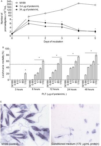
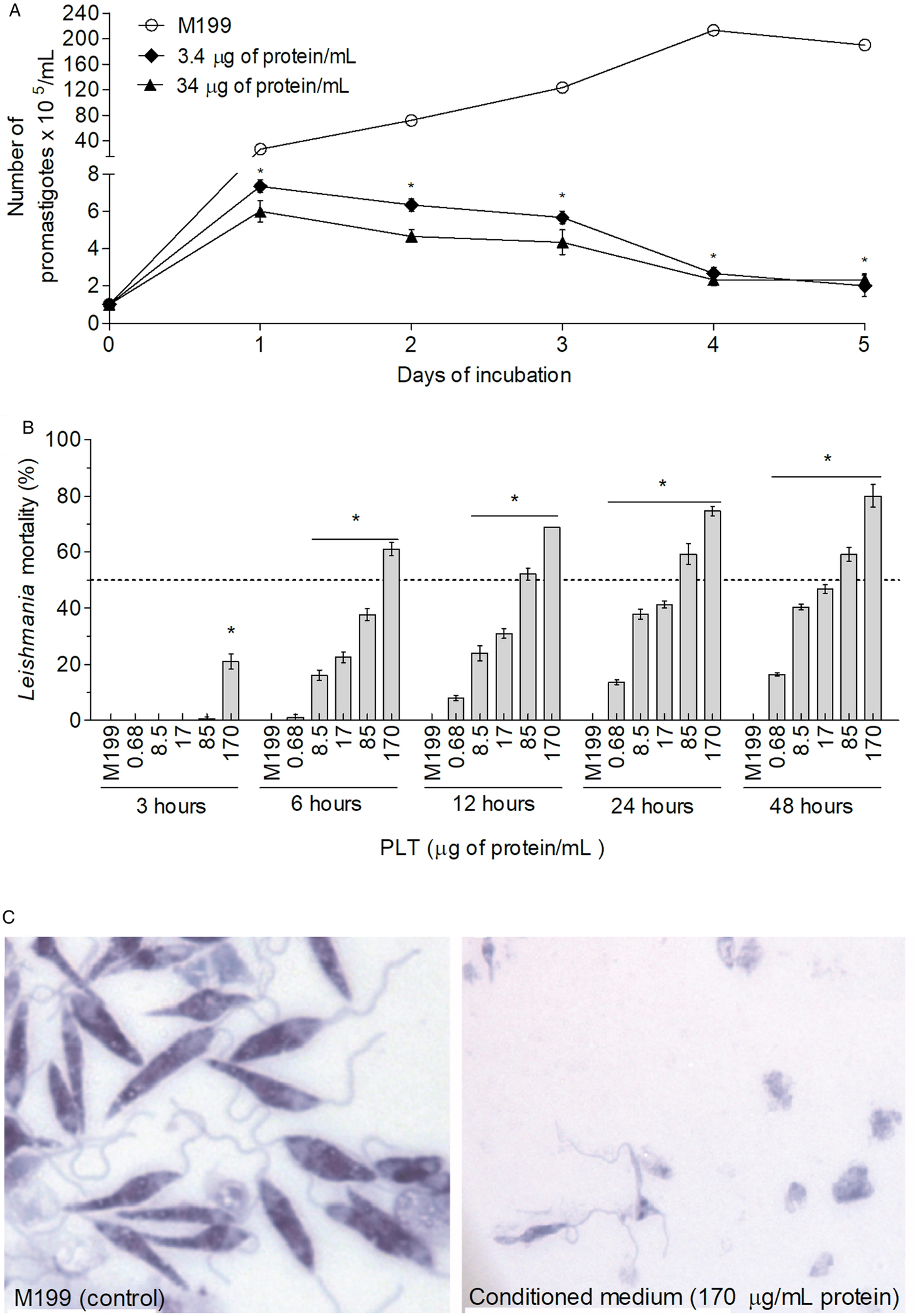
To test the toxicity of bacterial conditioned medium on parasites as first step, L. amazonensis promastigotes growth was analyzed in vitro in the presence of bacterial culture fluid. Our data show that metabolites secreted by P. luminescens in culture broth lead to a significant inhibition of parasite growth in both concentrations tested (Fig. 1A). Therefore, we will refer to the putative Photorhabdus-derived leishmanicidal toxin(s) as PLT in the following.
Effect of PLT on L. amazonensis promastigotes. (A) Growth kinetics of promastigotes treated with PLT. Promastigote forms of L. amazonensis (1 × 10⁵ mL−1) in M199 medium (control) or M199 plus PLT (at 3·4 and 34 µg of protein mL−1) were incubated at 26 °C and the parasite growth determined using a hemocytometer. (B) Leishmanicidal activity of PLT. Leishmania amazonensis (3 × 10⁶ well−1) were incubated in M199 medium (control) or M199 plus PLT in different concentrations of protein for 48 h. Leishmania survival was determined using a hemocytometer. Data are reported as means ± s.e.m. (n = 4) and are representative of three independent experiments. *P < 0·05 compared with control (M199 medium). (C) Effect of PLT on L. amazonensis integrity. Promastigote forms of L. amazonensis were treated with M199 medium or PLT at concentration of 170 µg of protein mL−1 for 48 h. L. amazonensis integrity after exposure to medium or PLT can be visualized in left and right panels, respectively (eosin-hematoxilin − 1000 × magnification). PLT, Photorhabdus-derived leishmanicidal toxin.
Leishmanicidal activity of P. luminescens conditioned medium on L. amazonensis promastigotes
PLT effectively killed the parasites in a concentration and time-dependent manner. It caused significant mortality even in the lowest concentration tested (0·68 µg mL−1 of protein: 16·4% of mortality within 48 h) (Fig. 1B), whereas culture fluids of pathogenic E. coli as well as E. faecalis had no significant effect (data not shown). The IC50 value of PLT on promastigotes of L. amazonensis was calculated to be 21·8 µg of protein mL−1 (Table 1). As illustrated in Fig. 1C, PLT at concentration of 170 µg of protein mL−1 led to total lysis of promastigotes.
Leishmanicidal activity and macrophages cytotoxicity of Photorhabdus-derived leishmanicidal toxin at 48 h

IC50 and CC50: concentration of bacterial culture fluids that causes 50% of L. amazonensis mortality and 50% of macrophage cytotoxicity, respectively.
95% CI: 95% confidence interval.
SIP and SIA selectivity index for promastigotes and amastigotes respectively, calculated as ratio of CC50 against mammalian cells/IC50 against L. amazonensis.
Proteinase K treatment, heating, acidification and alkalization affect the leishmanicidal activity of photorhabdus-derived leishmanicidal toxin
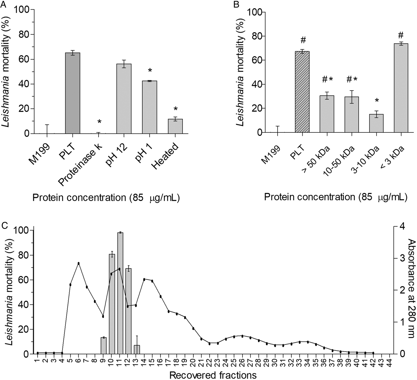
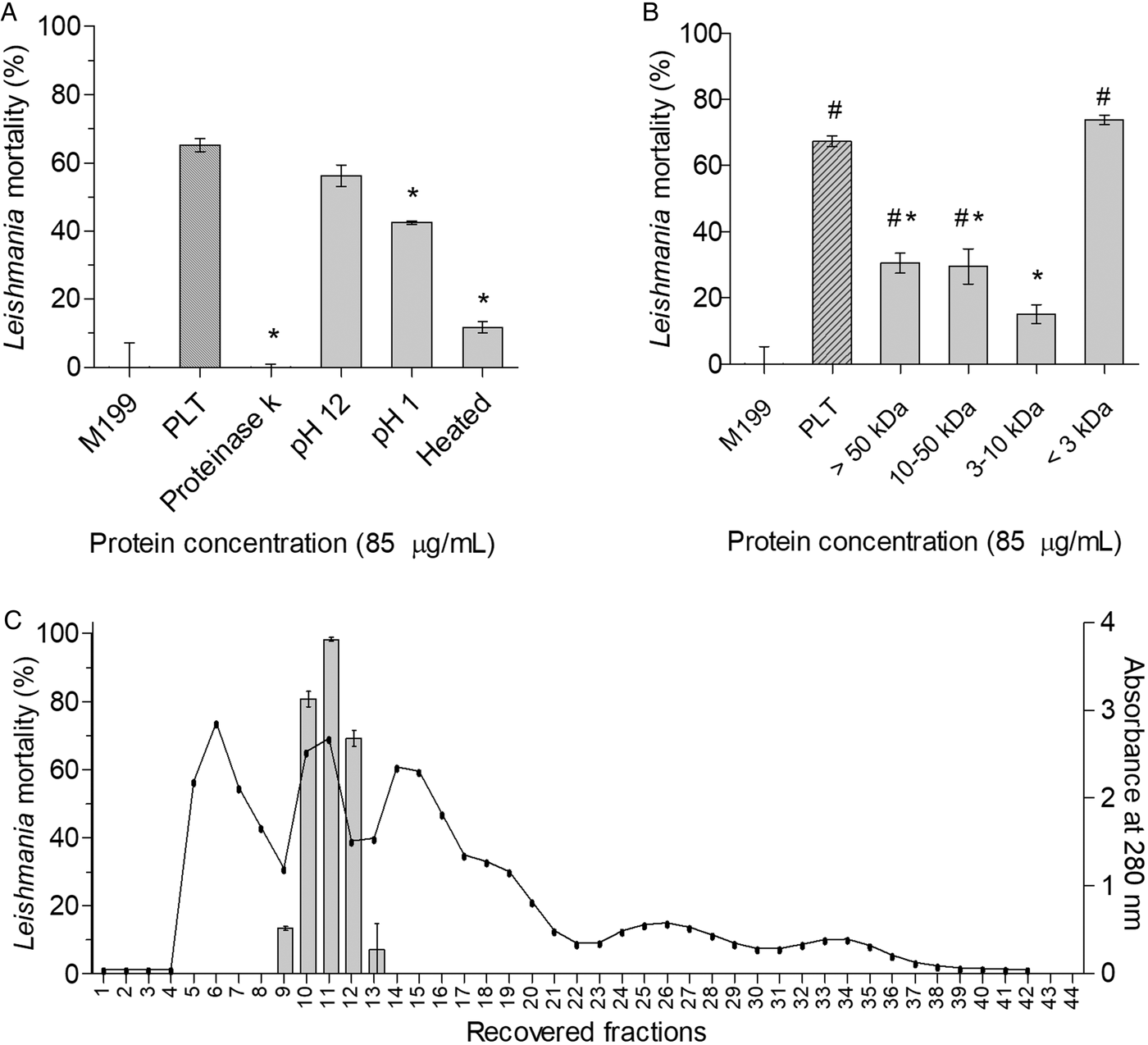
To pre-characterize the chemical nature of the PLT, the conditioned medium was pre-treated by heating, with proteinase K, acidification and alkalization, respectively, before tested for leishmanicidal activity. As we can see in Fig. 2A, the leishmanicidal activity of PLT was drastically reduced after the treatment with proteinase K or heating (Fig. 2A). This reveals that the chemical nature of the leishmanicidal compound(s) in the conditioned medium is similar to a protein or a peptide. Regarding the effect of pH variation on leishmanicidal activity, the acidification of the bacterial fluid to pH 1·0 also caused a significant decrease in Leishmania mortality compared with respective non-treated P. luminescens conditioned medium (pH 8·0), whereas alkalization to pH 12·0 had no significant effect on leishmanicidal activity of P. luminescens culture fluids (Fig. 2A).
Characterization and enrichment of PLT. (A) Effect of proteolysis, heating and pH changing on leishmanicidal activity of PLT. Leishmania amazonensis (3 × 10⁶ promastigotes well−1) were incubated for 24 h with M199 (control), untreated PLT (85 µg of protein mL−1), PLT treated with proteinase K, submitted to alkalinization or acidification following to restoring to initial pH = 8·0, or with heated PLT. After 24 h of incubation, Leishmania mortality was determined using a hemocytometer. Data are reported as means ± s.e.m. (n = 4) and are representative of three independent experiments. *P < 0·05 compared with untreated conditioned medium. (B) Anti-leishmanial activity of ultrafiltered PLT. Cell-free conditioned medium of P. luminescens culture was ultra-filtrated through membranes of 50, 10 and 3 kDa exclusion size (Ultrafree CL, Millipore). After restoring each fraction to its initial volume, promastigote forms of L. amazonensis were incubated with 85 µg of protein mL−1 of the conditioned medium of each fraction during 24 h. Leishmania survival was determined using a hemocytometer. Data are reported as means ± s.e.m. (n = 4) and are representative of three independent experiments. *P < 0·05 compared with P. luminescens conditioned medium. #P < 0·05 compared with M199 medium. (C) Protein determination (line) and leishmanicidal activity (bars) of fractions eluted from Sephadex G-25 column. The smaller than 10 kDa ultra-filtrate fraction (total of 224 µg of protein) was eluted through size exclusion chromatography column and the amount of protein monitored at absorbance of 280 nm (line) and the leishmanicidal activity (mortality) of each recovered fraction was assessed at 24 h. Data are reported as means ± s.e.m. (n = 2). PLT, Photorhabdus-derived leishmanicidal toxin.
Enrichment of P. luminescens protein-based leishmanicidal compound
To characterize the size of the PLT, the cell-free culture of P. luminescens was initially submitted to ultrafiltration using 50, 10 and 3 kDa cut-off membranes. Higher levels of leishmanicidal activity of PLT were observed in the fraction corresponding to molecular weight smaller than 3 kDa (Fig. 2B). Therefore, we will refer to this fraction as <3 kDa-PLT in the following.
To further enrich the PLT, the ultra-filtrate from 10 kDa membrane (smaller than 10 kDa fraction) was fractionated by size exclusion chromatography yielding 42 fractions of 1 mL each which were tested against promastigotes of L. amazonensis. After separation using a Sephadex G-25 column, three major protein peaks were observed. From the 42 fractions, only the 10, 11 and 12 (Fig. 2C) showed potent leishmanicidal activity, causing 80·7, 98·2 and 69·2% of mortality, respectively. In the bioactive fractions 10, 11 and 12, the amount of protein recovered was 20, 15 and 20 µg mL−1, respectively (55 µg mL−1 in total), corresponding to 24·67% of protein recovered.
PLT induces depolarization of the mitochondrial transmembrane potential of L. amazonensis
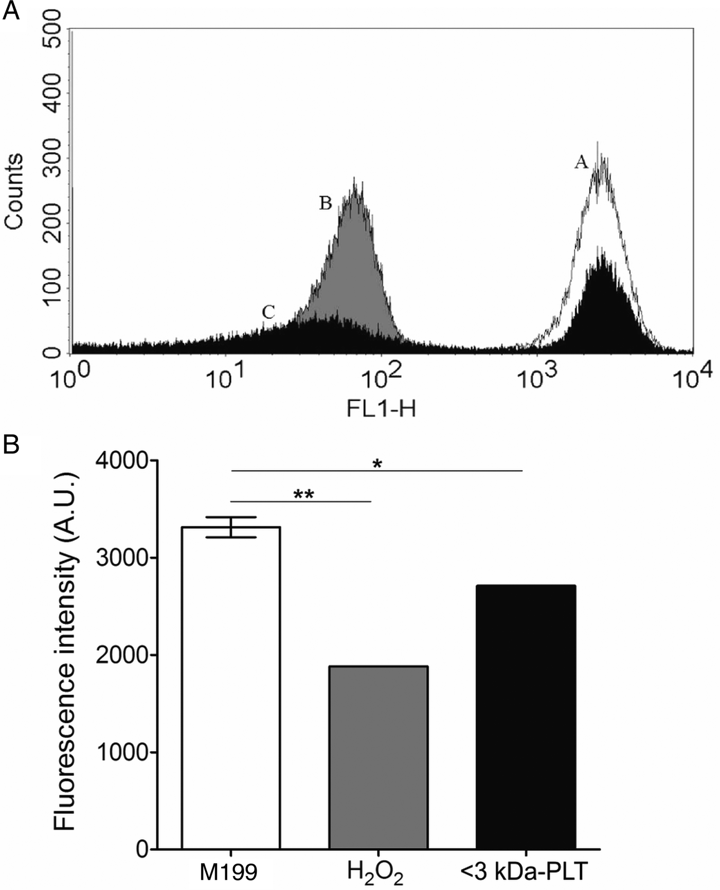
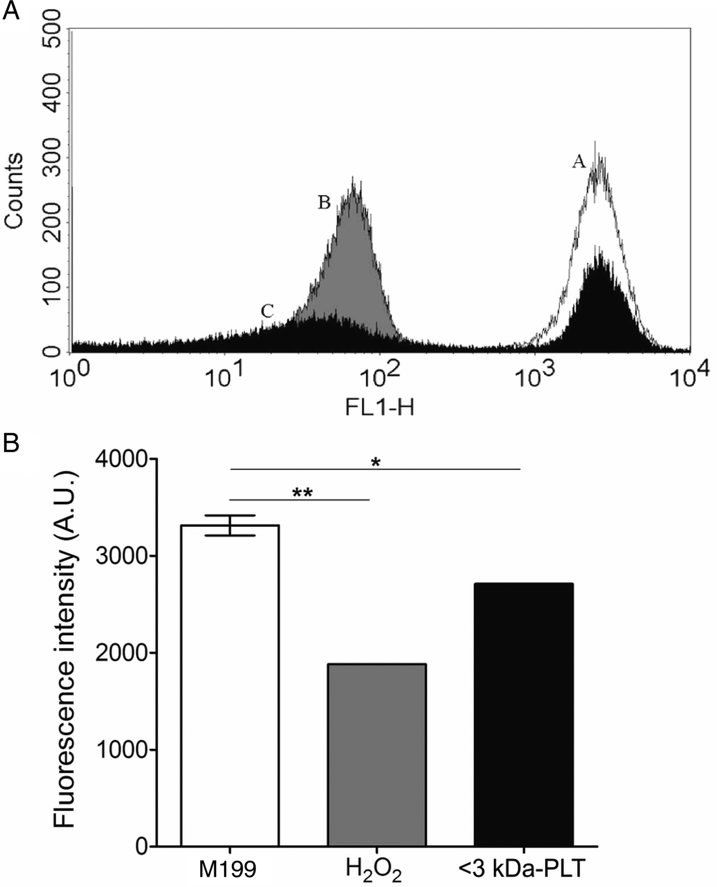
The effect of the PLT on the mitochondrial membrane potential was investigated. The treatment of parasites with <3 kDa-PLT at 3·25 µg of protein mL−1 led to a significant decrease in the Rh 123 fluorescence after 12 h of incubation (Fig. 3A and 3B). Promastigotes treatment with <3 kDa-PLT induced ΔΨm depolarization with IV value of – 0·18. As expected, the incubation of parasites with H2O2 at 2 mm caused potent depolarization of ΔΨm (IV = – 0·43).
Effect of <3 kDa PLT on mitochondrial membrane potential measured by flow cytometry. Histograms (A) and graphic representation of mean fluorescence intensity (B) in arbitrary units (A.U.) of L. amazonensis promastigotes untreated control (A – white), treated with 2 mm H2O2 (B – grey) or with <3 kDa-PLT (ultra-filtered fraction) at concentration of 3·25 µg of protein mL−1 (C – black) for 12 h. Data represent mean ± s.e.m. and are representative of two independent experiments. *P < 0·05 compared with M199 (control); **P < 0·001 compared with M199. PLT, Photorhabdus-derived leishmanicidal toxin.
Photorhabdus luminescens conditioned medium present low cytotoxicity on macrophages and erythrocytes
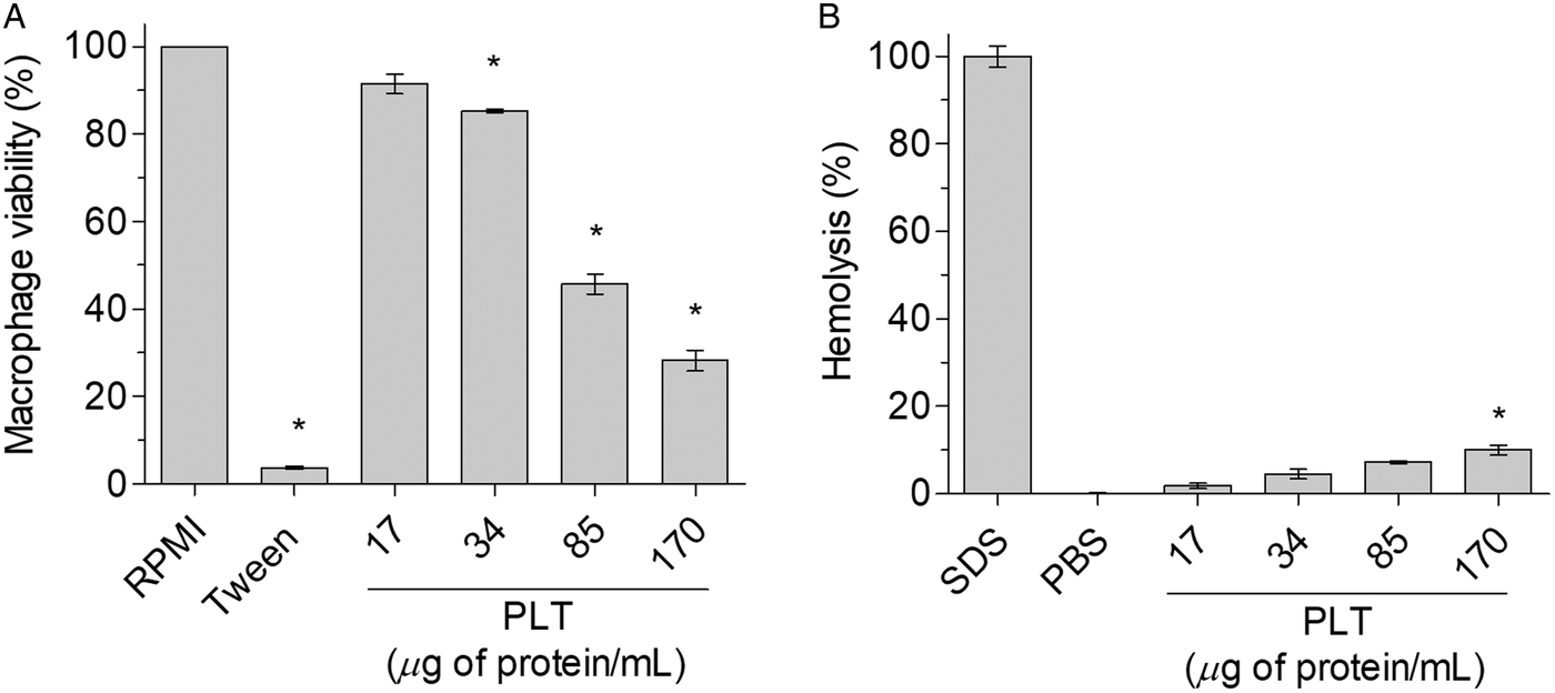
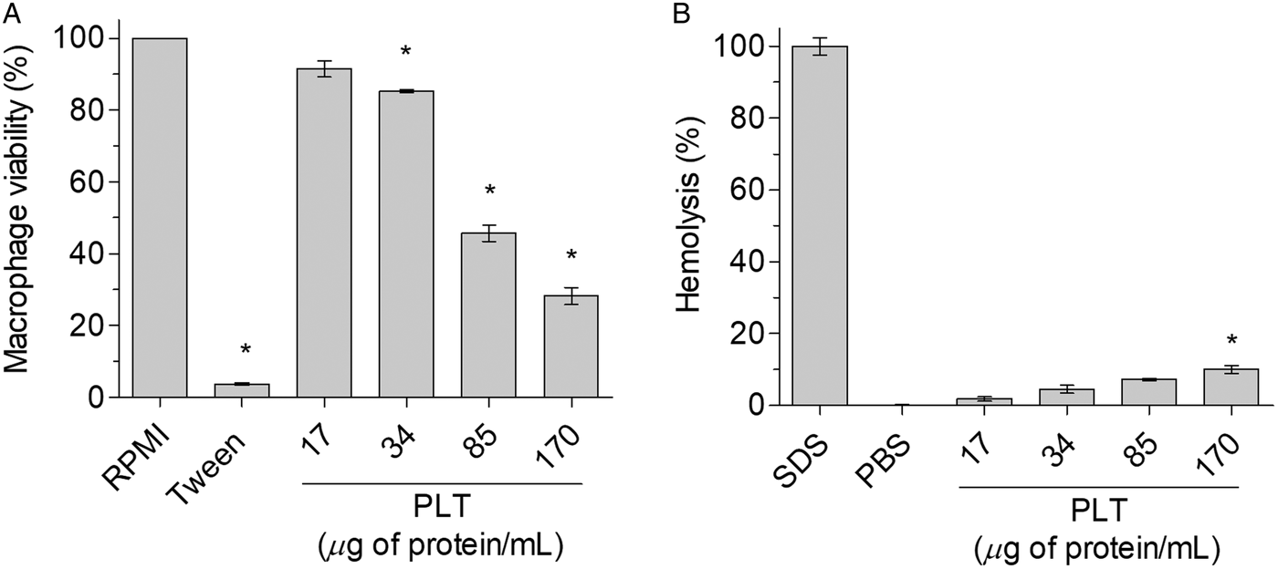
To assess the cytotoxic effect of PLT, we tested its toxicity toward macrophages and erythrocytes. As depicted in Fig. 4A, PLT showed low cytotoxicity against macrophages. Only the highest protein concentrations (85 and 170 µg mL−1) caused high levels of macrophage mortality. The concentration of PLT that causes 50% of macrophages cytotoxicity was determined as 85·48 µg mL−1 (Table 1). Moreover, PLT also showed low cytotoxicity against human erythrocytes (Fig. 4B).
Cytotoxic effects of P. luminescens conditioned medium on macrophages (A) and erythrocytes (B). (A) Macrophages were treated with RPMI medium (control) or bacterial conditioned medium (8·5–170 µg of protein mL−1) and the cell viability were determined by MTT assay after 48 h of incubation. (B) Hemolytic activity was performed using human erythrocytes incubated with P. luminescens conditioned medium, PBS (negative control) or SDS 0·01% (positive control) for 60 min. Hemolysis was determined by measuring the absorbance of the cells supernatants at 540 nm. Data are expressed as means ± s.e.m. of four replicates and are representative of three independent experiments. *P < 0·05 compared with RPMI medium (panel A) or PBS (panel B). PBS, phosphate buffered saline; SDS, sodium dodecyl sulphate.
PLT stimulates the leishmanicidal activity of macrophages by mechanisms independent of NO and TNF-α production
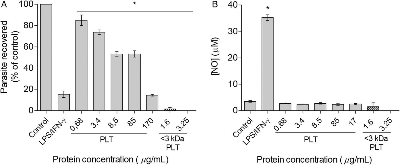
Since Leishmania parasites survive and proliferate inside infected cells as amastigotes, we tested the effects of the bacterial culture fluids on intracellular Leishmania. Based on IC50 value found for P. luminescens against promastigotes, the biological activity of PLT on intracellular amastigotes was investigated at concentrations ranging from 0·68 to 34 µg of protein mL−1. It was verified that the PLT stimulated the leishmanicidal activity of macrophages reducing the amastigotes survival in an order of 15–85·5% (Fig. 5A). The IC50 value obtained was 8·85 µg of protein mL−1 and the selectivity index calculated as the ratio of CC50 (macrophages cytotoxicity)/IC50 (anti-amastigote activity) was close to 10 (SI = 9·66), indicating a moderate to high selectivity to amastigotes (Table 1). Comparatively, the enriched <3 kDa-PLT at concentration of 1·6 µg of protein mL−1 caused almost 100% of amastigote mortality (Fig. 5A).
Effects of PLT on the viability of amastigotes of L. amazonensis and nitric oxide production in vitro. Macrophages were infected with L. amazonensis and incubated in the presence of M199 medium (control), LPS (10 ng mL−1) plus IFN-γ (10 ng mL−1), PLT (0·68–34 µg of protein mL−1) or <3 kDa PLT (1·6 and 3·25 µg of protein mL−1) After 48 h cells were lysed for the parasite viability determination as described in materials and methods (A) and the supernatant was used for NO measurement using Griess method. Data are expressed as means ± s.e.m. of four replicates and are representative of three independent experiments. *P < 0·05 compared with control (M199 medium). PLT, Photorhabdus-derived leishmanicidal toxin; IFN-γ, interferon γ.
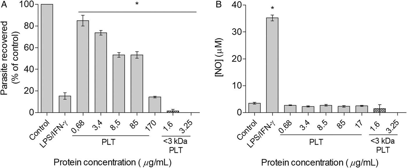
Unlike the leishmanicidal effect induced by stimulation with LPS/IFN-γ (85·2% mortality) that was NO-dependent, PLT or <3 kDa-PLT did not stimulate the NO production by L. amazonensis-infected macrophages (Fig. 5B). Moreover, the anti-amastigote activity induced by <3 KDa PLT fraction was not associated with the augment of costimulatory CD80 (control macrophages: Mean Fluorescence Intensity = 695·66 ± 0·57; L. amazonensis infected-macrophages: MFI = 696·3 ± 7·23; L. amazonensis-infected and <3 KDa-PLT-treated macrophages: MFI = 691·3 ± 6·02) and CD86 (control macrophages: MFI = 696·1 ± 8·71; L. amazonensis infected-macrophages: MFI = 696·53 ± 6·62; L. amazonensis-infected and <3 KDa-PLT-treated macrophages: MFI = 694·66 ± 5·50) molecules. On the other hand, in contrast to the stimulation of peritoneal macrophages with LPS (10 ng mL−1) plus IFN- γ (10 ng mL−1), the incubation of L. amazonensis-infected macrophages with <3 kDa-PLT at 3·25 µg of protein mL−1 did not increase the TNF- α production (data not shown).
Discussion
Photorhabdus bacteria contain a high number of genes that are assumed to encode enzymes involved in biosynthesis of novel secondary metabolites or bioactive compounds. Some of those novel metabolites have already been investigated and found to be involved in symbiosis with the nematodes, insect pathogenicity or showed antimicrobial activity (Tobias et al. Reference Tobias, Mishra, Gupta, Sharma, Thines, Stinear and Bode2016). In the last decades, the chemical diversity of Photorhabdus metabolites has been explored resulting in different classes of compounds. Furthermore, these bacteria have many gene clusters that encode enzymes apparently involved in secondary metabolism (Chaston et al. Reference Chaston, Suen, Tucker, Andersen, Bhasin, Bode, Bode, Brachmann, Cowles, Cowles, Darby, de Leon, Drace, Du, Givaudan, Herbert Tran, Jewell, Knack, Krasomil-Osterfeld, Kukor, Lanois, Latreille, Leimgruber, Lipke, Liu, Lu, Martens, Marri, Medigue, Menard, Miller, Morales-Soto, Norton, Ogier, Orchard, Park, Park, Qurollo, Sugar, Richards, Rouy, Slominski, Slominski, Snyder, Tjaden, van der Hoeven, Welch, Wheeler, Xiang, Barbazuk, Gaudriault, Goodner, Slater, Forst, Goldman and Goodrich-Blair2011). To the best of our knowledge, this is the first study reporting the leishmanicidal activity for P. luminescens secondary metabolites on both stages of Leishmania parasites. We demonstrated that PLT is effective to kill both promastigote and amastigote forms of L. amazonensis, one of the main agent of cutaneous leishmaniasis (Carvalho et al. Reference Carvalho, Barral, Costa, Bittencourt and Marsden1994; Franca-Costa et al. Reference Franca-Costa, Wanderley, Deolindo, Zarattini, Costa, Soong, Barcinski, Barral and Borges2012). Our data showed that P. luminescens secretes a small protein or a peptide with significant leishmanicidal activity (IC50 promastigote = 21·87 µg mL−1; IC50 amastigote = 8·85 µg mL−1). It is important to point out that we found an IC50 value for the anti-Leishmania drug pentamidine of 16·85 µg mL−1 (95% CI = 11·95–23·80) for promastigote of L. amazonensis.
It is well known that P. luminescens produces a huge number of secondary metabolites, including lipases, phospholipases, proteases, which are active against insects, as well as bacteria and fungi, which makes them a promising source of novel therapeutics (Herbert and Goodrich-Blair, Reference Herbert and Goodrich-Blair2007; Bode, Reference Bode2009; Waterfield et al. Reference Waterfield, Ciche and Clarke2009; Kronenwerth et al. Reference Kronenwerth, Brachmann, Kaiser and Bode2014; Tobias et al. Reference Tobias, Mishra, Gupta, Sharma, Thines, Stinear and Bode2016). Insecticidal activity of P. luminescens has been reported including Aedes aegypt larvae (LC50 = 21·18% v/v) (Nielsen-LeRoux et al. Reference Nielsen-LeRoux, Gaudriault, Ramarao, Lereclus and Givaudan2012; da Silva et al. Reference da Silva, Prado, da Silva, Silva, da Costa and Heermann2013, Reference da Silva, Undurraga Schwalm, Eugenio Silva, da Costa, Heermann and Santos da Silva2017) and Galleria mellonella larvae (LD50 of 28 bacteria per larvae) (Wu et al. Reference Wu, Zhao, Liu and Qiu2014). Moreover, Orozco et al. (Reference Orozco, Molnar, Bode and Stock2016) reported that metabolic crude extracts from P. luminescens sonorensis (Caborca and CH35 strains), at 40 µg mL−1, presented variable antibacterial activity against Bacillus subtilis and Pseudomonas syringae (radius of inhibition = 4·6–4·5 mm and 2·8–6·1, respectively for Caborca and CH35 strains). Additionally, in another study with different bacterial genera (Staphylococcus, Micrococcus, Paenibacillus, Escherichia, Salmonella, Klebsiella, and Bacillus), an inhibition zone ranging from 7·7 to 14·3 mm was found depending on bacterial genus and P. luminescens strain (El-Sadawy et al. Reference El-Sadawy, Forst, Abouelhag, Ahmed, Alajmi and Ayaad2016). Moreover, it was reported that P. luminescens crude extract from strains CH35 and Caborca, at 40 µg mL−1, also presented effect on Fusarium oxysporum with radius of inhibition of 3·6 and 4·0 mm, respectively (Orozco et al. Reference Orozco, Molnar, Bode and Stock2016). Shi et al. (Reference Shi, An, Zhang, Zhang and Yu2017) tested 7 compounds extracted from Photorhabdus temperate SN259 against phytopathogenic fungi, and the compound 7,2-isopropyl-5-[(E)-2-phenylethenyl]benzene-1,3-diol presented the best activity against mycelial growth (IC50 Pythium aphanidermatum = 2·0 µg mL−1; IC50 Rhizoctonia solani Kuhn = 6·3 µg mL−1; IC50 Exserohilum turcicum = 8·1 µg mL−1; IC50 F. oxysporum = 5·3 µg mL−1). Although a number of reports on insecticidal and antimicrobial activities, few studies have investigated the antiparasitic effects of Photorhabdus species.
Our data indicate that the PLT are heat-labile peptide-based molecule(s), since heating almost completely abolished the leishmanicidal activity of PLT against promastigotes. In addition, the peptide nature was confirmed by the inactivation of its leishmanicidal activity by proteinase K treatment. In an attempt to characterize the chemical nature of the molecule(s) responsible for the leishmanicidal activity, we performed an enrichment of the PLT from the bacterial culture fluid. Our data suggest that PLT(s) are small peptide(s) with a molecular weight smaller than 3 kDa. However, the exact chemical nature of the leishmanicidal compound(s) remains to be elusive. It is known that Photorhabdus species produce a variety of linear peptide antibiotics, as mevalagmapeptides and possibly carbapenem-like antibiotics, nonribosomal peptide synthetases (NRPS)-derived, as GameXPeptides, that showed anti-Plasmodium activity, and also polyketide synthase (PKS)-derived peptide, as stilbenes or anthraquinones (Brachmann and Bode, Reference Brachmann, Bode and Vilcinskas2013; Challinor and Bode, Reference Challinor and Bode2015). NRPSs are multifunctional enzymes involved in the production of drugs as cyclosporine, penicillin and vancomycin for example. They modify side chains of amino acids into linear or cyclic amino acid derivatives (Sieber and Marahiel, Reference Sieber and Marahiel2005), due to these modifications and different amino acids, several different non-ribosomally derived peptides are known (Cai et al. Reference Cai, Nowak, Wesche, Bischoff, Kaiser, Fürst and Bode2017). Naturally occurring peptide libraries include the cyanobactins (Donia et al. Reference Donia, Ravel and Schmidt2008), polylysine (Maruyama et al. Reference Maruyama, Toyoda, Kato, Izumikawa, Takagi, Shin-ya, Katano, Utagawa and Hamano2012) and streptothricin (Yamanaka et al. Reference Yamanaka, Maruyama, Takagi and Hamano2008) derivatives from different microorganisms. Cai et al. (Reference Cai, Nowak, Wesche, Bischoff, Kaiser, Fürst and Bode2017) also describe rhabdopeptide/xenortide class of non-ribosomally derived peptides (RXPs) in entomopathogenic bacteria as the largest class of peptides derived from NRPSs, composed of 2–8 amino acids with an overall molecular weight range between 395 Da and 1054 Da. Bode et al. (Reference Bode, Brachmann, Kegler, Simsek, Dauth, Zhou, Kaiser, Klemmt and Bode2015) described the bioactivity of mevalagmapeptides (from P. luminescens) and others RXPs against different protozoa (IC50: Trypanosoma brucei rhodesiense 129·7 µ m; Trypanosoma cruzi Tulahuen C4 118·0 µ m; L. donovani 60·7 µ m; Plasmodium falciparum NF 54 38·4 µ m) and mammalian cells (IC50 Rat L6 cells >150 µ m).
Depending on the biological environment, these secreted compounds are thought to have different effects, as isopropylstilbene that can act as an antibiotic against fungi and bacteria as well as being cytotoxic to insect cells. Kronenwerth et al. (Reference Kronenwerth, Brachmann, Kaiser and Bode2014) demonstrated that stilbene derivatives 13 and 14 were active against L. donovani with IC50 values of 3·71 and 7·47 µ m, respectively and T. cruzi (IC50 values of 16·3 and 8·80 µ m, respectively). However, in contrast to our data, these compounds presented high cytotoxicity on L6 cells, a myoblast cell line and they are therefore not suitable as a potential drug against leishmaniasis. The authors did not describe the parasite stage used in the study, but they highlighted the potential of stilbene compounds on pathogenic protozoa.
To evaluate more precisely the leishmanicidal potential of the PLT we also investigated the effect on intracellular amastigote forms. We showed that PLT reduces the intracellular survival of L. amazonensis in a dose-dependent manner by a mechanism independent of NO. Some authors, including our group, have demonstrated that reactive oxygen and nitrogen species plays important role in the parasite control (Fonseca et al. Reference Fonseca, Romao, Figueiredo, Morais, Lima, Ferreira and Cunha2003; Degrossoli et al. Reference Degrossoli, Arrais-Silva, Colhone, Gadelha, Joazeiro and Giorgio2011; Novais et al. Reference Novais, Nguyen, Beiting, Carvalho, Glennie, Passos, Carvalho and Scott2014). Moreover, activated macrophages produce several proteolytic enzymes in the phagolysosome that destroy microorganisms (Houghton et al. Reference Houghton, Hartzell, Robbins, Gomis-Ruth and Shapiro2009; Weiss and Schaible, Reference Weiss and Schaible2015) and also produces peroxynitrite a highly reactive oxidizing agent that destroy Leishmania (Giorgio et al. Reference Giorgio, Linares, de Capurro, de Bianchi and Augusto1996).
Considering that in mammalian host, the promastigotes of Leishmania inoculated by sandflies during the bite infect immune cells and differentiate into amastigotes inside the phagolysosomal vacuoles (Sacks and Kamhawi, Reference Sacks and Kamhawi2001; Chappuis et al. Reference Chappuis, Sundar, Hailu, Ghalib, Rijal, Peeling, Alvar and Boelaert2007), the cytotoxic effects of PLT on macrophages was evaluated. Our data showed that the metabolite(s) secreted by P. luminescens presented a slight toxicity on macrophages (more than 85% of viability at 34 µg mL−1 and IS = 9·66) and human erythrocytes (less than 10% of hemolysis at highest concentration tested), indicating a moderate to good safety profile of cytotoxicity (Oh et al. Reference Oh, Kim, Kong, Yang, Lee, Han, Goo, Siqueira-Neto, Freitas-Junior and Song2014). In this regard, we found that the SI of PLT for amastigotes was higher than those calculated for promastigotes, indicating a higher selectivity of PLT for amastigotes. Moreover, the <3 KDa ultra-filtered PLT at concentration that inhibited the amastigote survival in almost 100% did not cause any cytotoxicity against macrophages.
The maintenance of mitochondrial membrane potential is vital for metabolic process as well as for cell survival. Thus, we investigated the effects of PLT fraction smaller than 3 kDa on mitochondrial membrane potential of L. amazonensis using rhodamine 123, which accumulates in energized mitochondria. In our study, the treatment of promastigotes of L. amazonensis for 12 h with <3 kDa PLT caused significant mitochondrial transmembrane depolarization. The decrease in Rh 123 fluorescence suggests an increase in proton permeability across the inner mitochondrial membrane, which can lead to parasite death due to decreased ATP synthesis (Rodrigues et al. Reference Rodrigues, Ueda-Nakamura, Corrêa, Sangi and Nakamura2014; Garcia et al. Reference Garcia, Henrique da Silva Rodrigues, Din, Rodrigues-Filho, Ueda-Nakamura, Auzely-Velty and Nakamura2017). Indeed, Leishmania mitochondria is a target extensively explored, being essential to its survival (Sen et al. Reference Sen, Bandyopadhyay, Dutta, Mandal, Ganguly, Saha and Chatterjee2007; de Souza and Rodrigues, Reference de Souza and Rodrigues2009). Thereby, experimental evidences showed that antileishmanial drugs such as amphotericin B and pentamidine causes ΔΨm decrease and collapses respectively (Lee et al. Reference Lee, Bertholet, Debrabant, Muller, Duncan and Nakhasi2002).
In summary, here we demonstrated that P. luminescens metabolite(s) inhibited the parasite growth, presented potent leishmanicidal activity against promastigote and amastigote forms of L. amazonensis and low cytotoxicity to the host cells. The enrichment and first characterization of the chemical nature of PLT suggest that it seems to be related to a peptide molecule, which can induce macrophages control of intracellular parasites by a mechanism independent of NO, as well as acts on parasite causing mitochondrial dysfunction. In summary, our results further indicate that these PLT are promising candidates for chemotherapeutics against leishmaniasis.
Acknowledgements
The authors thank Dr Marisa da Costa for technical support with bacterial cultures, Dr Tiana Tasca, Dr Marilise Brittes Rott and Dr Adriana Seixas for scientific advices and Dr Josiane Somariva Prophiro for technical and scientific support with parasite cultures. They also thank Dr Vivian de Oliveira Nunes Teixeira, MSc. Gilson Pires Dorneles and Raísha Costa Martins for technical support with bioassays.
Financial support
The authors are grateful to the Brazilian agencies Coordenação de Aperfeiçoamento de Pessoal de Nível Superior (CAPES), Conselho Nacional de Desenvolvimento Cientifico e Tecnológico (CNPq) and Fundação de Amparo a Pesquisa do Estado do Rio Grande do Sul (FAPERGS) for financial support. PRTR and AB also thank CNPq for fellowships. Research of R.H. was financially supported by the Deutsche Forschungsgemeinschft (HE 5247/4-2).
Author disclosure statement
The authors declare that this article content has no conflict of interest.