Introduction
Computed tomography (CT) has become a standard imaging modality for the identification of target volumes in treatment planning due to the high incidence of cancers. Reference Khan, Gibbons and Sperduto1 Target volume contouring on CT images during radiotherapy is the first and vital step towards proper treatment. This CT modality is used not only for volume contouring but also for dose calculations. The position and volume of the tumour and surrounding tissues should be accurately identified during radiotherapy. Reference Khan, Gibbons and Sperduto1,Reference Mahmoudi, Jabbari and Khalkhali2 Nowadays, with the introduction of precise radiotherapy techniques such as intensity-modulated radiation therapy (IMRT), image-guided radiation therapy (IGRT) and stereotactic body radiotherapy (SBRT), the demand for high-quality CT images for accurate contouring has increased, although CT imaging suffers from poor soft tissue contrast. Reference Khan, Gibbons and Sperduto1,Reference Chen, Farwell, Luu, Donald, Perks and Purdy3–Reference Chen, Noid and Tai5 To overcome this limitation, the patient has to undergo two imaging modalities, one with high soft tissue contrast such as magnetic resonance imaging (MRI) Reference Mortezazadeh, Gholibegloo, Khoobi, Alam, Haghgoo and Mesbahi6,Reference Gholibegloo, Mortezazadeh and Salehian7 or positron emission tomography (PET) for an accurate identification of target volumes, and other method such as CT imaging for dose calculation. However, image fusion is rarely performed and sometimes can itself cause errors in target volume identification. Reference Farace, Giri and Meliado8 The long duration of MRI scanning and its uncomfortable conditions could lead to artifacts. Besides, MRI is not allowed in patients with cardiac pacemakers, metallic cochlear implants or aneurysm. Reference Chandarana, Wang, Tijssen and Das9 In PET, on the other hand, there is still no consensus regarding accuracy in the identification of tumour region and its relation to radionuclide uptake volume. Moreover, there is still debate over the appropriate radionuclide for each lesion and the proper technique for an accurate recognition of active target volumes. Reference Zaidi and El Naqa10–Reference Mortezazadeh, Gholibegloo, Riyahi Alam, Haghgoo, Musa and Khoobi12 Furthermore, as a general rule, as the number of steps between imaging and the beginning of treatment increases, more error is added to each subsequent step. Reference Khan, Gibbons and Sperduto1 Therefore, it seems that the improvement of CT image quality alone might reduce the need for additional imaging modalities. For radiotherapy of brain tumours, CT images are often fused with MRI images because of a low contrast of CT images, which has its challenges.
In CT scanning and other digital imaging techniques, there is a direct correlation between image quality and radiation dose. Excessive doses reduce noise and eventually decrease image quality. Reference Zaidi and El Naqa10 Although CT dose reduction strategies have been used for diagnostic purposes, they may not apply to all patients who need CT scans. Reference Hevezi and Mahesh14–Reference Maricle, Mathews and Yakoubian16 The radiation dose to patients should be as low as reasonably achievable (ALARA principle) while still providing adequate image quality to enable an accurate diagnosis and treatment. Reference Valentin17 The ALARA principle does not necessarily mean the lowest radiation dose to the patient. Under some clinical conditions, it may be necessary to slightly increase the dosage to obtain appropriate image quality. Reference Maricle, Mathews and Yakoubian16,Reference Ding, Alaei and Curran18,Reference Seibert19 The anti-scatter grid in radiography is a device that improves the contrast of images, but it might increase the image receptor dose by a factor of 2–4. Reference Seibert19,Reference Bor, Birgul, Onal and Olgar20 Moreover, patients who undergo CT imaging for radiotherapy planning may not benefit from dose reduction strategies. The radiation dose to these patients during treatment is much higher than the CT imaging dose (50 Gy vs. 10 mGy). Reference Hevezi and Mahesh14,Reference Alaei and Spezi21 The IGRT technique uses additional portal imaging approaches to modify the patient’s position during radiotherapy. Hence, CT imaging doses cannot be of much concern to patients even though the scan parameters are optimised to a high-dose scanning protocol in order to improve image quality. Reference Hevezi and Mahesh14,Reference Maricle, Mathews and Yakoubian16,Reference Alaei and Spezi21 For image quality improvement, parameters such as contrast-to-noise ratio (CNR) and signal-to-noise ratio (SNR) should be modified. Reference Hernandez-Giron, Calzado, Geleijns, Joemai and Veldkamp22–Reference Tomic, Papaconstadopoulos, Aldelaijan, Rajala, Seuntjens and Devic24
Previous studies related to CT imaging in radiotherapy have focused on the impact of scan parameters on the level of Hounsfield unit (HU) alterations that can eventually vary dose calculations. Reference Davis, Palmer and Nisbet25,Reference Samei, Bakalyar and Boedeker26 Few studies have investigated the effect of image quality improvement on target volume identification. Tomic et al. Reference Tomic, Papaconstadopoulos, Aldelaijan, Rajala, Seuntjens and Devic24 studied image quality parameters such as SNR, CNR and resolution in various CT simulators. Davis et al. assessed CT image quality using various scan parameters. They concluded that collimation and reconstruction algorithms have considerable impact on CNR. Reference Davis, Palmer and Nisbet25 In IGRT, according to the guidelines of the American Association of Physicists in Medicine (AAPM) TG-75 report, CT dosage should be taken into account with regard to the total treatment dose, and efforts should be made to avoid erroneous delivery of a high dose to normal tissues and a low dose to the tumor. Reference Hevezi and Mahesh14,Reference Murphy, Balter and Balter15 Furthermore, the high-quality image of IGRT allows fast contouring to be performed accurately during treatment. Reference Chen, Noid and Tai5,Reference Murphy, Balter and Balter15 Li et al. introduced a new strategy to optimise CT simulation images based on patient size, treatment planning task and radiation dose. Reference Li, Yu and Anastasio27 Westerly et al. concluded that an increase in CT imaging dose could lead to amplified CNR, and hence contouring can be accomplished with considerable accuracy. Reference Westerly, Schefter and Kavanagh28
In order to accurately identify a brain tumour border as well as to reduce dose delivery uncertainties in radiation therapy, the present study is an attempt to optimise CT scan protocols to accurately identify gross tumour volumes (GTVs) of the brain. The reason for choosing brain tumours is that diagnostic CT scans hardly produce acceptable soft tissue contrast, so most patients are referred to MRI fusion. The fusion has its own several challenges. In this regard, the aims of this study was to optimise CT scan protocols to accurately identify GTVs and detect and contour the soft tissue as well as reduce dose delivery uncertainties in radiation therapy
Method
In this study, measurements were undertaken on a similar head-and-neck phantom. A CT accreditation phantom (ACR Gammex 464; Sun Nuclear Corporation, Melbourne, Florida, USA) was used to optimise CT scan parameters and create image quality enhancement (IQE) protocols. This phantom consists of four modules that can test positioning and alignment, CT number accuracy, slice thickness, low-contrast detectability, image uniformity and spatial resolution. Reference Mansour, Mokhtar, Sarhan, Ahmed and El-Diasty29 Optimisation was performed on a Toshiba Alexion 16-row multislice CT scanner (Canon Medical Systems, Los Angeles, California, USA). This system had already been calibrated according to quality control instructions at local and international levels. Reference Samei, Bakalyar and Boedeker26
Among a wide range of soft tissue CT scan protocols, five IQE protocols produce high-contrast CT images of the soft tissue with a slightly higher dose compared to standard clinical protocols. Details of these protocols are summarised in Table 1. For all protocols, the matrix size and slice thickness were 512 × 512 and 3 mm, respectively, and all scans were done in a spiral mode with a soft tissue FC26 kernel. An Adaptive Iterative Dose Reduction 3D (AIDR 3D) algorithm was applied for all protocols. The automatic exposure control system (CARE Dose4D) was turned off in the IQE protocols. CT dose index (CTDI), dose–length product (DLP) and effective dose for each protocol were recorded. Reference Li, Yu and Anastasio27 The standard protocol had the lowest CTDI (11.1 mGy) and effective dose (0.5 mSv), while protocol 5 had the highest values (50 mGy and 2.5 mSv, respectively). Each CT scan was repeated three times to reduce statistical uncertainties. Furthermore, soft tissue convolution kernels (FC26), with a window width of 100 HU and a window level of 100 HU, were selected for use in the protocols.
Parameters of different protocols used in the ACR phantom

The image quality of IQE protocols was compared with the standard protocol to identify the protocol with most optimal scan parameters. Low-contrast resolution, percentage of contrast and high-contrast spatial resolution were directly extracted from the Automated CT Software (ACTS) v.21, and CNR, SNR and noise level were obtained from the images. CNR was measured from two low-contrast regions of interest (ROIs) in objects using Equation (1), Reference Verdun, Racine and Ott13,Reference Thitaikumar, Krouskop and Ophir31 where A and SDinside are the mean signal and standard deviation of ROIs inside the object, and B and SDoutside are the mean signal and standard deviation of ROIs outside the object. For both ROIs, a circle with 20 mm diameter was used for marking the area.
 $${\rm{CNR }} = {{2{\rm{}}\left( {{A^2} - {\rm{}}{B^2}} \right)} \over {SD_{inside}^2 + {\rm{}}SD_{outside}^2}}$$
To measure SNR and noise level, a series of ROIs in the centre and peripheral areas (top, bottom, left, right, centre) were selected and then measured by Equations (2) and (3), in which
 $\overline {CT} }_i}}\$
and SDidenote the CT number and standard deviation of peripheral and central ROIs, respectively.
Reference Verdun, Racine and Ott13,Reference Thitaikumar, Krouskop and Ophir31
 $${\rm{SNR }} = {1 \over 5}\sum\nolimits_i {{{{{\overline {CT} }_i}} \over {S{D_i}}}} $$
 $${\rm{Noise }} = {1 \over 5}\mathop \sum \limits_i S{D_i}$$
To determine the accuracy of low-contrast object identification, the border of low-contrast circular objects with 25 mm diameter and 0.6% (6 HU) difference from the background density were contoured using the standard and optimised high-contrast protocols by an expert radiation oncologist who was blinded to the research objectives. Finally, the contouring areas in the standard and optimised protocols were compared with each other.
Results
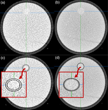
Among the five high-contrast scan protocols, low-contrast resolution was 2 mm at 0.6 HU difference from the background, and high-contrast resolution was 0.7 lp/cm for all. Changes in mean CNR and SNR for the protocols are illustrated in Figures 1 and 2, respectively. The highest mean CNR (76.7%) for protocol 5 was more than that of the standard protocol, while the highest mean SNR for protocol 4 was slightly higher than that of protocol 5 (0.69 vs. 0.59) whose value was approximately 66.5% higher than the standard protocol. Furthermore, as depicted in Figure 3, maximum noise reduction (59.2%) was also observed in protocol 4, although the noise level of protocols 4 and 5 was approximately identical (13.26 vs. 13.53). Figure 4 shows a comparison of image quality of protocols 4 and 5 with that of the standard protocol, and Figure 5 shows the identification of low-contrast objects in protocol 5 and the standard protocol, and their contouring pattern. The real area of the objects was about 490.5 mm2, while the areas identified by the standard and No. 5 protocols were about 370 and 485 mm2, respectively. Thus, the maximum error rates in the identification of low-contrast objects by the standard and No. 5 protocols were about 25% and 0.1%, respectively.
Mean contrast-to-noise ratios of protocols using the low-contrast detectability module of the ACR phantom.

Mean signal-to-noise ratios of protocols using the low-contrast detectability module of the ACR phantom.

Mean noise levels of protocols using the image uniformity module of the ACR phantom.

Reconstructed images with kernel FC26 using the low-contrast detectability module of the ACR phantom. (a) Standard protocol, (b) protocol 5 with CT dose index (CTDI) = 60.8 mGy, and (c) protocol 4 with CTDI = 59.5 mGy.

Image quality of a low-contrast object for (a) the standard protocol and (b) protocol 5. The solid line in (c) and (d) represents the real border of the object. The dashed line in (c) represents contouring of the object using the standard protocol. The dotted line in (d) represents contouring of the object using protocol 5.

Discussion
To increase the accuracy of GTV contouring, CT scan parameters in radiotherapy could be optimised to improve the overall treatment response. Reference Shuryak, Hall and Brenner32 In routine CT scanning for brain tumours, the surrounding normal tissue and oedema are barely discriminated from the tumour, so in treatment planning for brain radiotherapy, the accurate identification of tumour border remains a challenging issue. To ensure sufficient irradiation inside the subclinical microscopic areas of regional infiltration, as well as taking into account patient positioning errors, clinical target volume and planning target volume are added to the GTV, where the volume depends on the type and anatomical site of the tumour, clinical systematic and random errors and also image quality. Any error in GTV contouring could lead to underdosing to the tumour and overdosing to normal tissues. As a result, the patient might suffer severe complications or recurrence after a few months. In the present study, the main aim was, firstly, to reduce dose delivery uncertainties in radiation therapy. For this purpose, the study was implemented in two steps. First, the effects of various CT scan parameters on low-contrast detectability were evaluated, and then the accuracy of tumour volume identification and contouring using a standard image quality protocol was compared with that of five IQE protocols capable of producing high-contrast CT images of soft tissues. In our study, among the IQE protocols, protocol 5 had the highest effective dose (2.5 mSv) compared to the standard protocol (0.5 mSv). Using high-dose parameters in CT simulation can improve not only tumour identification but also fusion accuracy of CT with MRI or PET images. Reference Chen, Noid and Tai5,Reference Hevezi and Mahesh14 In IGRT where fast modification of volume changes may be necessary during treatment, a high-quality image plays a vital role in rapid, real-time tumour contouring. Reference Chen, Noid and Tai5 At first glance, it may seem that we barely increased the standard scan parameters to higher dose parameters, but it should be noted that the ALARA principle considers imaging and treatment simultaneously in radiotherapy. Reference Murphy, Balter and Balter15 Therefore, when the total dose of both imaging and radiotherapy is considered, the benefits of a slight increase in imaging dose (which is negligible in a very high-dose radiotherapy process) may be clearly justified. Reference Chen, Noid and Tai5
Recently, studies have concluded that CNR and SNR are critical image quality parameters for contouring accuracy and precision. Reference Chen, Noid and Tai5 However, few studies have reported CNR improvement in high-dose scan parameters for radiotherapy, and the effect of enhanced CNR and SNR on GTV contouring has not been clearly demonstrated. In our study, the maximum CNR of 2.8 was reported in protocol 5, while the SNR of protocol 4 was slightly higher than that of protocol 5. Noise level is another important parameter that can affect the details of low-contrast objects, eventually leading to a decline in overall image quality. Reference Ween, Kristoffersen, Hamilton and Olsen33 In the present study, the noise level of protocol 4 was reduced by 59.2% compared to the standard protocol, and spatial resolution in all the protocols was 0.7 lp/mm. In general, it is clear that the noise level barely affects spatial resolution and contrast, which can consequently ruin the diagnostic perception of the image. It has been reported that image noise decreases inversely by the square root of the dosage, Reference Verdun, Racine and Ott23 but in our study, we observed that spatial resolution was rarely affected by dosage. It could be concluded that an objective evaluation of image noise effect on spatial resolution in phantom studies is deficient. The soft tissue reconstruction kernels of CT algorithms eliminate information concerning high spatial frequency and thus create images with low levels of noise and spatial resolution. Reference Verdun, Racine and Ott23 The low tube voltage and high tube current–time product of protocol 5, compared to those of the standard protocol, caused a high increase in CNR. Chen et al. proposed a moderate tube voltage of 100–120 kVp and an extremely high tube current–time of 1000 mA to improve CNR for abdominal and pelvic CT scans, but high tube loadability (high milliamperes) is not possible for some CT systems and, if implemented, is rarely applicable in clinics due to early depreciation of the system. Reference Chen, Noid and Tai5 The maximum SNR in our study was achieved using protocol 4, which may have been due to a slight increase in tube voltage leading to noise reduction and, eventually, SNR improvement. Consistent with this result, Karmazyn et al. found that noise level increased from 0.08 to 0.11 when tube voltage was decreased from 120 to 100 kVp. Reference Karmazyn, Liang, Klahr and Jennings34 In the study of Boas and Fleischmann who focused on CT artifacts, by increasing the tube current from 60 to 440 mA, the noise level reduced 2.7 times. Hence, the authors concluded that by gradually increasing the noise level, the images become increasingly blurred, obscuring soft tissue boundaries. Reference Boas and Fleischmann35 Hence, increasing the tube current could be a useful way to cope with image blurring, because of a possible reduction in Poisson noise. Hernandez-Giron et al. investigated the effect of different tube voltages and demonstrated that a tube voltage of 80 kVp could greatly improve low-contrast detectability. Reference Hernandez-Giron, Calzado, Geleijns, Joemai and Veldkamp22 The drastic effect of tube voltage on low-contrast detectability improvement was also observed in our study. Although the noise levels of protocols 4 and 5 were approximately the same, the CNR of protocol 5 with low tube voltage was about 42% higher than that of protocol 4.
The identification of GTV border and distinguishing it from OARs, especially in soft tissues such as the brain, is one of the serious challenges in treatment planning. In the present study, the contouring performance of protocol 5 was decided to be evaluated because of its highest CNR. In the subjective analysis, the accuracy of low-contrast circular object contouring was tested using the images created by the standard protocol and protocol 5. The error rate in contouring using the standard protocol was 25%, while for protocol 5 it was only 1%. This may be because of the high CNR and low noise level of images produced by protocol 5. Although CT plays a big role in cancer diagnosis, Reference Farhood, Raei and Ameri36 extensive research is needed to address and eliminate its potential faults.
This pilot study has some limitations. First, image quality parameters were assessed by a phantom based on an objective analysis. Then, a subjective analysis was performed to evaluate the contouring accuracy of low-contrast circular objects. Tube loadability, in order to create high-exposure conditions, is another limitation of this study. Exposure conditions were selected so that they can be practically implemented in the clinic. We used an iterative algorithm in this study. Although a filtered back-projection algorithm is known to improve CNR significantly, the procedure may be time-consuming. For a patient immobilised because of fixation, long-term CT imaging may not be possible. For verification of our findings, further studies should be conducted using the filtered back-projection algorithm.
Conclusion
CT image quality can be optimised using high-dose parameters to provide better soft tissue contrast for treatment planning in radiotherapy. We demonstrated an improvement in the accuracy of GTV identification by this approach.
Acknowledgement
This study was approved by the Research Ethics Committee of Tabriz University of Medical Sciences (code: IR.TBZMED.VCR.REC.1398.202).
Conflict of Interest
None.