Overview
There are a wide variety of therapeutic biomedical devices that require reliable, stable, chronic interfaces between inert, abiotic, engineered electronic components, and living tissue. Examples include cochlear implants,[Reference Wilson and Dorman1] retinal prostheses,[Reference Weiland, Cho and Humayun2] pacemakers,[Reference Norlin, Pan and Leygraf3] deep brain stimulators,[Reference Gimsa, Habel, Schreiber, van Rienen, Strauss and Gimsa4] and microfabricated cortical electrodes.[Reference Drake, Wise, Farraye, Anderson and BeMent5] These bionic devices have already had an extraordinary impact on the quality of life for hundreds of thousands of individuals. They have been able to restore some measure of hearing to the deaf[Reference Wilson and Dorman1] and sight to the blind.[Reference Weiland, Cho and Humayun2] They can also help manage the debilitating tremor associated with Parkinson's disease.[Reference Kringelbach, Jenkinson, Owen and Aziz6] Other medical conditions that might one day be treated with electronic implants include drug-addiction, obsessive-compulsive disorder, obesity, and depression.[Reference Schlaepfer, Bewernick, Kayser, Hurlemann and Coenen7]
Efforts are also actively underway to develop next-generation prosthetic limbs that will be directly integrated with peripheral nerves and muscles.[Reference Kung, Langhals, Martin, Cederna and Urbanchek8] One strategy recently proposed for this purpose is the hybrid Regenerative Peripheral Nerve Interface (RPNI) device (Fig. 1) that involves the use of an extracted piece of free muscle tissue that acts as a local target for the sprouting nerve endings. As shown in Fig. 1, the overall design of the RPNI takes into account the dramatic changes in mechanical properties and mechanisms of charge transport between living neurons and myocytes (soft, ionic conductors), through an ionically and electronically conducting polymer [such as poly(3,4-ethylene dioxythiophene), or PEDOT]-coated regenerative matrix, and then on to metal electrodes (stiff, electron conductors). The RPNI promotes the formation of new neuromuscular junctions between the nerves and muscle, leading to a stable, functional, and biological interface. This has the extra benefit of dramatically suppressing neuroma formation near the limb stump, a common problem for nearly all amputees.[Reference Kung, Langhals, Martin, Cederna and Urbanchek8] The RPNI approach is essentially a micro-scale version of the targeted muscle reinnervation surgery developed by Kuiken.[Reference Kuiken9] Given the rate of recent progress, it is reasonable to anticipate that amputees will soon be able to directly manipulate prosthetic devices through bionic interfaces with their efferent (motor) nerves and to extract information about the environment through interfaces to their afferent (sensory) nerves.[Reference Kung, Langhals, Martin, Cederna and Urbanchek8]
Schematic diagram of the design of a RPNI.[8] The RPNI has been proposed as a means for more reliably interfacing metallic biomedical electrodes with living neural and muscle tissue. As shown in the schematic, the properties of the materials at the interface are between those of the metal electrode and the biological tissue.

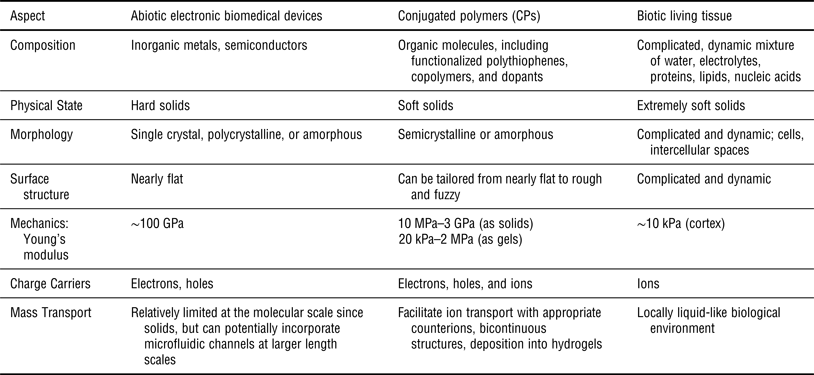
The general concept of integrating various types of engineered electronic components with living systems to achieve desirable therapeutic outcomes has been described as “bioelectronic medicine”.[Reference Birmingham, Gardinaru, Anikeeva, Grill, Pikov, McLaughlin, Pasricha, Weber, Ludwig and Famm10] In all of these applications, there is a need to reliably interface an electrically active, solid, abiotic device with an ion-rich, living, water-laden, dynamic biological environment. The optimization of novel materials that might improve the performance of these components therefore needs to take many different design factors into consideration (Table I). The active components of electronic biomedical devices are constructed from single-crystalline semiconductors (silicon) or polycrystalline metals such as stainless steel, platinum, gold, or iridium.[Reference Merrill, Bikson and Jefferys11] These are all hard inorganic solids, with typical elastic moduli of the order of ~100 GPa. Their surfaces are usually relatively flat, and charge transport is accommodated by solid-state conduction of electrons or holes. Local mass transport through a conventional metal or semiconductor is relatively limited since these are fully dense solid substances, although it is possible to fabricate microfluidic channels for local fluid delivery or sampling near the device surface.[Reference Chen, Wise, Hetke and Bledsoe12] In contrast, living tissue consists of a complex, dynamic, wet mixture of both anionic and cationic electrolytes, along with a rich variety of organic biomolecules, including proteins, nucleic acids, and lipids. Most tissues are extremely soft when compared with conventional engineered materials, although the mechanical properties of course vary widely by tissue type and location, and they can also be quite anisotropic.[Reference Meyers, Chen, Lin and Seki13] As one example the typical elastic modulus of cortical brain tissue has been estimated to be of the order of a few kPa.[Reference Subbaroyan, Martin and Kipke14]
Materials design considerations for using conjugated polymers for interfacing abiotic, engineering electronic biomedical devices with biotic, living tissue. The chemistry, structure, and properties are intermediate to those of the devices and tissue.
As shown in Table I, conjugated polymers (CPs) are excellent candidate materials to consider using at the abiotic–biotic device–tissue interfaces inevitable in bioelectronic medical devices. Their composition, surface structure, and mechanisms of charge transport allow them to integrate well with both the solid substrate as well as the living tissue. Furthermore, the flexibility of organic materials chemistry and processing methods make it possible to precisely tailor CP composition, structure, and properties. The Young's modulus of CPs has been reported to vary over a large range depending on whether the material is in bulk solid or thin-film form (10 MPa–3 GPa),[Reference Lang, Naujoks and Dual15–Reference Yang and Martin17] or if it is embedded into a gel matrix (20 kPa–2 MPa).[Reference Aouada, Guilherme, Campese, Giroto, Rubira and Muniz18–Reference Cho, Wu, Shim, Kuan, Mastroianni, Young, Kuo, Epps III and Martin21]
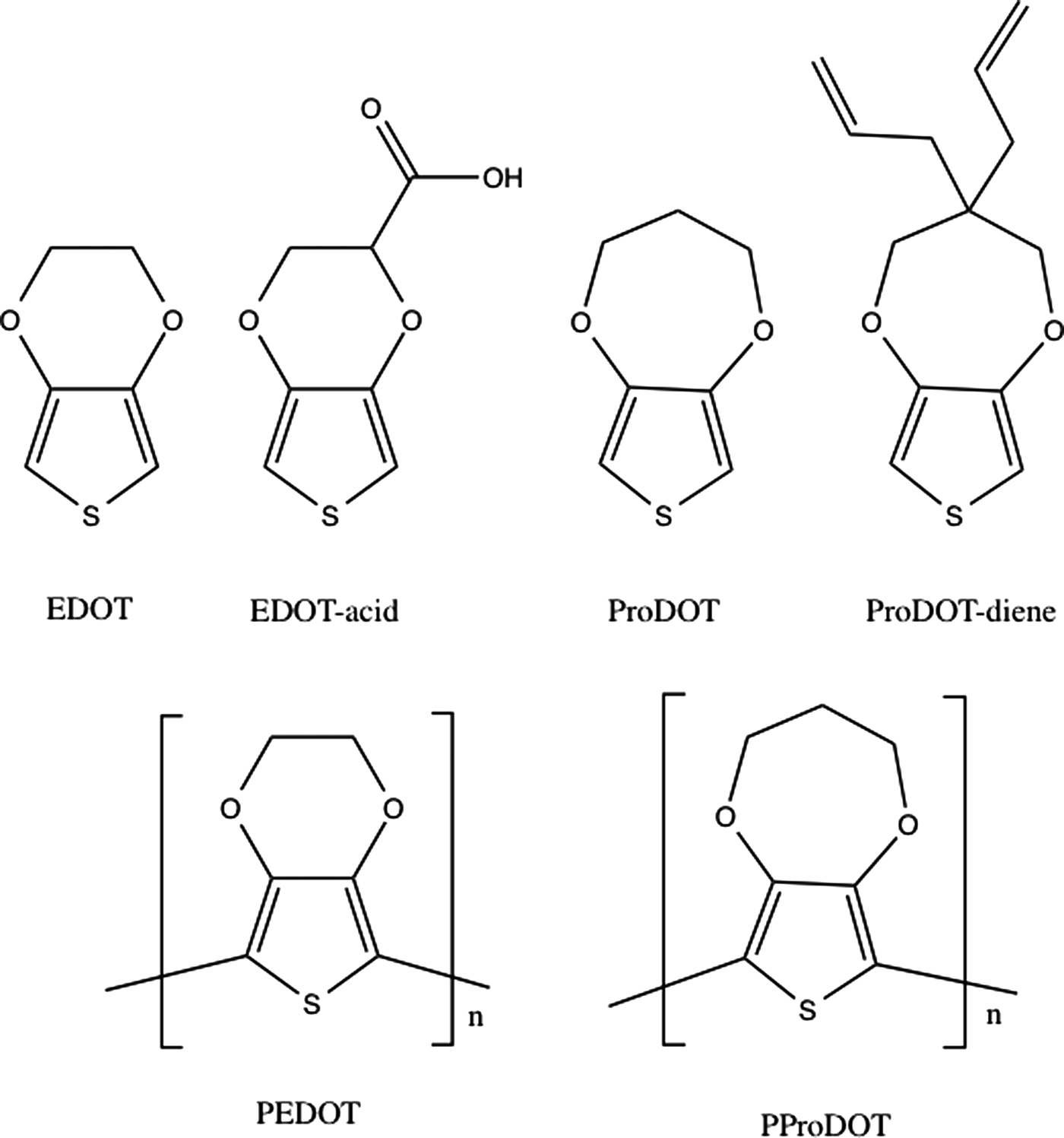
CPs have alternating single and double bonds along their molecular backbone; this leads to the extended pi-bond formation that facilitates charge conduction along the chain as well as relatively easy lateral charge hopping between molecules, particularly if they can locally arrange themselves in a face-to-face stacking geometry that accommodates pi–pi orbital overlap. Common examples of CPs include poly(aniline), poly(pyrrole), and poly(thiophene).[Reference Skotheim and Reynolds22] Much recent interest has focused on the alkoxy-substituted poly(thiophenes) such as PEDOT and poly(3,4-propylene dioxythiophene) (PProDOT) (Fig. 2), because they have been shown to have extremely high chemical stabilities and excellent charge transport properties. The chemistry of CPs can be readily tailored to optimize performance by introducing specific functionality onto the molecular backbone. They can also be processed into a variety of interesting morphologies that make it possible to refine their properties to meet specific design requirements.
Chemical structures of EDOT, carboxylic-acid functionalized EDOT (EDOT-acid), ProDOT, and di-propylene functionalized ProDOT (ProDOT). The chemical structures of the corresponding polymers formed from EDOT (PEDOT) and ProDOT (PProDOT) are shown at the bottom of the figure.
In this review paper, we describe the molecular design, synthesis, and characterization of conjugated poly(thiophene) copolymers that have shown particular promise for use at biomedical device—tissue interfaces. The EDOT and ProDOT monomers each have only two reactive hydrogens located in the 2,5 positions on opposite sides of the sulfur atom in the conjugated thiophene ring (Fig. 2). This insures that the resulting PEDOT and PProDOT polymer chains remain fairly linear. It also means that neither PEDOT nor PProDOT have residual hydrogen atoms remaining directly adjacent to the conjugated molecular backbone, providing improved chemical stability. Finally, the two oxygen atoms on the thiophene ring from each monomer unit contribute additional electron density to the conjugated molecular backbone, leading to relatively high levels of charge conductivity.
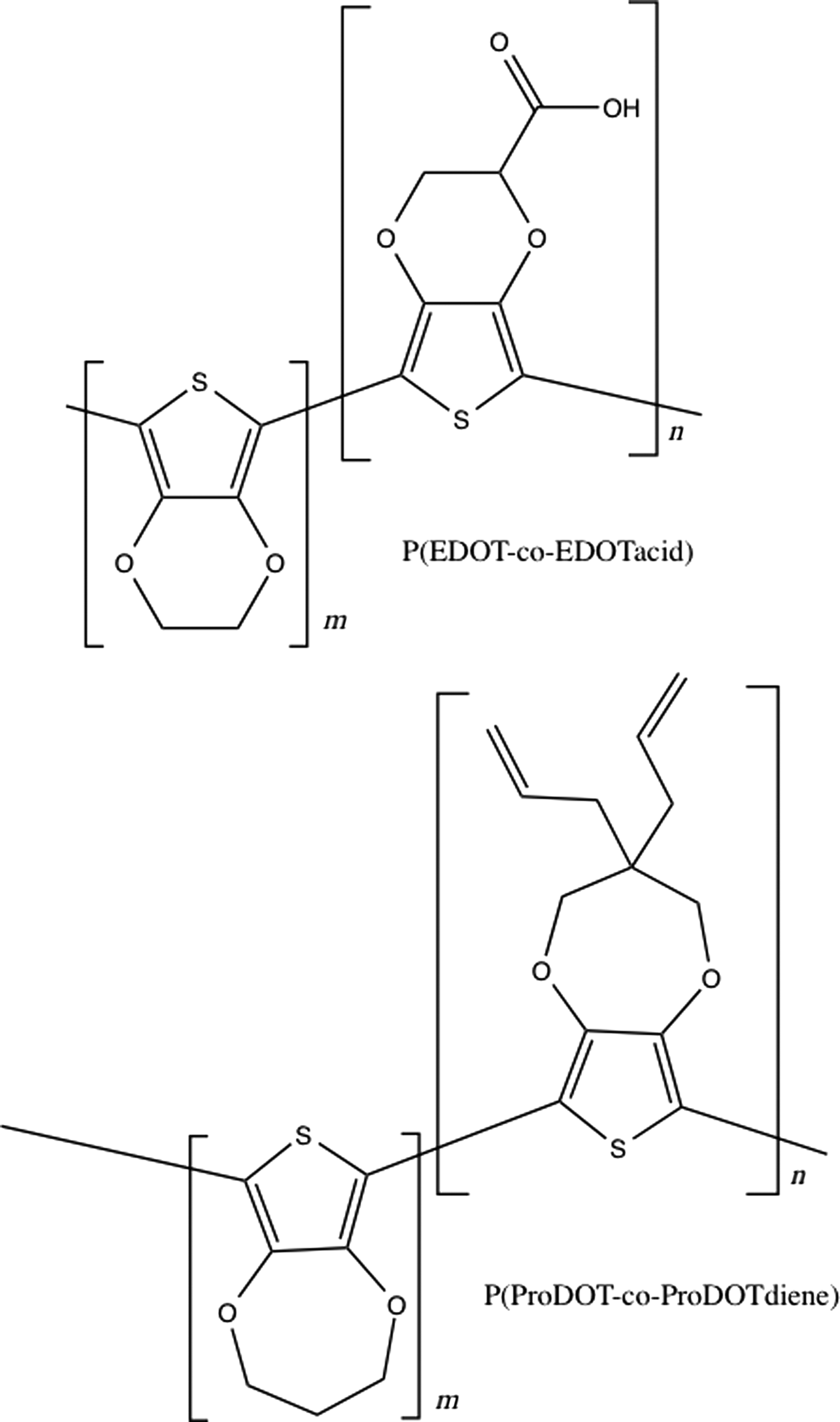
Although the PEDOT and PProDOT homopolymers themselves have already shown considerable potential for use in a variety of biomedical devices, there are some important technical issues that remain, including their relatively low resistance to fracture, limited adhesion to solid substrates, and lack of specific interactions with particular types of tissue. The chemistry of PEDOT and PProDOT can be systematically manipulated by the copolymerization of the EDOT and ProDOT monomers with suitably functionalized comonomers. Specific examples of these include EDOT-acid, a carboxylic functionalized EDOT that can be used to make P(EDOT-co-EDOTacid) copolymers, and ProDOT-diene, a dialkene functionalized ProDOT that can be used to make P(ProDOT-co-ProDOTdiene) copolymers (Fig. 3). The ability to systematically tailor the chemical composition makes it possible to tailor interactions with both solid substrates and living systems. These monomers also make it feasible to conduct post-polymerization modifications on thin films of these materials after they have been deposited onto solid substrates. We will describe some recent experimental results in the design, synthesis, and characterization of these and other functionalized poly(thiophene) copolymers. We will also discuss some of the newest molecular designs of other comonomers and their corresponding copolymers that are of continued current interest to us and other research groups.
Copolymers prepared by mixing together EDOT and EDOT-acid: PfEDOT-co-EDOT-acid); and ProDOT and ProDOT-diene: P(ProDOT-co-ProDOT-diene).
Background
CPs are either being actively considered for or are already used in a number of important technological applications, including organic photovoltaics, light-emitting diodes, batteries, corrosion mediation, and electrochromic displays.[Reference Kirchmeyer and Reuter23] The oxidation reaction required to convert conjugated organic monomers into polymers may be performed either with appropriate reagents such as Lewis acids (chemically) or by with an externally applied current (electrochemically). The resulting polymers show charge conductivity when they are slightly oxidized; this is achieved by introducing negatively charged counter ions during synthesis. Typical counter anions that have been used include synthetic polymers (PSS), small molecules (LiClO4), or anionic biomolecules (heparin).[Reference Skotheim and Reynolds22, Reference Asplund, von Holst and Inganäs24]
There are a number of excellent reviews that give additional background on conjugated thiophene copolymers and their use in specific applications. Groenendall et al. described the synthesis and characterization of alkoxy-functionalized thiophene copolymers such as PEDOT and PProDOT in two comprehensive manuscripts.[Reference Groenendall, Jonas, Freitag, Pielartzik and Reynolds25, Reference Groenendaal, Zotti, Aubert, Waybright and Reynolds26]
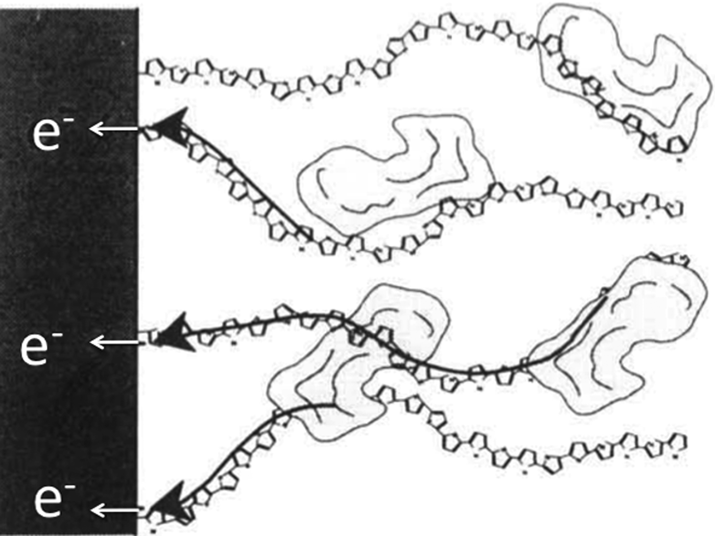
The potential of CPs for interfacing electronic biomedical devices with living systems was discussed by Gopel and Heiduschka in 1994.[Reference Göpel and Heiduschka27] Some of the fundamental limitations for the use of these materials in chemical sensor applications were also described.[Reference Göpel28] These relatively early conceptual papers considered the importance of both the electronic and ionic mechanisms of charge transport, and the need to interface the polymer to the solid metal electrode as well as functional molecules such as proteins present in the biological media. The authors described the possibility of CPs acting as “molecular wires” that could facilitate communication between a fabricated sensor and the surrounding environment (Fig. 4).
Schematic showing polythiophene chains acting as “molecular wires”, connecting biological media with a metal or semiconducting solid electrode. From Göpel.[Reference Göpel28] Reproduced with permission.
The use of the CPs for stimulating neurite outgrowth on electrochemically deposited polypyrrole was investigated by Schmidt et al.[Reference Schmidt, Shastri, Vacanti and Langer29, Reference Shastri, Schmidt, Langer and Vacanti30] They found that PC-12 neural cells showed enhanced neural outgrowth when grown on the electrically active substrates. The use of biomimetic design strategies for interfacing electrically active materials with the nervous system were discussed by Winter and Schmidt.[Reference Winter, Schmidt, Dillow and Lowman31]
Cui investigated the electrochemical deposition of both pyrrole and PEDOT on microfabricated neural probes intended for chronic implantations in cortex, and found substantially improved chemical stability of PEDOT as compared with polypyrrole.[Reference Cui, Hetke, Wiler, Anderson and Martin32–Reference Cui34] The PEDOT-coated electrodes showed a dramatic reduction (~two orders of magnitude) in the impedance at biologically important temporal frequencies of 1 kHz and less; this was associated with formation of an open, fuzzy surface morphology that accommodates rapid ion transport.[Reference Cui and Martin35] It was also demonstrated that bioactive peptides, including the YIGSR adhesion sequence from laminin could be incorporated into the films during deposition.[Reference Cui and Martin35]
Corey et al. showed that oriented assemblies of electrospun nanofibers directed the neurites from dorsal root ganglia to regenerate in a directionally controlled manner.[Reference Corey, Lin, Martin and Feldman36] Abidian et al. showed that PEDOT could be formed into nanotubes by electrochemically polymerizing around electrospun nanofibers. The insides of these nanotubes could be loaded with specific drugs of therapeutic interest such as dexamethasone.[Reference Abidian, Kim and Martin37] An external bias was used to control the release of this inflammatory agent from the resulting PEDOT nanotubes. Oriented nanotubes of PEDOT were created by incorporating EDOT monomer into the nanofibers during processing, and then exposing the fibers to a chemical oxidant after the fibers were oriented into a controlled assembly.[Reference Feng, Wu, Cho, Leach, Franz, Naim, Gu, Corey and Martin38] These oriented PEDOT nanotubes proved to be highly effective at controlling the directed regeneration of neurites (Fig. 5).
(a) Confocal image of DRG neurites cultured on aligned electrospun PEDOT nanofibers. Beta-tubulin (green), a structural component of neurons, indicates that the DRG neurites followed the horizontal direction of fiber alignment, (b) Schwann cells are identified by co-localization of s100 (red) and DAPI (blue). From Feng et al.,[Reference Feng, Wu, Cho, Leach, Franz, Naim, Gu, Corey and Martin38] Reproduced with permission.
The use of conjugated organic materials in biomedical devices has been the subject of a number of recent reviews. Kim et al. described the use of CPs in biomedical devices, with a particular emphasis on cortical microelectrodes.[Reference Kim, Richardson-Burns, Povlich, Abidian, Spanninga, Hendricks, Martin and William39] Rozlosnik reviewed the use of PEDOT and its derivatives in biomedical sensors.[Reference Rozlosnik40] Povlich et al. described the development and use of electroactive polythiophene copolymers as biomaterials.[Reference Povlich, Ducheyne, Healy, Hutmacher, Grainger and Kirkpatrick41] The use of various organic bioelectronic materials in nanoscale medical components was reviewed by Svennersten et al.[Reference Svennersten, Larsson, Berggren and Richter-Dahlfors42] and more recently by Rivnay et al.[Reference Rivnay, Owens and Malliaras43] The development and implementation of organic materials for bionic devices was the focus of a monograph by Wallace et al.[Reference Wallace, Moulton, Higgins and Kapsa44] Otero and Martinez discussed the similarities between swollen systems of CPs and intracellular matrix, and how they could be exploited to make actuators and sensors.[Reference Otero, Alfaro, Martinez, Perez and Martinez45] Fattahi et al. recently reviewed the use of CPs at neural interfaces.[Reference Fattahi, Yang, Kim and Abidian46] Liao et al. described the use of flexible organic electronic components for interfacing devices with biological systems.[Reference Liao, Zhang, Yao, Hua, Li and Yan47] Balint et al. discussed the use of CPs in tissue engineering.[Reference Balint, Cassidy and Cartmell48] Recent developments in materials chemistry and processing for organic bioelectronics applications were described by Molino and Wallace.[Reference Molino and Wallace49]
For materials that interface electronic components with tissue, the ability to accommodate charge transfer between solid metals and semiconductors and soft electrolytic tissue is critical. While solid-state electronics, microelectronics, and nano-electronic devices are now well developed, there has much less attention paid to analogous devices prepared from ionically transporting materials, or “iontronics”. Several organic electronic, iontronic devices have now been designed, including ion pumps for controlling delivery of pharmacological agents to cells[Reference Isaakson, Kjäll, Nilsson, Robinson, Berggren and Richter-Dahlfors50] and logic gates.[Reference Tybrandt, Forchheimer and Berggren51] We expect that future efforts will reveal many interesting analogies between systems that facilitate charge transport ionically, and their more well-established electronic counterparts. Some of these issues have been addressed in a recent monograph.[Reference Leger, Berggren and Carter52]
Morphology control
The regular, linear structure of the PEDOT molecule leads to a reasonably ordered, semicrystalline morphology in the solid state.[Reference Martin, Wu, Shaw, King, Spanninga, Richardson-Burns, Hendricks and Yang53] The PEDOT crystals have the conjugated chains oriented and planar, organized into individual layers with face-to-face packing between molecular backbones. However, the degree of order varies considerably depending on the detailed method of synthesis, processing, and choice of counterion. The detailed relationship between solid-state structure and transport properties of CPs is under active investigation.[Reference Salleo, Kline, DeLongchamp and Chabinyc54–Reference Noriega, Rivnay, Vandewal, Koch, Stingelin, Smith, Toney and Salleo57] It has been generally thought that higher degrees of order are important because of the need to promote π – π interactions between stacks of the conjugated molecular backbones. However, it has been found that in high molecular weight polymer materials, only small amounts of local aggregation are sufficient for obtaining efficient charge transport.[Reference Noriega, Rivnay, Vandewal, Koch, Stingelin, Smith, Toney and Salleo57] Recent efforts have found that with synthetic designs that keep the molecular backbone conformation relatively planar and torsion-free, even highly disordered CPs can show excellent transport properties.[Reference Venkataraman, Nikolka, Sadhanala, Lemaur, Zelazny, Kepa, Hurhangee, Kronemeijer, Pecunia, Nasrallah, Romanov, Broch, McCulloch, Emin, Olivier, Cornil, Beljonne and Sirringhaus58]
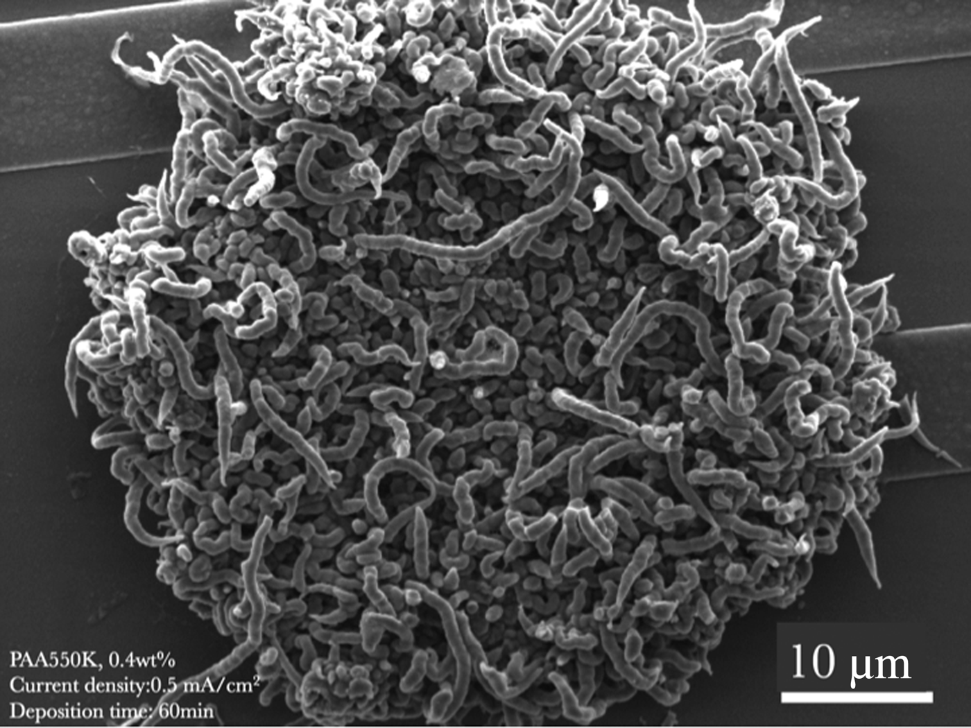
At somewhat larger (100–1000 nm) length scales, the structure of PEDOT and other CPs can be precisely controlled by manipulating the composition of the reaction mixture and the methods of deposition. For example, nanofibrils of PEDOT can be created by introducing high molecular weight poly(acrylic acid) (PAA) during deposition. Figure 6 shows an example using 550 kg/mol PAA at a concentration of 0.4 wt%. The fuzzy morphology leads to an extremely high effective surface area, resulting in low impedances and soft mechanical properties.[Reference Yang and Martin17] Although reproducible, the mechanism of nanofibril formation is not yet firmly established, but is likely due to coating of growing PEDOT filaments with PAA during the electrodeposition process.[Reference Yang, Lipkin and Martin59]
Nanofibrils of PEDOT prepared by electrochemical polymerization with 0.4 wt% 550 kg/mol polvlacrylic acid) (PAA). From Yang et al.[Reference Yang, Lipkin and Martin59] Reproduced with permission.
More regular nanoscale structures can be obtained using removable template systems that are able to self-assemble into ordered arrangements with a higher degree of structural perfection. For example, polystyrene latex spheres with diameters from 30 nm up to several microns or more are now readily commercially available.[Reference Sharp and Beard60] These can be deposited into crystalline aggregates from aqueous suspensions onto surfaces, and the PEDOT then grown out electrochemically into the interstitial spaces between the particles (Fig. 7).[Reference Yang and Martin61, Reference Yang and Martin62] The more recent development of the so-called particle replication in non-wetting templates (PRINT) process by DeSimone and co-workers has led to a rich set of complicated, asymmetric shapes, including rectangular prisms, toroids, and boomerangs.[Reference Euliss, DuPont, Gratton and DeSimone63–Reference Merkel, Herlihy, Nunes, Orgel, Rolland and DeSimone65] These new particle geometries could readily be used in a similar manner to template CP materials into even more intricate and potentially useful microstructures.
Porous PEDOT structures made by polymerizing around PS latex spheres with diameters of (a) 89 nm, (b) 100 nm, (c) 300 nm, and (d) 485 nm.
At somewhat smaller length scales (1–10 nm), ordered phases can be created from non-ionic surfactants and block copolymers. Hulvat and Stupp showed that these liquid crystalline ordered phases could be used to template the structure of CPs.[Reference Hulvat and Stupp66, Reference Hulvat and Stupp67] Yang et al. used a similar approach to deposit ordered structures of PEDOT on microfabricated neural electrodes.[Reference Yang, Kim, Hendricks, Leach, Northey and Martin68] Cho et al.[Reference Cho, Wu, Shim, Kuan, Mastroianni, Young, Kuo, Epps III and Martin21] recently described the formation of bicontinuous cubic structures of PEDOT with the gyroid symmetry using a non-ionic surfactant (NP-10) (Fig. 8). The bicontinuous nature of these ordered phases has been shown to be important for applications such as electroactive actuators since different types of electron and ionic transport can be accommodated simultaneously.[Reference Kim, Kim, Park, Hwang and Park69]
Schematic diagram of bicontinuous phases with gyroid (GYR) symmetry. The original NP-10 surfactant phase (top) is loaded with EDOT monomer (middle) and then polymerized to PEDOT with a mild oxidant (bottom). Reproduced from Cho et al.[Reference Cho, Wu, Shim, Kuan, Mastroianni, Young, Kuo, Epps III and Martin21] with permission from the PCCP Owner Societies.
Designer materials with open, regular, interconnected networks having regular crystal structures controlled at molecular length scales (0.1–1 nm) can be created from hybrid combinations of metals and rigid organic linkers. These materials are known as metal-organic frameworks (MOFs).[Reference Chen, Eddaoudi, Hyde, O'Keeffe and Yaghi70] The open structure of the MOF reticulated network is of considerable interest for catalysis, gas storage, and drug delivery. Recent studies have incorporated conjugated moieties into MOF crystals, suggesting that may also be able to facilitate charge transport.[Reference Feng, Chen, Honsho, Saengsawang, Liu, Wang, Saeki, Irle, Seki, Dong and Jiang71–Reference Feng, Ding and Jiang73]
In the future, it will likely be necessary to precisely control the morphology of CPs for biomedical devices at several different length scales simultaneously. The ability to create hierarchical structures at several dimensions at once has already been demonstrated for certain types of oxides.[Reference Yang, Deng, Zhao, Feng, Pine, Chmelka, Whitesides and Stuckey74] An interesting example of a processing method that can simultaneously control CP morphology at two different length scales is surface self-assembly of silica spheres in combination with laser interference patterning.[Reference Yuan, Lasagni, Hendricks, Martin and Das75–Reference Lasagni, Hendricks, Shaw, Yuan, Martin and Das77] The combination of the laser interference pattern with the near-field focusing of the radiation provided by the spheres leads to controlled structures in the PEDOT at both micron and sub-micron length scales (Fig. 9). For some applications, it is also important to create structures that scale in a self-similar, branched manner, so that mass transport and charge transport can both be accommodated effectively across the interface.[Reference Hong, Hwang, Kye, Myung, Won and Huh78] Another area ripe for future exploration is the opportunity to control the length scale of the microstructure at different points in space, perhaps leading to films with large pores at the surface to accommodate integration with tissue, with smaller holes near the solid substrate to promote adhesion.
Controlling surface morphology of PEDOT at two different length scales using a thin film of self-assembled silica nanospheres and a laser interference pattern with variable fluence. The damage threshold fluence was 300 mJ/cm2. At high fluence (top), the resulting pattern consisted of thin lines of spheres, with gaps containing holes from the focused laser. At lower fluence, the gaps were smaller and less well defined. From Yuan et al.,[Reference Yuan, Lasagni, Hendricks, Martin and Das75] Reproduced with permission.
PEDOT in hydrogels
Hydrogels are crosslinked, water-swollen gels of hydrophilic polymers that have found widespread use in tissue engineering and drug delivery.[Reference Peppas and Langer79, Reference Peppas, Bures, Leobandung and Ichikawa80] CPs can be grown into hydrogels[Reference Kim, Abidian and Martin81] (Fig. 10), creating materials systems that are both electrically and ionically conductive, as well as mechanically robust.[Reference Green, Hassarati, Goding and Baek20, Reference Poole-Warren, Lovell, Baek and Green83–Reference Green, Matteucci, Hassarati, Giraud, Dodds, Chen, Byrnes-Preston, Suaning, Poole-Warren and Lovell85] It has recently been shown that by carefully controlling the rates of chemical polymerization and precipitation from solution of CPs during synthesis, swollen gels with a high degree of elasticity (with strains to failure over 70%) can be obtained, even without the addition of a hydrophilic polymer matrix.[Reference Lu, He, Cao, Guo, Zhang, Li, Shao, Cui and Zhang86]
PEDOT grown through a hydrogel, through an electrospun mat of fibers deposited on a cortical microelectrode. (a) top view. (b) side view. (c) SEM image. (d) high magnification SEM image. From Abidian et al.,[Reference Abidian and Martin82] reproduced with permission of John Wiley & Sons.
There are a wide variety of other synthetic options for hydrogels that could be functionalized with CPs in future studies. Examples include self-assembled peptides[Reference Nowak, Breedveld, Pakstis, Pine, Pochan and Deming87] and naturally derived polysaccharides such as hyaluronic acid.[Reference Jia, Yeo, Clifon, Jiao, Kohane, Kobler, Zeitels and Langer88, Reference Jha, Hule, Jiao, Teller, Clifton, Duncan, Pochan and Jia89] Considerable future potential is likely with the development of efficient, bioorthogonal chemistries that can readily functionalize these materials without disrupting the biologically active components.[Reference Zhang, Dicker, Xu, Jia and Fox90] Some initial results have shown that designer peptides can be used to control the spacing and orientation of conjugated chromophores, providing insight about the local molecular mechanisms of energy transport.[Reference Rothberg, Paquette, Rhinehart, McCamant, Kas, Charati, Galvin and Kiick91]
PEDOT and ProDOT copolymers
The electrochemical deposition of copolymers makes it possible to locally control their chemical composition, as shown schematically in Fig. 11. For example, consider polymerizations involving both EDOT and EDOT-acid (Fig. 2). Depending on the composition of the reaction medium, the deposited polymer could be a PEDOT homopolymer (blue), a PEDOT-acid homopolymer (pink), or a PEDOT-co-PEDOT-acid copolymer with some intermediate, tailorable composition (purple). The composition could also be changed during the progress of the reaction, leading perhaps to a systematic change in color (from blue to purple to pink) or even alternating layers (pink and blue) (Fig. 11).
Schematic diagram of the possibilities provided by the ability to electrodeposit PEDOT copolymers with various compositions at specific points on a given device. Consider a single comonomer such as the EDOT-acid molecule shown in Fig. 2. The polymer deposited on an individual electrode could be PEDOT homopolymer (blue), the PEDOT-acid homopolymer (pink), or some intermediate composition (purple). The composition could also be changed during deposition, perhaps leading to a systematic gradient in composition or a series of alternating compositions (top).
The PEDOT and ProDOT homopolymers are relatively hydrophobic due to the non-polar substituents on side of the thiophene ring. It is possible to tailor the surface chemistry of the final CP by introducing comonomers with other functional groups.[Reference Groenendall, Jonas, Freitag, Pielartzik and Reynolds25] Copolymers of PEDOT with sulfonatoalkoxy- and hydroxymethylated-PEDOT were shown to be compatible with cells, and able to significantly reduce the impedance of microfabricated electrodes.[Reference Xiao, Cui and Martin92, Reference Xiao, Martin, Cui and Shenai93] They also made it possible to incorporate biofunctional peptides, such as the CDPGYIGSR sequence from laminin.[Reference Cui, Lee, Raphael, Wiler, Hetke, Anderson and Martin33, Reference Xiao, Martin, Cui and Shenai93]
Luo et al. examined a number of functionalized EDOT monomers derived from methyl alcohol functionalized (EDOT-OH), including carboxylic acids, acetates, azides, N-hydroxysuccinimide, and found that they could all be readily deposited on surfaces using electropolymerization from aqueous microemulsions.[Reference Luo, Ali, Tansil, Yu, Gao, Kantchev and Ying94] Zhang showed that the carboxylic acid functionalized C4-EDOT-COOH monomer could be used to create high-quality films of interest for chemical and biological sensing applications.[Reference Zhang, Wen, Yao, Xu, Duan and Zhang95]
Povlich et al. described the synthesis and characterization of an EDOT monomer with the carboxylic acid moiety directly attached to the thiophene ring (EDOT-acid).[Reference Povlich, Cho, Spanninga, Martin and Kim96] Bhagwat et al. have recently shown that the EDOT-acid monomer makes it possible to precisely tailor the wetting properties of the resulting P(EDOT-co-EDOT-acid) copolymers in a systematic manner[Reference Bhagwat, Kiick and Martin97] (Fig. 12). It was found that copolymers of EDOT and EDOT-acid could be surface-functionalized with arginyl-glycyl-aspartic acid (RGD) peptides from extracellular binding domain of the integrin protein fibronectin[Reference Povlich, Cho, Leach, Kim, Corey and Martin98] (Fig. 13). Primary motor neurons exposed to the RGD functionalized surfaces showed enhanced adhesion and more advanced states of differentiation[Reference Povlich, Cho, Leach, Kim, Corey and Martin98] (Fig. 13).
By changing the composition of the copolymer, the surface wetting properties can he systematically tuned from hydrophobic to hydrophilic. From Bhagwat et al.[Reference Bhagwat, Kiick and Martin97] Reproduced with permission.
The number of primary neurons per square millimeter on conjugated polymer samples with various compositions. More cells attached to PEDOTacid with covalently bound ROD, and these cells were more highly differentiated. The PEDOT-acid films were found to be more resistant to cell adhesion than unmodified PEDOT, RGD blended into PEDOT, or the non-adherent peptide sequence RGE. From Povlich et al.,[Reference Povlich, Cho, Leach, Kim, Corey and Martin98] reproduced with permission from Elsevier.
Functionalized ProDOT monomers containing alkene side groups were described by Feldman and Martin.[Reference Feldman and Martin99] These alkenes make it possible to perform highly efficient “click” reactions when exposed to thiols in the presence of ultraviolet (UV) radiation.[Reference Feldman and Martin99] This synthetic route opens up many convenient opportunities for making new thiophene monomers, and for the surface functionalization of the corresponding CPs after deposition. To further increase the surface density of the alkenes, a difunctional ProDOT was synthesized and has been shown to allow a wide variety of different post-polymerization modifications with various thiols, including branched alkanes, ethylene glycols, and ferrocenes[Reference Wei, Ouyang, Liu and Martin100] (Fig. 14).
Chemical structure of ProDOT-diene monomer (1) and the PProDOT-diene polymer (PI). Various thiols (2a–c) including a branched ethyl-hexyl (2a), a hexyl ethylene glycol (2b), and alkyl-pentyl ferrocene (2c) were reacted with the ProDOT-diene polymer to give functionalized copolymers (P3a–c). From Wei et al.,[Reference Wei, Ouyang, Liu and Martin100] Reproduced with permission.
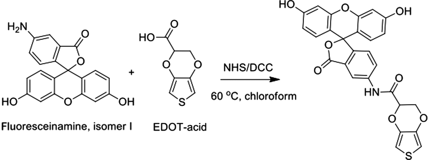
Another possibility we have investigated is the incorporation of fluorescent substituents. Here, our goal was to create a functionalized EDOT monomer that would have a strong visible light absorption, so that its diffusion could be monitored by optical microscopy. An example of a fluorescein functionalized EDOT-acid molecule is shown in Fig. 15.[Reference Ouyang101] Several other EDOT derivatives were also created in a similar fashion, including substituents corresponding to coumarin, Cascade Blue, and Alex Fluor 350.[Reference Ouyang101] Unfortunately, none of these monomers were able to be electrochemically polymerized. This is evidently because the large, bulky substituents dominate the resulting chemistry, rendering the monomer unreactive. Another issue is that these large, often polar side groups may make the corresponding polymers too soluble in water. The usual methods of electrochemical polymerization take advantage of the dramatic change in solubility as the conjugated monomers are converted from oligomers to polymers. If the side groups are too hydrophilic, then the resulting polymers will be too soluble to form solid precipitates on the anode surface. This might be overcome by changing solvents, but in many cases it is of interest to remain in an aqueous or even biological environment.
EDOT monomer modified with a fluorescent moiety. The EDOT-acid derivative was reacted with a fluoresceinamine isomer, leading to the funcationalized derivative shown at right. From Ouyang,[Reference Ouyang101] used with permission.
Polymerizations in living tissue
Given our observations that the electrochemical polymerization of PEDOT could be readily templated around a wide variety of both synthetic and biological materials, we investigated whether it would be feasible to grow the polymer around living cells. Indeed, we found that the electrochemical polymerization of PEDOT was possible both in vitro[Reference Richardson-Burns, Hendricks, Povlich, Foster, Kim and Martin102] and as well as in tissue slice culture.[Reference Richardson-Burns, Hendricks and Martin103] We then demonstrated that the process could be extended to living animals, using an electrode next to a microcannula to locally deliver the precursor monomer[Reference Ouyang, Feldman, Green and Martin104, Reference Wilks, Wooley, Ouyang, Martin and Otto105] (Fig. 16). We found that essential requirement to obtain best results was to precisely balance the rate of reactant delivery to the rate of polymerization.[Reference Ouyang, Feldman, Green and Martin104] The rationale behind this approach was to try and create a pathway for charge transport that would span across the reactive glial scar that is found after devices are implanted in tissue for extended periods of time.[Reference Biran, Martin and Tresco106]
Schematic diagram of the in situ polymerization of conjugated polymers into living neural tissue. The monomer (yellow) is delivered to the site with a microcannula. An insulated wire is used to electrochemically polymerize the PEDOT (blue) across the activated scar layer of glia and microglia (green), establishing electrical contact with the surrounding neural tissue (pink).
Recent results have shown that the in vivo deposition of PEDOT in the living rat hippocampus is indeed feasible and reproducible, and the treated animals do not lose a significant performance in a delayed alternation task that evaluates memory retention.[Reference Ouyang, Shaw, Liu, Griffin and Martin107] These results clearly demonstrate that the approach is technically possible, but several issues remain. Although the impedance of the electrodes was substantially decreased initially, the effect did not persist for a long periods of time. It was also observed histologically that there was still a secondary scarring effect from the locally deposited PEDOT.[Reference Ouyang, Shaw, Liu, Griffin and Martin107] Further studies are now needed to determine the optimal deposition conditions. Presumably slower reaction rates would be beneficial, but there are practical limits on how much time is available for a given animal between the initial surgery, PEDOT polymerization, and recording of neural activity. The use of functionalized EDOT or ProDOT comonomers is also possible; to date studies have only examined unmodified EDOT. The EDOT monomer is known to be cytotoxic at sufficiently high concentrations and extended exposure times.[Reference Richardson-Burns, Hendricks, Povlich, Foster, Kim and Martin102, Reference Richardson-Burns, Hendricks and Martin103] Presumably alternative functionalized EDOTs and ProDOTs (or other polymers) could be found that remain sufficiently chemically reactive, are able to facilitate charge transport, however, are also compatible with the eventual tissue target.
Controlling reaction conditions
During the electrochemical deposition of poly(thiophenes), a strongly oxidative environment develops around the working electrode (anode), while a reducing environment forms around the counter electrode. The local pH changes can be easily visualized by putting a pH-sensitive indicator dye into the reaction chamber. Figure 17 shows an example of one such experiment in a system where the reaction medium included an alginate hydrogel (0.6 wt%). The pH indicator was orange (strongly acidic, pH < 4) around the working electrode, and purple (strongly basic, pH > 10) region around the counter electrode (Fig. 17). It is remarkable to note the spatial extent of these changes, with the local region of pH change having a diameter of the order of 1 cm around each electrode.
Electrochemical polymerization of PEDOT from DI water with an alginate gel (0.6 wt%), 0.01 M EDOT, and 0.1 wt% PSS. Universal pH indicator shows strongly acidic (pH ~ 4) conditions near the working electrode, and basic conditions (pH ~ 10) near the counter electrode. Images taken by Chin-Chen Kuo.
While water is a convenient and inexpensive reaction medium for the electrodeposition of polythiophenes, there are often advantages with other solvents. For example, PEDOT films deposited from propylene carbonate show significantly improved adhesion to solid substrates and resistance to cracking. Propylene carbonate can also be used to deposit both EDOT and EDOT-acid copolymers,[Reference Bhagwat, Kiick and Martin97] and has shown advantages in improving the integrity of CP films used in chronic biomedical device applications.[Reference Kung, Langhals, Martin, Cederna and Urbanchek8] Ionic liquids have shown promise for the electrochemical deposition of polythiophenes, both as solvents.[Reference Lu, Fadeev, Qi, Smela, Mattes, Ding, Spinks, Mazurkiewicz, Zhou, Wallace, MacFarlane, Forsyth and Forsyth108, Reference Luo and Cui109] and as moieties incorporated into EDOT monomers directly.[Reference Döbbelin, Pozo-Gonzalo, Marcilla, Blanco, Segura, Pomposo and Mecerreyes110]
Failure modes
Although CPs have mechanical properties that are intermediate to those of inorganic solids and biological tissue, for long-term applications it will be important to establish that they are able to resist failure due to delamination, cracking, and repetitive loading (fatigue). Previous studies have shown that failure can occur by adhesive delamination at the interface between the CP and inorganic substrate, as well as internal cracking due to limited intrinsic strength. Hence, modifications that will increase the interactions of the film with the substrate, as well as its inherent stress to failure continue to be of interest.
Examples of the mechanical failure of CPs on biomedical devices that have been seen in previous studies include film cracking and delamination associated with increases of impedance and loss of charge storage capacity.[Reference Cui and Zhou111] Evidence for delamination was also seen near the edges of PEDOT films electrodeposited onto microfabricated cortical electrodes.[Reference Abidian, Corey, Kipke and Martin112] Some evidence for film cracking and delamination was seen after extended in vivo animal studies of PEDOT-coated stainless steel electrodes in RPNI devices.[Reference Kung, Langhals, Martin, Cederna and Urbanchek8] Future studies will need to focus additional attention on these possible failure modes and their potential impact on device performance and biological response. They also point out the need to continue to design mechanically robust materials systems for improved chronic performance.
Crosslinking conjugated polymers
To improve the mechanical stiffness, strength, and adhesion of CPs to solid substrates, it is possible to consider methods for introducing controlled amount of chemical crosslinking. Khodagholy et al. described the use of 3-glycidoxypropyltrimethoxysiale (GOPS) to crosslink films of solution cast PEDOT suspensions.[Reference Khodagholy, Doublet, Gurfinkel, Quilichini, Ismailova, Leleux, Herve, Sanaur, Bernard and Malliaras113] The crosslinker lead to improved mechanical stability, but somewhat decreased ion transport performance.[Reference Stavrinidou, Leleux, Rajaona, Khodagholy, Rivnay, Lindau, Sanaur and Malliaras114] GOPS crosslinked PEDOT films have been shown to be suitable for interfacing with neural stem cells.[Reference Pires, Ferreira, Rodrigues, Morgado and Ferreira115]
Another method for creating crosslinked films is to introduce multifunctional EDOT monomers during synthesis. Figure 18 shows one such candidate crosslinking agent: the trifunctional monomer 1,3–5-tri[2-(3,4-ethylene dioxythienyl)]-benzene, or EPh. Copolymers of PEDOT-co-EPh show dramatic changes in their color as the EPh content increases, corresponding to a shift of the UV/vis absorption from the near-infrared (leading to a blue color), to the blue side of the visible (leading to a red color). The charge transport properties also change dramatically, with small amounts of EPh addition leading to much less conductive films. Extensive studies of cytotoxicity reveal that the EPh containing copolymers remain highly compatible with PC12 neural cells.
(a) Chemical structure of EPh, a tri-functional variant of EDOT. (b) Optical images of PEDOT-co-EPh copolymers with increasing amounts of EPh. As the EPh content increases, the color of the film shifts from blue to red. Reproduced from Ouyang co-workers (2015), with the permission of the Royal Society of Chemistry.
Simultaneous electronic and ionic transport
A key requirement of materials to be used at the interface between an electronically active solid device and living tissue is that they must accommodate both electronic charge transport (to facilitate communication with the substrate) and ionic charge transport (to facilitate communication with the tissue). Materials design criteria must therefore consider the need to optimize both of these properties at the same time. Anions and cations are much more massive than electrons, and thus ion transport in liquids is much slower than electrical currents in solid-state metals or semiconductors. Furthermore, in polar liquids (such as water) solvated ions always have a shell of solvent molecules surrounding them at equilibrium, so their net motion in an external field becomes all that more difficult. Because of these reasons, the conductivity of ionic solutions (electrolytes) is strongly dependent on the chemistry of the ionic species as well as their concentration.
There are certain inorganic ceramic solids can exhibit both high electron transport and ionic transport, and are therefore referred to as mixed ionic–electronic conductors (MIECs).[Reference Riess116] Ceria, or cerium oxide (CeO2) is a well-known example of such a material. Inorganic MIECs are of considerable interest for a variety of applications such as solid oxide fuel cells (SOFCs) and Li-ion batteries.[Reference Riess116, Reference Litzelman and Tuller117] The relative importance of role of ionic and electronic transport can be quite sensitive to the details of the sample morphology, since there can be significant differences in the rate of transport through the bulk or at grain boundaries.[Reference Tuller118] Such considerations are particularly important in determining the influence of nano-sized grain dimensions.
Since the dynamics of charge transport are much different for electrons and holes in the solid state than they are for ionic transport in the solvated state, electrochemical impedance spectroscopy (EIS) is often used to extract information about the dynamics of the response of a given system.[Reference Barsoukov and Macdonald119] By comparing the measured response over a broad range of temporal frequencies (typically 0.1–106 Hz) with microscopic studies of the sample morphology, it is possible to get information about the influence of specific contributions to the observed behavior.[Reference Cui, Hetke, Wiler, Anderson and Martin32, Reference Abidian and Martin120] It is also possible to combine spectral measurements of charge transport (in time) with surface roughness measurements over a range of length scales, and use these results to extract information about the characteristic diffusion coefficients that control ion motion near the electrode–electrolyte interface.[Reference Cui, Hetke, Wiler, Anderson and Martin32] The usual means for extracting the relative contributions of ionic and electronic conductivity in a mixed conduction system is to use ion-blocking or electron blocking layers, also known at the Hebb–Wagner method.[Reference Litzelman and Tuller117]
Pickup and co-workers have examined the ionic and electronic contributions to charge transport in both PPy and PEDOT using impedance spectroscopy and correlated their observations with the two-phase, porous microstructure of the film as well as the oxidation state of the polymer backbone.[Reference Ren and Pickup121–Reference Li and Pickup124] CPs can undergo dramatic changes in optical absorption (color) when they are switched from the oxidized to reduced state.[Reference Sotzing, Reynolds and Steel125, Reference Pei, Zuccarello, Ahlskog and Inganäs126] This electrochromic phenomenon was exploited by Wang et al. to directly monitor ion currents in PPy.[Reference Wang, Shapiro and Smela127] Stavrinidou et al. employed a similar method to monitor ion drift mobilities in PEDOT,[Reference Khodagholy, Doublet, Gurfinkel, Quilichini, Ismailova, Leleux, Herve, Sanaur, Bernard and Malliaras113] and used this information to develop a model for ion injection and transport.[Reference Stavrinidou, Leleux, Rajaona, Fiochhi, Sanaur and Malliaras128]
Considerations for biomedical devices
Biomedical devices are typically operated capacitively (with the external bias kept near 0 V) to avoid doing any irreversible electrochemistry at the electrode–tissue interface.[Reference Cogan129] The local current densities are carefully monitored to insure the safe operation of the implanted device.[Reference Merrill, Bikson and Jefferys11] It is remarkable that certain components, like deep brain stimulation (DBS) electrodes implanted in the subthalamic nucleus for treating Parkinson's disease, are routinely operated at quite high voltages (between 2 and 3 V).[Reference Li, Cao, Zhang, Zhan, Chen and Sun130] These relatively large potentials are similar in magnitude to the dissociation energies of typical bonds found in organic molecules,[Reference Blanksby and Ellison131] suggesting that there are likely some inevitable electrochemically induced changes, particularly near the electrode surfaces where the spatial gradients in the electric fields (V/m) are the highest.
Structural and physical characterization
Developing materials for interfacing solid, inorganic devices with living tissue requires characterization methods that span the broad range of structure and properties from traditional hard materials through to soft biomaterials. Techniques that provide corroborative information across several length scales are particularly valuable. One of the most powerful high-resolution methods currently available is the focused ion beam/scanning electron microscope (FIB/SEM), particularly when used in conjunction with an optical microscope.[Reference Blanksby and Ellison131] Correlative stages that make it possible to easily image the same area of the same sample on both types of instruments help provide complimentary information about structure and chemical composition.
Developments in the structural analysis of biological materials can also have important implications for materials development. The ability to map out biological function in space and time using immunochemistry and fluorescent optical microscopy has allowed for detailed examinations of the tissue response to implanted devices.[Reference Biran, Martin and Tresco106] Recently, it has been shown that the relatively opaque lipids can be removed from brain tissue using a technique called CLARITY, resulting in a nearly transparent organ that can be readily imaged by light microscopy.[Reference Chung and Deisseroth133, Reference Tomer, Ye, Hsueh and Deisseroth134] Most recently, an expansion microscopy technique was described using swelling induced by hydrophilic acrylate copolymers.[Reference Chen, Tillberg and Boyden135] This approach made it possible to overcome some of the resolution limits inherent in conventional optical imaging. Such a technique might have somewhat limited applicability to systems where inorganic or even CPs have been implanted, but they nevertheless show some of the possibilities available for investigators working at the interface between biomaterials and materials characterization methods.
Improved methods for mechanical characterization are also of continued interest. Yang et al. used nanoindentation methods to show PEDOT films show a decrease in effective stiffness with increasing thickness, and that this correlated well with their charge transport properties as measured by impedance spectroscopy.[Reference Yang and Martin17] A convenient technique for estimating the shear strength of thin coatings on ductile substrates is to examine the fracture behavior in tension, and compare the results to a shear-lag model similar to that used to estimate interfacial strengths in fiber composites.[Reference Tang, Foran and Martin136] We are actively examining this method for estimating the shear strength of PEDOT and PEDOT copolymer coatings on metal surfaces. We are also measuring the mechanical response of individual polymer samples in the FIB/SEM using a specially designed in situ deformation stage.
Direct observations of the electrochemical deposition of CPs have provided interesting information about the process, and there are still many unanswered scientific questions that remain. One recent example from our own laboratory is shown in Fig. 19. Here, the PEDOT electrodeposition was done in the presence of an agarose hydrogel. As the polymerization was monitored in the optical microscope, the characteristic blue color of the PEDOT oligomers was seen to develop around the working electrode. However after a period of time (~30 min), discrete particles of what turned out to be highly birefringent crystals were observed to nucleate in the area around the anode. The precise identity of these crystals has yet to be established, but transmission electron microscopy and electron diffraction have revealed them to be highly ordered with a single predominant crystal orientation throughout.
Optical micrographs of the electrochemical deposition of PEDOT with a 25 µm diameter gold wire in the presence of an agarose gel under constant current (galvanostatic) conditions. The reaction is shown at the start and after 10 min (upper right), 20 min (lower left), and 30 min (lower right). After 20–30 min, there is evidence for birefringent, needle-shaped crystals forming in the reaction mixture. Electron diffraction and SEM confirm the high degree of orientation and perfection of the precipitates. Optical microscope images taken by James Gaillard and Chin-Chen Kuo.
The highest resolution images are obtained in the transmission electron microscope (TEM).[Reference Wu, Shaw, Martin, Hashimoto and Thomas132, Reference Martin and Thomas137, Reference Martin, Chen, Yang, Drummy and Kübel138] Certain ordered CP and organic molecular thin films, such as polydiactylenes and pentacene derivatives, have been examined in considerable detail using low-dose techniques.[Reference Wu, Shaw, Martin, Hashimoto and Thomas132, Reference Martin, Chen, Yang, Drummy and Kübel138] However, TEM imaging requires extremely thin (~100 nm) samples, and exposure to the high vacuum of the microscope column. Recently, specially designed sample stages have been developed that encapsulate liquids between thin films of silicon nitride membranes. These designs make it possible to place thin films of liquids to be placed directly in the beam.[Reference de Jonge and Ross139] Electrochemical sample holders have also been designed and commercialized, now making it possible to image the electrodeposition process directly. Future studies may demonstrate the ability to image the electrodeposition of PEDOT directly in the TEM, perhaps providing new insights into the nature of the process at near-molecular resolution. Any such studies will of course need to take into account the potential impact of the high-energy electron beam.[Reference Schneider, Norton, Mendel, Grogan, Ross and Bau140]
Theoretical modeling of conjugated polymer electrodeposition
Shibuta et al. examined the electrodeposition of copper in water using phase field modeling.[Reference Shibuta, Okajima and Suzuki141, Reference Shibuta, Okajima and Suzuki142] Their results show the development of rough, branched structures similar to that seen in experiments (Fig. 20). Their work also provided some predictions about the relationship between reaction conditions and film microstructure. Higher applied voltages lead to a few thick branches, whereas the branches were thin and more numerous at lower voltages (Fig. 20). At lower copper cation concentrations in solution the structures became more complicated and intricate. Preliminary studies and discussions have shown that similar approaches can be readily adapted to the electrodeposition of PEDOT.[Reference Yu and Thornton143, Reference Thornton, Agren and Voorhees144]
Phase field modeling of the electrodeposition of copper. The morphology of the resulting film depends on the concentration of reactants (y-axis) and strength of the electric field (x-axis). From Shibuta et al.[Reference Shibuta, Okajima and Suzuki141] Used with permission.
Commercialization and technology transfer
To transition developments from an academic research laboratory into a technology that can be demonstrated as safe effective, considerable efforts are required to show the efficacy and reliability of the candidate materials. There are well-established international standards (such as ISO-10993) that describe detailed methods for confirming the biocompatibility of materials that are under development for contact with the body.[145] It is also important to appreciate that the United States Food and Drug Administration does not approve the use of specific materials per se, they approve the use of specific devices. Once a new material has demonstrated its ability to function safely and effectively in a particular biomedical device, it might then be considered for a similar purpose in a related component.
Biomimetic materials and structures
The molecular design of functionalized thiophenes includes an extended conjugated ring structure, with oxygen atoms (and no hydrogen atoms) located directly adjacent to the molecular backbone. This design leads to high chemical stability and charge transport properties. It is worth noting that these same molecular design principles are also true for melanin, a biological molecule that is found in skin and hair, where its ability to strongly absorb light means it serves as a pigment.[Reference Kim, Richardson-Burns, Povlich, Abidian, Spanninga, Hendricks, Martin and William39] Natural melanin is also found in certain active tissues, such as the cochlea (inner ear) and the substantia nigra (brain).
There is current interest in using natural melanin for a number of different devices, including energy storage devices[Reference Kim, Wu, Chun, Whitacre and Bettinger146] and neural interfaces.[Reference Bettinger, Bruggeman, Misra, Borenstein and Langer147, Reference Bettinger, Misra, Langer and Borenstein148] We have examined the behavior of a methoxy-functionalized melanin derivative that is electrochromic and highly ordered in the solid state.[Reference Povlich, Le, Kim and Martin149] Another natural conjugated biomolecule of current interest for making active devices is indigo, the dye that is used in denim fabrics.[Reference Irimia-Vladu, Sariciftci and Bauer150, Reference Irimia-Vladu, Glowacki, Troshin, Schwabegger, Leonat, Susarova, Krystal, Ullah, Kanbur, Bodea, Razumov, Sitter, Bauer and Sariciftci151]
In addition to natural conjugated molecules, recent studies have shown that there are higher level structures in nature that facilitate charge transport as an integral part of their function. Studies have shown that certain bacteria such as Shewanella oneidensis can develop extended nanowires that facilitate electronic charge transport over macroscopic length scales of centimeters or more.[Reference Pirbadian, Barchinger, Leung, Byun, Jangir, Bouhenni, Reed, Romine, Saffarini, Shi, Gorby, Golbeck and El-Naggar152–Reference El-Naggar, Wanger, Leung, Yuzvinsky, Southam, Yang, Lau, Nealson and Gorby154] The detailed mechanisms of how this is accomplished are still under active investigation, but this concept has already revolutionized the way energy transport is accommodated in living systems. It seems reasonable that the lessons learned from these studies of materials and processes used natural organisms will continue to motivate and inspire the development of new materials and devices.
Alternative and complimentary approaches
There are a number of interesting approaches under development that have implications for future generations of biomedical devices. One concept is to minimize the effective size of the electrode by putting the active sites off to the side of a larger needle.[Reference Seymour and Kipke155, Reference Seymour and Kipke156] This open architecture has shown significant reductions in the reactive response to implanted needle electrodes. A similar strategy is to create a porous, permeable substrate that presumably allows for local transport of biological components near the device surface.[Reference Skousen, Bridge and Tresco157, Reference Skousen, Merriam, Srivannavit, Perlin, Wise and Tresco158] Transient devices have been developed with components that can dissolve away after implantation.[Reference Kim, Viventi, Amsden, Xiao, Vigeland, Kim, Blanco, Panilaitis, Frechette, Contreras, Kaplan, Omenetto, Huang, Hwang, Zakin, Litt and Rogers159]
Most microfabricated devices have planar or at best multilayered structures because they build on conventional microelectronics processing methods. However, it has been shown that by precisely controlling the geometry of two-dimensional (2D) structures, mechanical forces can cause these systems to buckle into a variety of 3D shapes including baskets, cages, and starbursts.[Reference Xu, Yan, Jang, Huang, Fu, Kim, Wei, Flavin, McCracken, Wang, Badea, Liu, Xiao, Zhou, Lee, Chung, Cheng, Ren, Banks, Li, Paik, Nuzzo, Huang, Zhang and Rogers160] It has been demonstrated that CPs can be used to create actuators that can drive the controlling folding of shapes including boxes and flowers.[Reference Smela161–Reference Smela, Inganäs and Lundström164] Presumably by combining these technologies, dynamic structures could be created that could pop open or closed into desirable architectures on command.
Several groups are also investigating the design of microelectrodes based on fiber geometry.[Reference Kozai, Jaquins-Gerstl, Vazquez, Michael and Cui165, Reference Kozai, Alba, Zhang, Kotov, Gaunt, Cui, De Vittorio, Martiradonna and Assad166] Smaller components should help to minimize the mechanical strains near the tissue–device interface,[Reference Subbaroyan, Martin and Kipke14] and help address the failures seen over extended implantations.[Reference Kozai, Catt, Gugel, Olafsson, Vazquez and Cui167] Multi-component fiber electrodes have been created using a thermal drawing technique.[Reference Canales, Jia, Froriep, Koppes, Tringides, Selvidge, Lu, Hou, Wei, Fink and Anikeeva168] These new geometries may make it possible to do electrical, chemical, and optical sampling in a device with minimal mechanical disruption to the tissue.
The ability to tailor mechanical properties to better integrate with tissue also remains of continued interest. A device created with mechanically compliant materials has recently been described for long-term interfacing with the spinal cord.[Reference Minev, Musienko, Hirsch, Barraud, Wenger, Moraud, Gandar, Capogrosso, Milekovic, Asboth, Torres, Vachicouras, Liu, Pavlova, Duis, Larmagnac, Vörös, Micera, Suo, Courtine and Lacour169] The use of materials that are mechanically stiff enough to insert into tissue, yet soften over time, has been described, and shown to significantly reduce the amount of inflammation seen around chronically implanted neural probes.[Reference Minev, Musienko, Hirsch, Barraud, Wenger, Moraud, Gandar, Capogrosso, Milekovic, Asboth, Torres, Vachicouras, Liu, Pavlova, Duis, Larmagnac, Vörös, Micera, Suo, Courtine and Lacour170, Reference Jorfi, Skousen, Weder and Capadona171]
In addition to CPs, there is also widespread interest in the use of predominantly carbonaceous materials for biomedical devices, including carbon nanotubes,[Reference Hu, Ni, Montana, Haddon and Parpura172, Reference Keefer, Botterman, Romero, Rossi and Gross173] graphene,[Reference Luo, Weaver, Tan and Cui174] and ultrananocrystalline diamond.[Reference Xiao, Wang, Carlisle, Mech, Greenberg, Freda, Humayun, Weiland and Auciello175] Manipulating the chemistry, structure, and functionality of these materials presents some technical challenges, many related to issues with processability and the potential for scaling up to commercially important volumes. There is also concern with the ability to tailor the mechanical properties to be as soft as those seen in biological tissue. However, given the extent of interest in the community, it seems reasonable that future progress is likely. Indeed, hybrid systems that take advantage of both CPs and carbon-based materials have been demonstrated.[Reference Luo, Weaver, Zhou, Greenberg and Cui176, Reference Mousavi, Bobacka, Lewenstam and Ivaska177]
Conclusions
The development of chemically stable, electrically active CPs and copolymers has opened up a wealth of new opportunities for materials to interface bionic devices with living tissue. These materials should help enable the directly biointegrated engineering technologies that have been proposed in many science fiction novels, television, and movies. Continued progress will require interdisciplinary collaborations between materials scientists, surgeons, biomedical engineers, biologists, electrical engineers, and neuroscientists. Ongoing improvements in our understanding of the mechanisms of long-term mechanical performance and failure, charge transport in biological materials and structures, and in the design and development of materials that can simultaneously transmit both electronic and ionic transport revealing will help to improve the performance of these and similar materials in the future.
Acknowledgments
DCM gratefully acknowledges financial support for this research activity from the National Institutes of Health (NINDS-N01-NS-1-2338, 1R01EB010892), the Defense Advanced Research Projects Agency (N66001-11-C-4190), the Army Research Office (MURI W911NF-06-1-0218), the National Science Foundation (DMR-1103027, DMR-0802655, DMR-0518079), the National Aeronautics and Space Adminstration (NCC04AA21A), the National Academies Keck Futures Initiative, the American Chemical Societies Petroleum Research Fund, and the University of Delaware. DCM is a Co-Founder and Chief Scientific Officer for Biotectix LLC (www.biotectix.com), a University of Michigan spin-off company that is actively working to develop electrically active conjugated polymer-based coatings for biomedical devices.