Introduction
The term depression defines a large group of psychopathological disorders characterized by the presence of depressed or irritable mood, somatic, and cognitive symptoms provoking maladjustment of family, work, or educational life, eating habits, sleep, and physical health, with a progressive impairment of quality of life. 1 , Reference Moussavi 2
Epidemiological data show that depression is a widespread disease affecting 5 out of 100 people (4.4%), about 322 million individuals worldwide, with a prevalence in the low- or medium-low income groups and higher incidence in women (5.1%) compared to men (3.6%). These rates also vary according to age with a peak between the elderly and adult people: in women aged 55 to 74, the figures go beyond 7.5% while for men they reach about 5.5%. 3
Over the years, an increasing number of studies showed that the immune system might be involved in the pathophysiology of depression and, more in general, of mood disorders. 4–17 Interestingly, individuals exposed to severe stressful life events may become more sensible to both immune system alterations and depressive disorder.Reference Pace 5 , 18–23 It has been proposed that depression is a systemic disease associated with a mild inflammatory state: indeed, depressed patients show increased levels of proinflammatory cytokines, such as interleukin 1 (IL-1), interleukin 6 (IL-6), tumor necrosis factor-α (TNF-α), increased acute phase reactants (haptoglobin, C-reactive protein, α-1-acid glycoprotein), and changes in innate and adaptive immune responses (decreased lymphocyte proliferation and NK cell activity). Such changes have also been related to hyperactivity of the aberrant hypothalamic–pituitary–adrenal (HPA) axis activity with consequent alterations of glucocorticoids and of monoamine neurotransmission.Reference Miller, Maletic and Raison 4 , Reference Sluzewska, Rybakowski, Bosmans, Sobieska, Berghmans and Maes 6 , Reference Miller 18 , 24–47 Pro-inflammatory cytokines show powerful stimulatory effects on the HPA axis by inducing peripheral resistance to glucocorticoids. In turn, this resistance would provoke an inhibition of the negative feedback at hypothalamic level with further release of corticotropin-releasing hormone (CRH) and, consequently, adrenocorticotropic hormone (ACTH), glucocorticoids, and pro-inflammatory cytokines, thus triggering a loop-forward circuit.Reference Pace, Hu and Miller 39 , Reference Rein, Ambree, Fries, Rappeneau, Schmidt and Touma 44 , 48–51
It is well known that platelets are not simple cytoplasts with the unique role of maintaining hemostasis. They contribute to different processes that occur over hours or days and changing both adjacent cells and their phenotype. 52–55 There is a general agreement that traditional platelet functions, including adhesion, aggregation, and secretion of preformed mediators, contribute to systemic inflammatory responses, making platelets central to systemic inflammatory responses. 56–59 The inflammatory environment provides a variety of signals, including increased platelet activation factor (PAF) and thrombin release, leading to platelet activation. 60–62 PAF and thrombin through G-protein coupled receptors induce rapid changes in the structure and activity of the platelet itself, determining the release of the secretome (the set of proteins expressed and secreted into the extracellular space), the translocation of the P-selectin on the surface of the cell membrane acting as a bond for the leukocytes, the expression of αIIbβ3 integrin that binds the fibrinogen, allowing the homotypic platelet aggregation that can potentially settle in the blood vessels, promoting the thrombogenic process. 63–66
Following their activation, in addition to the rapid release of inflammatory mediators stored in intracellular granules, platelets also synthesize proteins in response to extracellular signals. 67–69 Indeed, platelets contain precursor mRNA (pre-mRNA) for different molecules, such as IL-1β and tissue factor (TF), cyclo-oxygenase-2 (COX-2), and matrix metalloproteinases (MMP-2 and MMP-9). 70–82
Platelets exhibit important biochemical similarities to presynaptic serotonergic neurons in the re-uptake, storage, and metabolism of serotonin (5-HT), 83–86 so that they have been widely used as a peripheral model of neuronal activity in neuropsychiatry. 87–103
Several findings highlighted changes in platelet structure and function in depressed subjects, such as altered platelet structure, dysfunctions of reuptake system or monoamine receptors, modified levels of basal and/or induced intracellular calcium, variations in the release of bioactive substances, exposure of pro-aggregating molecules on the cell surface, and, in some cases, increased response to pro-activating stimuli. 104–112
It is interesting to note that some studies identified alterations of amyloid-β (Aβ) levels and other amyloid-precursor protein (APP) derivatives in the plasma of subjects with depressive disorder.Reference Laghrissi-Thode 113 , Reference Pollock, Laghrissi-Thode and Wagner 114 Considering that platelets are the main source of Aβ in plasma, it is possible to hypothesize that subjects with depressive symptoms might also show an altered metabolism of APP and the formation of Aβ deriving from an altered platelet responsiveness to certain pro-activating stimuli. 115–121 This hypothesized hyperreactivity also partially explains the link between depression and increased susceptibility to Alzheimer’s disease (AD) and other neurodegenerative disorders. 122–124
The supposed changes in APP metabolism and of Aβ formation could suggest that platelets in depressed patients are hyperactive and/or hypersensitive and, together with eventual inflammatory state and HPA axis system dysfunctions, might constitute a potential link between depression and increased risk of metabolic syndrome, cardiovascular events, and strokes.Reference Marazziti 45 , Reference von Kanel 111 , Reference Williams 112 , 125–131
The aim of this paper was to identify and analyze the current state of the art regarding the possible relationships between depression and eventual impaired platelet function in order to better understand the pathophysiological processes that could underlie the depressive symptomatology.
Methods and Materials
Search strategy
According to the PRISMA guidelines, we manually searched eligible studies in the literature for this systematic review.Reference Liberati 132 We carried out this work through PubMed up to January 2020 with the search terms:
(1) platelets in depression[Title/Abstract]”; (2) “(platelets[Title]) AND depressive disorder[Title/Abstract]”; (3) “(Platelets[Title]) AND Major Depressive Disorder[Title]”; (4) (platelets[Title]) AND depressed[Title]”; (5) (platelets[Title]) AND depressive episode[Title]”; (6) (platelets[Title]) AND major depression[Title]”; (7) platelet activation in depression[All fields]”; and (8) platelet reactivity in depression[All fields].”
We screened the titles and abstracts of all possible relevant papers on the basis of the following criteria. Furthermore, we added manually to the selection of other articles by screening the references of the eligible articles.
Selection criteria
Exclusion criteria
Articles were excluded because they were: (1) animal or biological studies, (2) review or meta-analyses, (3) the subjects included in the depressed sample were also suffering from a comorbid cardiovascular disease, and (4) lack of platelet parameters and biomarkers of the subjects selected in the study.
Inclusion criteria
Articles were included if they synchronously satisfied the following criteria: they (1) were published in English; (2) were one of the following study design: clinical study, clinical trial, comparative study, controlled clinical trial, multicenter study, observational study, randomized-controlled trial (3) contained a sample of patient with a diagnosis of depressive disorder and a control cohort; (4) used the authoritative diagnostic criteria for depressive disorder, such as the Diagnostic and Statistical Manual of mental disorders (DSM), international classification of disease (ICD) and evaluation tools for severity illness, such as diagnostic manual disorder of Hamilton depressive rating scale (HAM-D), Montgomery-Åsperg depression rating scale (MADRS), Beck depression inventory (BDI), inventory of depressive symptomatology (IDS), for distinguishing depressed patients to controls, (5) had an original data in cross-sectional study or in longitudinal study, (6) included a sample size of ≥10 for each group.
Data extraction
Data were abstracted using a predefined data extraction form: first author, publication year, study design, sample size, diagnosis, specific platelet changes, and impact on the platelet function.
Results
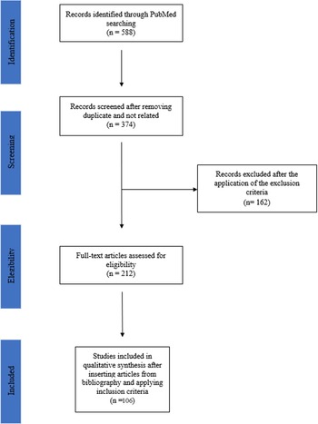
By using the above keywords in the scientific literature search method, we found 588 studies. After a detailed screening analysis and the exclusion of duplicate and noninherent articles, we selected 374 articles. Applying the exclusion criteria, we considered 212 articles as eligible. As a final step, we included other inherent articles studying the bibliography of the eligible articles and applied the inclusion criteria for a total amount of 106 articles for qualitative synthesis (Figure 1).
Flow chart of the systematic review according to PRISMA guidelines.

The article selection included 60 analytical, observational, cross-sectional studies, and 46 longitudinal studies, of which 45 clinical trials and 1 prospective cohort study. The overall subjects were 8124, of whom 3320 were classified as depressed and 4804 healthy controls.
A prompt consideration should be done herein. From a psychopathological and diagnostic point of view, the depressed groups were very heterogeneous and not such specific, as some studies classified as depressed those subjects who presented a current depressive episode during the recruiting time, and used different criteria to assess both diagnosis and illness severity. Apparently, taking into consideration only a limited period without considering lifetime patient’s history, that episode might also be ascribed to different psychiatric disorders, while being quite unspecific. Therefore, for this reason, several findings should not be considered reliable, and we indicated in the specific tables of each paragraph whether the authors specified which psychiatric disorder the current depressive episode belonged to.
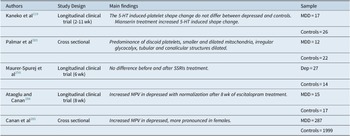
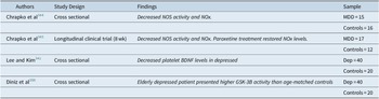
The articles were grouped according to the specific topic they referred to. We found a total number of 16 studies concerning platelet 5-HT2A receptor function: 7 cross-sectional and 9 clinical trials; the total sample includes 498 depressed patients and 395 control subjects. The 5-HT uptake studies were 36 in total, of which 24 cross-sectional studies and 12 longitudinal clinical trials; the sample size includes 1392 depressed and 1252 control subjects. Sixteen studies regarding platelet 5-HT concentration sorted in 6 cross-sectional studies and 10 longitudinal studies (8 clinical trials and 1 observational prospective cohort study), for a total of 553 depressed and 558 control subjects. The articles dealing with platelet α2-adrenergic receptor function were 23, separated in 10 cross-sectional studies and 13 clinical trials with a total sample of 492 depressed subjects and 532 control subjects. As far as MAO platelet activity, we selected a total of 5 articles, 1 cross-sectional study, and 4 clinical trials; the total amount of the included subjects is 115 depressed and 134 control subjects. About platelet biomarkers, we found 7 articles, of which 4 cross-sectional studies and 3 clinical trials for a total of 133 depressed and 120 control subjects. Platelet altered responsivity: 24 articles divided into 18 cross-sectional studies and 6 clinical trials consisting of a total sample of 586 depressed and 492 healthy individuals. Five articles about platelet morphological changes were included (2 cross-sectional studies and 3 clinical trials, 358 depressed and 2078 healthy control subjects). Finally, we examined four studies about other platelet function indices. Two studies deal with platelet nitric oxide synthase activity (eNOs) and nitric oxide metabolites (NOx), one study analyzes the brain-derived neurotrophic factor (BDNF) concentration in platelets and 1 takes into consideration Glycogen synthase kinase 3-beta (GSK-3B). Globally, we included 3 cross-sectional studies and 1 clinical trial for a total 112 depressed and 68 healthy individuals.
Discussion
Serotonergic pathway
Platelets, alike neurons, express at the membrane level both 5-HT2A receptors and the 5-HT reuptake transporter (SERT).
The SERT binds a 5-HT molecule, after its release in the synaptic cleft, along with one sodium ion, transporting them to the intracellular space and thus reducing the neurotransmitter concentration.Reference Owens and Nemeroff 133 The dense granules contain the majority of our body’s serotonin.Reference Skop and Brown 134 Upon 5-HT release from dense granules at endothelial damage sites, platelet aggregation is promoted.
The 5-HT2A receptors, once the 5-HT molecule is bound, promote the coupling of the G-protein, activating the membrane-bound phospholipase C that provokes the phosphorylation of phosphatidylinositol 4,5-bisphosphate into diacylglycerol and inositol 1,4,5-triphosphate. This cascade of events leads to the mobilization of intracellular calcium reserves that induces transglutaminase (the activated form of coagulation factor XIII) and utilizes 5-HT to transamidate Rab4. RhoA leads to cytoskeletal rearrangements, while Rab4 is a small guanosine-5 ‘triphosphate binding protein that regulates vesicular trafficking.Reference Walther 135 The final result of receptor stimulation is platelet activation.Reference Schins 136 , Reference Heger and Collins 137
As previously mentioned, due to the structural and functional similarities between neurons and platelets and the importance attributed to the disturbances of the serotonergic system in the pathophysiology of depression, great attention has been paid over the years to the study of any eventual alterations in serotonergic functionality in platelets. Most of the studies have been performed in the past decades and focused their attention on SERT and 5-HT2A receptor parameters and, to a lesser extent, on platelet 5-HT concentration.
Platelet 5-HT2A receptors
A short premise should be made herein. In pharmacology, the binding parameters are expressed as maximum binding capacity (B max, corresponding to the number or density of binding sites), and constant (K d), that is reciprocal of the affinity constant (K a), and indicates concentration of the drug necessary to saturate 50% of all the sites present. Furthermore, the Michaelis–Menten equation, generally, employed to assess the enzyme kinetics, is also used to calculate the Michaelis–Menten constant (K M) of the reuptake, while representing the substrate concentration necessary for the reaction to reach a velocity equal to half the maximum one (or semi-maximum speed). The rate of reaction when the reuptake proteins are saturated with substrate represents the maximum rate of reaction (V max). The relationship between rate of reaction and concentration of substrate depends on the affinity of the ligand for its substrate.
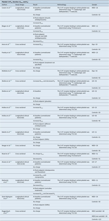
Out of the total 16 studies selected, nine were longitudinal and seven cross-sectional.
Longitudinal studies, assessing receptor affinity parameters at baseline and after drug treatment, reported controversial results. At baseline, five studies showed no change in B max, 138–142 three increased B max, 143–145 and one decreased B max,Reference Markovitz 117 with no changes in K d value.
At the final endpoint, no change was observed in B max in five studies,Reference Sheline 139 , Reference Bakish 140 , Reference Stain-Malmgren 142 , Reference Pandey 144 , Reference Hrdina 145 increased B max in three studiesReference Markovitz 117 , Reference Cowen 138 , Reference Alvarez 141 and decreased B max in one study.Reference Biegon 143 Furthermore, one study with 6 months of sertraline treatment showed a decreased K d.Reference Markovitz 117
In one of the first studies, no statistically significant differences emerged in B max or K d in a sample of 26 patients with a diagnosis of major depressive disorder (MDD) and 25 age-matched controls at baseline. In this case, the 5-HT2A binding properties were evaluated using the [125I]-LSD. However, after 6 weeks of tricyclic antidepressants (TCAs, amitriptyline, dothiepin, and desipramine) B max values resulted significantly increased.Reference Cowen 138 It should be highlighted that the impact of TCA treatment on the binding parameters was tested only on 11 subjects. Furthermore, it is very difficult to ascertain the possible pharmacological characteristics of each compound (amitriptyline, dothiepin, and desipramine). In another longitudinal study, comparing depressed placebo responders (PR = 10), depressed placebo nonresponders (PNR = 23), and controls (C = 14), no statistically significant differences at baseline and at final endpoint were detected using [125I]-LSD as ligand.Reference Sheline 139 The results of this study could be influenced by the numerical intergroup heterogeneity and by the short observational time (10 days). In an 8-week, double-blind trial with two selective 5-HT reuptake inhibitors (SSRIs, paroxetine versus fluoxetine), using [3H]-LSD, no changes in B max were found at baseline and at final endpoint. Nevertheless, dividing the depressed group into suicidal and nonsuicidal, the B max resulted higher at baseline in suicidal depressed.Reference Bakish 140 These findings should, however, be interpreted in the context of some limitations: the relatively small sample size and the treatment with two different drugs. In a sample of 54 subjects (27 unipolar depressed [UD] and 27 controls), using [3H]-LSD, in a 12-week clinical trial, there were no significant differences at baseline. However, a statistically significant increase in B max was observed after clomipramine treatment.Reference Alvarez 141 The small number of patients represents the main limit of the study. Similarly, Stain-Malmgren et al,Reference Stain-Malmgren 142 in a longitudinal study with a sample of 30 MDD patients and 30 controls, described no baseline intergroup differences in B max and K d ([3H]-LSD) and no changes after 6 months of treatment (paroxetine or sertraline). It should be stressed that this is a long observational study compared with the previous ones, and that the many outpatient check-ups led to a high therapeutic compliance.
In a small sample of 12 unmedicated depressed patients, the mean B max value was twice as higher as that of 12 age- and gender-matched control individuals. After 4 weeks of antidepressant treatment, B max values were normal. The 5-HT2A-receptor binding was determined using [3H]-ketanserin. The authors hypothesized that the increased B max might be an adaptive mechanism due to a reduction in 5-HT availability, readily restored by drug treatment.Reference Biegon 143 However, given the small number of patients, it was not possible to distinguish the effects of clomipramine and trazodone.
Others reported similar findings at baseline with [125I]-LSD in 23 depressed patients, underlying how the increase in B max was more represented in those depressed subjects with a recent history of suicide attempts or suicidal ideation. No correlations were identified between B max, K d, illness severity, and drug response. In this study, no changes were detected after 4 to 6 weeks of treatment.Reference Pandey 144 However, it must be said that the authors did not specify the drugs used and did not show the B max and K d values. Another 8-week longitudinal study, comparing 60 UD with 40 control subjects, supported the increased density of 5-HT2A receptors, calculated with [3H]-LSD, mainly enhanced in women and in the suicidal subgroup. According to these authors, the persistence of this difference after the antidepressant treatment would suggest that 5-HT2A up-regulation is a trait rather than a state-dependent marker.Reference Hrdina 145 Nevertheless, it should be noted that the authors treated the depressed subjects with different antidepressant classes (SSRIs, TCIs, SNRIs, placebo, etc.) without analyzing them separately. Only one study, comparing 21 MDD patients and 21 age-matched control subjects, showed a reduced B max at baseline using [3H]-LSD as a ligand. In this case, sertraline treatment not only restored receptor density of 5-HT2A but also reduced K d.Reference Markovitz 117 The small sample size, the short duration of treatment (6 weeks), and the lack of a placebo control group represent the main limitations of the study.
Even the cross-sectional studies report conflicting results: four studies do not describe statistically significant differences between the depressed and the control groups regarding B max 146–148 and three studies showed an increase in B max in depressed subjects. 149–152 The receptor affinity showed no differences between the two groups, except two studies reporting reduced K d value.Reference Sheline 151 , Reference Rosel 153
By employing the [3H]-LSD ligand, a sample of 30 MDD patients and 30 age and matched controls showed similar B max and K d, as well as a positive correlation between anxiety rate, MADRS scores, suicidal behaviors, and B max.Reference Neuger 147 The same results were obtained again with [3H]-LSD in 88 MDD subjects (of which 60 with suicidal behavior and 28 nonsuicidal) and 123 controls, with no difference between patients with and without suicidal behavior had no differences compared with non-suicidal depressed patients.Reference Roggenbach 148 No difference in B max was also found in other two studies. In the first, 5-HT2A receptors were labeled with [125I]-LSD and one group of 43 MDD patients was compared to 42 healthy controls.Reference McBride 146 In the second study, [3H]-ketanserin was used and 51 MDD patients were compared with 31 control subjects.Reference Rosel 153 It should be noted that, in all these studies, patients and healthy subjects were not well matched, while being quite heterogeneous in terms of sex, age, and size.
One study labeling 5-HT2 receptors using [3H]-LSD, reported increased B max values in a sample of 29 depressed patients, which are more evident in women.Reference Arora and Meltzer 150 The depressed group was not homogeneous from a psychopathological point of view, as it included subjects with BD, UD, and schizoaffective disorder experiencing a current major depressive episode (MDE). Furthermore, it should be added that the groups were not well age- and sex-matched. Confirmations of the increased density of 5-HT2A came from two different cross-sectional studies comparing UD and control subjects. In one study, 25 depressed patients, especially women, showed also increased K d values with the receptors labeled by([3H]-LSD.Reference Hrdina 152 In the second case, using the [125I]-LSD, a group of 35 MDD patients showed a positive correlation between the cognitive symptoms of depression and B max.Reference Sheline 151
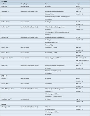
As already underlined, most of the findings are probably affected by methodological limitations, such as the neglect of psychiatric or medical comorbidity, or of confounding factors in the evaluation of single serotonergic parameters (such as cigarette smoking), differences in the diagnostic evaluation (considering, eg, different affective disorders belonging to distinct psychopathological spectra as a unique depressed group), the severity of the disease, not considering the possible impact of the previous pharmacological treatment on platelet function, and different laboratory methodology as the use of dissimilar ways of labeling the receptor (Table 1).
Studies Focused on Platelet 5-HT2A Function

Abbreviations: BD, bipolar disorder; MDD, major depressive disorder; Dep, indicates a diagnosis of major or minor depressive episode, so it can be included in the context of a BD, a unipolar depression or a Schizoaffective disorder; UD, unipolar depression
Through the analysis of these studies, it is not possible to draw definitive conclusions. The results are controversial and sometimes conflicting. However, it is possible to identify some trends: within the whole sample of depressed patients, women, and subjects with suicidal behaviors would seem to have slightly higher B max values at baseline, and the treatment with TCAs would seem to cause a greater increase in B max values compared to other ADs.
In any case, it should be highlighted that the majority of these studies was carried out some decades ago and the ensuing findings would require to be replicated in larger samples and within the frame of the latest and more comprehensive pathophysiological models of depression. With no doubt, the platelet 5-HT2A receptors in depressed subjects appear to be overexpressed and to have a greater sensitivity and reactivity. This would imply a hyperresponsivity to platelet activation factors. It is possible to hypothesize that neuroendocrine and neuroinflammatory alterations may affect platelet reactivity in subjects suffering from depression.
Platelet 5-HT reuptake transporter (SERT)
Serotonin transporter, SERT (SLC64A for solute carrier family 6, member A4), is a 12 transmembrane domain (TMDs) protein, containing two sites of N-linked glycosylation. It performs the uptake of 5-HT through dissipation of the Na+ gradient established by the electrogenic pump Na/K ATPase. Serotonin is transported from the synaptic cleft back to presynaptic neuron where it terminates its action. This transporter is mainly located in cholesterol-rich membrane microdomains, also called lipid-rafts that act as platforms for the regulated assembly and functioning of signaling receptors and transporters.Reference Allen, Halverson-Tamboli and Rasenick 154 The N- and C-terminal regions of SERT dip into the cytosol and interact with several proteins. Cytoplasmic domains, located between TMDs, also contain sites of posttranslational modifications. Regulating extracellular 5-HT, SERT influences the magnitude and duration of the 5-HT response. The SERT encoding gene was first cloned from rat brain and basophilic leukemia cells in 1991,Reference Blakely 155 , Reference Hoffman, Mezey and Brownstein 156 while the human SERT gene was cloned 2 years later; it is located on chromosome 17q11.2 and contains 14/15 exons spanning around 40 kb.Reference Ramamoorthy 157
Due to limitations in access to study the function of brain SERT directly in humans, it has been difficult to make inferences about the activity of neuronal SERT in clinical studies. Platelet membranes contain SERT and are easily accessible, so that they have been and are widely used to explore SERT that is one of the major biological parameters studied in neuropsychiatry. Although several studies suggested a high genetic and functional affinity, it is still whether platelet SERT fully correlates with brain SERT.Reference Pletscher and Laubscher 85 , 157–165 The kinetic characteristics of the SERT have been investigated throughout the decades by means of [3H]-imipramine ([3H]-IMI), [3H]-paroxetine ([3H]-Par), or [3H]-citalopram binding, in parallel with development of SSRIs, or through [3H]-5-HT and [14C]-5-HT reuptake parameters. Only a few studies used other specific ligands to test the pharmacokinetic characteristics of the platelet SERT because the data are meager and they did not comply with the selection criteria, they will be not reviewed in this paper.
Platelet SERT assessed by [3H]-IMI binding
The potential role of [3H]-IMI binding to platelets as a biological marker of depression has been much discussed over the years, since Briley et alReference Briley 166 reported that such a binding was decreased in the platelets of a group of depressed patients compared with the ones of a healthy control group. Platelet and brain [3H]-IMI binding sites consist of two components, with different levels of affinity (high and low affinity sites). Only the high-affinity fraction is Na+ dependent, protease-sensitive, saturable, and, therefore, correlated with the regional distribution of endogenous 5-HT levels.Reference Backstrom and Marcusson 167 , Reference Marcusson 168 The site inhibited by [3H]-IMI binding corresponds to the 5-HT recognition site of the transporter, thus the [3H]-IMI binding and the 5-HT recognition sites may be not overlapping, albeit associated through a Na+-dependent allosteric mechanism. The [3H]-IMI shows a K d value of 0.79 nM at 0 °C for platelet SERT.Reference Mann 169
Through our search criteria, we identified 3 longitudinal studies and 14 cross-sectional. It is interesting to note that the results of the clinical trials are discordant both at the baseline and after treatment.
One study compared [3H]-IMI binding values in 46 depressed patients, divided into endogenous and non-endogenous, and two groups of control subjects, as well as patients with schizophrenia or senile dementia. No significant differences were found in either B max, or in K d, or after treatment with electroconvulsive therapy (ECT), TCAs, or IMAOs.Reference Plenge, Mellerup and Gjerris 170
Another study, although reporting no significant difference in B max at baseline between 20 unmedicated depressed patients and 10 healthy volunteers, detected a mean K d value significantly higher in the first than in the second group. Furthermore, nonresponders (after 2 weeks of treatment with different antidepressants) showed significantly lower initial B max values than responders or controls, while suggesting that it might be a predictor of early response to antidepressants.Reference Hrdina 171 However, these results are severely limited by the short observational time (2-4 weeks), the reduced sample size, and the use of different classes of antidepressants. Contrary to the first two studies, Maj et alReference Maj 172 found a significantly lower baseline B max values in 19 MDD patients than in normal controls. After four and eight sessions of ECT, B max resulted slightly increased, but remained significantly lower than that of the control group. At the time of complete clinical recovery, B max values were restored to the normal range in responder patients only. No effect of ECT on mean K d values was observed.
Four of the cross-sectional studies reported no statistically significant differences between depressed patients and healthy controls.Reference Arora and Meltzer 150 , 173–175 One study analyzed the [3H]-IMI binding values in 12 untreated children and adolescents (aged 11-17 years), who met the DSM-III criteria for MMD, deserves to be mentioned. The affective patients were compared to 13 non-affective patients and 15 normal controls of similar ages, and no intergroup significant differences in B max and K d values were found.Reference Rehavi 174 Even if it failed to discriminate between patients with MDD and those with bipolar disorder (BD) or other disorders, this was the first study examining [3H]-IMI binding kinetic parameters in a population of children and adolescents.
Other authors measured platelet [3H]-IMI binding in 51 depressed patients and 43 normal control subjects before and after dexamethasone administration. At baseline, depressed women (n = 32) showed a significantly lower B max than control women (n = 25). Among the total group of depressed patients, significant negative correlations were observed between the B max values and plasma cortisol levels at 4 PM (n = 41) and 11 PM (n = 41) following dexamethasone administration, mainly among melancholic patients.Reference Roy 173 These data, although suggesting a link between hypercortisolemia and serotonergic affinity, were not confirmed in other studies.Reference Nemeroff 176
All the other cross-sectional studies reported similar results, with B max decreased in the group of depressed patients, compared with control subjects, with no change in K d.
Once more, the [3H]-IMI binding was assessed in 63 depressed inpatients, drug-free for at least 1 month, and in 53 age- and sex-matched healthy control subjects. The B max was significantly lower in depressed subjects and especially in those who had attempted suicide by violent means.Reference Wagner 177 Similarly, lower B max values were detected in 28 patients with MDD, 11 with BD, and 28 healthy subjects. A significant and negative correlation was noted in the patients between B max and the 17-item HDRS, while suggesting a potential relationship with depression severity.Reference Nankai 178 Another study used a least-square computer-assisted analysis in 46 untreated depressed patients and 35 healthy controls. The results revealed a clear and highly significant 22% decrease in B max, however, only dysthymic patients showed B max values significantly associated to symptom severity, as assessed by the HRSD.Reference Jeanningros 179 This study unfortunately is affected by an important limitation, specifically, it included patients diagnosed with MDD, BD, dysthymic, and schizoaffective disorder, irrespective of medical comorbidities, and compared with not well-matched groups.
Other authors, on the contrary, did not provide further supports for the view that the reduced B max of [3H]-IMI binding should be considered a trait marker for susceptibility to depression, and they casted doubt on its specificity as a state-dependent marker for depression. In 63 depressed, 33 nondepressed psychiatric patients and 40 healthy control subjects, B max was found significantly lower in both patient groups, but unequivocal associations between binding parameters and individual symptoms, or groups of symptoms, were not established.Reference Ellis 180
In different studies, Nemeroff et alReference Nemeroff 176 , Reference Nemeroff 181 , Reference Nemeroff 182 evaluated [3H]-IMI binding in young, middle-aged and geriatric depressed patients. Every group of depressed patients (under 50 and over 60 years of age) exhibited significant reduction of B max (42%) with no change in K d, when compared with their age-matched control subjects, with no relationship between post-dexamethasone plasma cortisol concentrations and binding parameters.Reference Nemeroff 176 These results were confirmed in a larger sample of 150 depressed patients and 100 controls subjects. The authors inferred that patients who tend to respond to antidepressants might exhibit lower B max than nonresponders, and that clinical recovery was associated with an increase in B max values.Reference Nemeroff 181 Moreover, 2 years later, in two consecutive experiments involving 40 MDD patients and 40 healthy controls, they demonstrated that the lower B max was not due to prior antidepressant drug exposure.Reference Nemeroff 182
An elegant study measured B max and B max in patients with MDD (n = 11), dysthymia (n = 9), generalized anxiety (n = 18), and panic disorder (n = 10), as well as in healthy individuals (n = 13). Apart from the finding of a decreased B max in all patient groups, the effects of examination stress on platelet binding in medical students were assessed as well. Compared to after vacation period, when binding parameters were similar to the control subjects, B max values were significantly decreased during examinations and similar to patient values. Examinations were also associated with an increase in plasma cortisol levels. They suggested a neurochemical link between depression, anxiety, and stress, and that disturbances in neurochemical functioning may be associated with specific symptomatology, independent from psychiatric diagnosis.Reference Iny 183
A single study showed increased K d values together with a decreased B max in 18 patients diagnosed with MDD with melancholia, as compared with a control group of 63 subjects.Reference Rosel 184 However, in a subsequent study, the same authors did not confirm these changes in K d.Reference Rosel 153
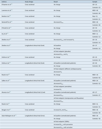
In conclusion, most of the available studies reported more or less marked lower B max values in depressed patients. It is, therefore, surprising that generally no clear relationship did emerge to date between a subgroup of depressive illness and decreased [3H]-IMI binding, except for a correlation with illness severity. In the same way, there are no clear and univocal data regarding the variations of receptor affinity after antidepressant treatments. It is difficult to compare the absolute values of binding in these studies, as the kinetic parameters in both depressed and control groups vary considerably. Probably, the heterogeneity of the results could be the direct consequence of the use of different subject selection criteria, sample sizes, and methodological approaches in the assay process, particularly the method of platelet preparation and the method employed to determine the protein concentration in the assay sample. It should be highlighted that the use of inappropriate displacing agents, such as desipramine, may have been responsible for the conflicting results in [3H]-IMI binding in platelets from depressed patients (Table 2).
Studies on 5-HT Uptake Measured Using [3H]-Paroxetine

Abbreviations: Dep, depressive episode so it can be included in bipolar disorder, unipolar depression, and Schizoaffective disorder; MDD, major depressive disorder; UD, unipolar depression.
Platelet SERT assessed by [3H]-Par
Both [3H]-Par and [3H]-IMI bind to the same macromolecular reuptake complex (although not to the same polymers). [3H]-Par binding appeared to include a homogeneous class of high-affinity binding sites with 90% of the binding being 5-HT-sensitive. [3H]-IMI binding is rather heterogeneous, because this compound labels both 5-HT uptake and non-5-HT uptake sites. [3H]-Par is a more specific inhibitor of 5-HT uptake than [3H]-IMI, which may indicate that it is a better candidate for labeling the SERT, allowing for detection of small binding densities. The K d of [3H]-Par has a value of 0.2 nM even at 37 °C.
We found 16 works divided into 5 longitudinal clinical trials and 11 cross-sectional studies.
Taken together, the results of the clinical trials are quite discordant. At baseline, three studies reported no change in B max Reference Stain-Malmgren 142 , Reference Hrdina 145 , Reference Hrdina 152 and two described a decreased B max values,Reference Alvarez 141 , Reference Sallee 188 with no change in K d. After 10 days of placebo treatment, placebo nonresponders (PNR) showed the lowest B max, placebo responders (PR) intermediate values and normal control subjects (C) the highest values, while PNR and PR had higher 5-HT K d.Reference Sheline 139 A treatment of 8 weeks with different antidepressants (mainly SSRIs) provoked an increase in K d that was related to decreased HRSD total scores.Reference Hrdina 145 In another study, the B max of [3H]-Par binding increased after paroxetine and decreased after sertraline treatment (with no baseline difference between depressed and healthy controls.Reference Stain-Malmgren 142
In a sample of 24 depressed children and adolescents, a statistically significant baseline decrease in B max value emerged, when compared with 22 healthy subjects. The decreased B max values were associated with nonresponse and suicide attempt history. The 8-week sertraline treatment did not influence the B max, but led to a significant decrease in K d.Reference Sallee 188 This study was methodologically and clinically well performed, the two samples examined were perfectly matched, medical and psychiatric comorbidities were considered, and the strict clinical monitoring ensured therapeutic compliance. In line with these findings, another work reported decreased B max in unmedicated patients.Reference Alvarez 141
Even considering the cross-sectional studies, conflicting results emerged. Eight studies reported no statistically significant differences in B max between depressed and healthy controls,Reference Hrdina 152 , Reference Rosel 153 , Reference Lawrence 175 , 183–187 two reported a reduced B max Reference Sheline 151 , Reference Nemeroff 182 and one reported an increased B max.Reference Neuger 147 The K d resulted unmodified in all studies, except that in two where it was increased.Reference Sheline 151 , Reference Sallee 188
Several research groups, by using similar laboratory methodologies, but different sample sizes, selection criteria, diagnostic assessments, or illness severity, reported no significant differences in B max between depressed and healthy subjects, and no correlation with either illness severity or course.Reference Hrdina 152 , Reference Rosel 153 , Reference Lawrence 175 , 183–187
Two different studies, although using different diagnostic tools and sample sizes, revealed decreased B max.Reference Sheline 151 , Reference Nemeroff 182 One of the two papers also detected an increased K d in depressed subjects with increased B max inversely correlated with the severity of cognitive symptoms of depression.Reference Sheline 151
Higher B max and K d also resulted more enhanced in male patients, with a correlation between B max and subscale scores for anxiety.Reference Neuger 147
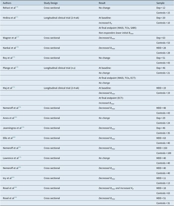
These discrepant findings have been attributed to differences among studies in depression severity (inpatient versus outpatient), platelet binding methods, diagnostic criteria, protein assay methods, and probably other unknown factors (Table 3).
Studies on 5-HT Uptake Measured Using [3H]-Imipramine

Abbreviations: Dep, depressive episode so it can be included in bipolar disorder, unipolar depression, and Schizoaffective disorder; MDD, major depressive disorder; UD, unipolar depression.
Functional Studies on Platelet 5-HT Reuptake
We reviewed 14 studies analyzing the functional reuptake parameters (the maximum velocity, V max and Michaelis constant, K m) with [3H]-5-HT used as a ligand in nine studies, [14C]-5-HT in four, and, finally, fluorescent transporter substrate IDT307 (f-IDT307) in one. The use of 5-HT radioligands ([3H]-5-HT and [14H]-5-HT) allows for greater specificity in the analysis of serotonergic uptake. On the other hand, IDT307 (APP+) is a fluorescent analogue of the dopaminergic neurotoxin MPP+ monoamine transporter substrate fluorescing after reuptaking into cells. IDT307 is a substrate for the dopamine transporter (DAT), norepinephrine transporter (NET), and SERT. IDT307 (APP+) was found to label catecholamine neuronal cell bodies with high selectivity in midbrain and was used to investigate SERT function in both platelets and lymphocytes.
Studies using [3H]-5-HT include four longitudinal and five cross-sectional studies. Longitudinal clinical trials are really controversial. An 8-week-clinical trial revealed that at baseline V max was significantly higher in depressed patients, particularly in women; the K m resulted increased, particularly in a subgroup of SSRI-treated patients (n = 15).Reference Hrdina 145 On the other hand, in another study at baseline, there were no statistically significant differences between the depressed and the control groups. However, in this case, the V max was significantly decreased at week 4 and week 8 and more with paroxetine than with fluoxetine, with a positive correlation with HRSD scores in responders only.Reference Bakish 140 Similarly, Fisar et alReference Fisar 189 found no significant baseline differences between 26 MDD patients and 30 control subjects. After 3 to 7 weeks of citalopram treatment, the K m significantly increased from baseline and the uptake efficiency (V max/K M) significantly decreased.Reference Fisar 189
Different results emerged from a 2-week-double-blind trial in 30 MDD patients (20 men and 10 women, mean age 41 years), randomized to receive amitriptyline (150 mg/d; 15 patients) or paroxetine (30 mg/d; 15 patients), and 49 controls. Untreated patients showed a significant decrease in V max, which persisted after antidepressant treatments, with no change in K m. In any case, the K m significantly increased during paroxetine treatment, whereas only a slight, nonsignificant increase was seen with amitriptyline.Reference Schlake 190 It should be noted that the two groups were quite different in terms of age and, more importantly, that 2 weeks of treatment represent insufficient time to fully evaluate the effects of a pharmacological treatment on any receptor affinity.
The analysis of cross-sectional studies also revealed strong discrepancies in the reuptake parameters. In two studies, no statistically significant differences in V max and K m were measured between depressed subjects and healthy controls.Reference Arora and Meltzer 150 , Reference Hrdina 152 The kinetic parameters of 5HT uptake and the platelet-5HT content (discussed below) were analyzed in 56 healthy subjects and 47 depressed patients who had not been taking psychotropic medications for several months. However, the V max/K m ratio resulted to be positively correlated with the platelet-5HT concentration in healthy subjects, with women showing a higher correlation coefficient than men. A marked deviation from the linear relationship between 5-HT content and the ratio V max/K m was observed in female depressed patients.Reference Franke 191 A trend toward higher values of reuptake efficiency was evident in patients with high net reuptake rate but the platelet-5HT content was similar to that of corresponding controls. The mean scores of the HRSD-D scale (total score and psychic anxiety item) were significantly higher in the low net reuptake rate group of patients than in those with a high net reuptake rate.Reference Franke 191 The same authors in 28 MDD patients explored whether the presence of gastrointestinal symptoms, as defined by item 12 of HRSD, might be related to kinetic characteristics of platelet-5-HT reuptake. The high frequency of relatively low V max and K m of 5-HT reuptake in this group (n = 12), all in the lower range of controls, resulted in significantly lower mean values compared with patients without gastrointestinal symptoms (n = 16; item 12 = 0) and with 57 healthy subjects. They postulated that the noticeable frequency of relatively low apparent V max and K m values of platelet 5-HT reuptake in depressed patients with severe appetite loss could be an indicator for a functional variation in 5-HT transport, possibly based on a low expression of SERT proteins in neuronal and nonneuronal tissues.Reference Franke 192 Opposite results emerged from another cross-sectional study, in which the affinity parameters were calculated in 60 acutely suicidal inpatients and compared to 28 nonsuicidal depressed patients and 123 healthy controls. Mean V max of 5-HT reuptake in washed platelets was significantly higher in suicidal patients than in healthy controls, but not when compared with nonsuicidal depressed patients. Nevertheless, considering depressed patients as a single group and comparing it with control subjects, no statistically significant differences in V max and K m were observed.Reference Roggenbach 148
Studies with [14C]-5-HT consist of one longitudinal and three cross-sectional. In a double-blind study, the platelet [14C]-5-HT uptake was investigated in 30 MDD patients at baseline and after 6 months of treatment with either paroxetine or sertraline. Baseline V max was significantly lower in patients than in controls. Twenty-three patients responded to treatment, as judged by a 50% or more reduction in MADRS total scores, after 6 months of treatment, with no differences between paroxetine and sertraline. A strong correlation was observed between K m and plasma drug concentration in patients at their first episode, but not in patients who had suffered from multiple episodes.Reference Stain-Malmgren 142 In another study, the [14C]-5-HT uptake was determined in platelets from 30 untreated MDD patients and compared with 30 matched healthy controls. The V max was significantly decreased in patients compared to control subjects, with no changes in K m. As the difference was more evident in women, the authors suggested that there might be a gender-related difference in serotonergic dysfunction in depression.Reference Neuger 147 The two remaining studies found no statistically significant differences between the depressed and the control group.Reference Roy 173 , Reference Uebelhack 193
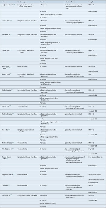
In a recent study, platelet 5-HT content and 5-HT reuptake capacity of SERT were measured by enzyme-linked immunosorbent assay (ELISA) and flow cytometry with IDT307 in depressed (n = 21) and anxious (n = 10) patients at baseline and after SSRI treatment for 4 weeks. Twenty-seven healthy age- and gender-matched subjects were used as controls. As compared with healthy subjects, patients showed higher levels of platelet 5-HT concentration and IDT307 fluorescence intensity. The SSRI administration for 4 weeks significantly decreased platelet 5-HT content, but did not change the IDT307 fluorescence intensity of platelets. However, after incubation with fluoxetine in vitro, the IDT307 fluorescence intensity of isolated platelets from both healthy subjects and patients decreased in a dose-dependent manner. These findings could provide further evidence supporting the role of platelet 5-HT content and SERT as peripheral biomarker in depression and anxiety patients and could help in understanding the delay of the onset of their full therapeutic effects. However, this study has several limitations: the small sample size, with different SSRIs, with heterogeneous pharmacokinetic and pharmacodynamic characteristicsReference Zhuang 194 (Table 4).
Studies on 5-HT Uptake Measured Using Other Ligands

Abbreviations: Dep, depressive episode so it can be included in bipolar disorder, unipolar depression, and Schizoaffective disorder; MDD, major depressive disorder; UD, unipolar depression.
To summarize, the platelet SERT has been widely used in biological psychiatry for about 40 years and still represents a main tool to investigate presynaptic serotonergic neurons. If, in the early 1980s, the labeling of the SERT was carried out by means of [3H]-IMI, a TCA that is not specific for the SERT, or by the direct assessment of the reuptake, the development of SSRIs provided more selective ligands, such as [3H]-Par. Altogether, the studies on platelet SERT, especially labeled with [3H]-Par, rather significantly promoted an astonishing bulk of data in the field, while strongly supporting the serotonergic hypothesis of depression and the key role of presynaptic mechanisms. As a result, throughout the years, the specific literature progressively grew until the point that it became evident that the SERT, one of the main targets of SSRIs, is involved not only in depression, but also in a broad range of several other psychiatric conditions all responding to SSRIs. These observations, on one side, confirmed the lack of nonsological specificity of the platelet SERT abnormalities in depression, and, on the other side, contributed to a great shift in psychiatry, while suggesting that they might be possibly related to symptom clusters or dimensions.Reference Marazziti 195
Platelet 5-HT concentration
Ten controversial longitudinal studies and six cross sectional studies were selected.
In a longitudinal study, platelet 5-HT levels were measured by liquid chromatography with electrochemical detection (LCEC) in MDD, dysthymic disorder, schizophrenia, and other psychosis patients. A significant reduction in platelet 5-HT levels was found in MDD patients and in female schizophrenic patients, with no significant differences between drug-free or medicated patients.Reference Le Quan-Bui 196 Similarly, two different clinical trials, using spectrofluorimetric method, reported significantly lower basal platelet 5-HT in drug-free, depressed patients than healthy subjects. In one of them, a treatment with amitriptyline or paroxetine provoked a further decrease in platelet 5-HT content.Reference Schlake 190 These findings are affected by a main bias, as the subjects were not matched for sex and age, with the healthy group composed generally by young subjects (mean age: 27.5 years) with a discrepancy of about 14 years, compared to the depressed group. In the second study, platelet 5-HT was determined after 4 and 24 weeks of sertraline treatment in 15 female non-suicidal, nonpsychotic MDD patients, compared with 15 drug-free healthy women. Depressed patients were subdivided according to the treatment response into remitters, responders, and nonresponders based on, respectively, the 70%, 50 to 69%, and <49% reductions in MADRS scores. Despite the small number of patients, sertraline induced a further decrease in platelet 5-HT concentration, while suggesting that pretreatment values of platelet 5-HT did not predict therapeutic outcome to sertraline treatment at least in female depressed patients.Reference Pivac 197 Another longitudinal study detected a baseline decrease of platelet 5-HT, as assessed by the HPLC, and showed a further decrease after both clomipramine and fluoxetine.Reference Alvarez 141 Similar results were obtained in an 8-week clinical trial with paroxetine carried out in 11 patients with postpartum depression, with platelet 5-HT determined with both immunocytochemical assay and HPLC. Although 5-HT levels were significantly lower in responders, compared with nonresponders, the small sample size makes this finding a mere observation.Reference Maurer-Spurej, Pittendreigh and Misri 198 On the other hand, four longitudinal studies reported no baseline intergroup differences. In one study, the mean platelet 5-HT level resulted normal in 30 depressed patients compared with 20 healthy subjects, and decreased after 2 to 4 weeks of antidepressant treatments (TCAs, SSRIs, and MAOIs), independently from the clinical outcome.Reference Karege 199 The same result was obtained in a 6 weeks open-label study with sertraline in 21 MDD patients,Reference Markovitz 117 or in 36 female MDD patients after 4 weeks of treatment with paroxetine or tianeptine (in this case, 5-HT content was assessed by spectrofluorimetric method).Reference Muck-Seler 200 As previously mentioned, the study by Zhuang et alReference Zhuang 194 also reported no significant baseline differences between the depressed and the control group, while using the enzyme-linked immunosorbent assay to determine the platelet concentration of 5-HT. Again, SSRI administration for 4 weeks significantly reduced platelet 5-HT content.
Taking cross-sectional studies into consideration, five reported no statistically significant differences between the depressed and the control subjects, with three using the same HPLC despite, but including heterogeneous samples.Reference Roggenbach 148 , Reference Franke 191 , Reference Uebelhack 193 One study, although underlying no difference between patients and controls, noted that higher platelet 5-HT concentrations were more evident in psychotic than in nonpsychotic patients. Furthermore, the lowest platelet 5-HT concentration was associated with the most severe self-aggressive behaviors (both suicidal attempts and completed suicides).Reference Muck-Seler 201 , Reference Muck-Seler, Jakovljevic and Pivac 202 An exploratory study examined associations between cortisol parameters, platelet 5-HT content, and platelet activity markers (CD40, CD40L, and CD62P) in 19 patients with type 2 diabetes (T2DM) and untreated comorbid MDD, 24 T2DM patients, 21 patients with untreated MDD, and 25 age- and sex-matched healthy control subjects, by using HPLC with UV detection. Subgroups did not differ in 5-HT or cortisol slope, but in patients with both T2DM and MD, 5-HT and CD62P were positively correlated.Reference Zahn 203 In any case, this study suffers from some limitations: the cross-sectional design, the relatively small sample size, so only medium to large effects could be detected with sufficient statistical power; the cortisol samples were measured on a single day, so they might reflect an actual state rather than chronic burden; the exclusion of patients with cardio- and cerebrovascular disease, or peripheral artery disease, might only refer to T2DM patients without macrovascular diseases.
Finally, one cross-sectional study detected reduced platelet 5-HT content. This study examined the relationship between platelet 5-HT, plasma cortisol, and PRL concentrations in 20 schizophrenic, 25 depressed, and 25 healthy women. At the time of blood sampling, the schizophrenic and depressed patients had been drug-free for at least 7 days. Platelet 5-HT, plasma cortisol, and PRL concentrations were determined by spectrofluorimetric, radioimmunoassay, and immunoradiometric methods, respectively. Platelet 5-HT concentration was higher in schizophrenic patients than in depressed patients or in healthy control subjects, while it was lower in depressed patients than in healthy subjects or in schizophrenic patients. Plasma cortisol levels were significantly increased in both schizophrenic and depressed patients compared with values in healthy controls. The values of plasma PRL were similar across groups. Unfortunately, this study did not determine any relationships between platelet 5-HT concentrations and plasma PRL and/or cortisol levels after a neuroendocrine challenge test, and reported no information on phases of the menstrual cycleReference Muck-Seler 204 (Table 5).
Studies on Platelet 5-HT Concentration

Abbreviations: Dep, depressive episode so it can be included in bipolar disorder, unipolar depression, and Schizoaffective disorder; MDD, major depressive disorder; UD, unipolar depression.
Taken together, the impact of studies on platelet 5-HT contents is limited, given the main contribution of peripheral 5-HT and no clear evidence of any relationship with CNS 5-HT. Not surprisingly, the literature on this topic dates back to some decades ago, with a few exceptions.
Adrenergic system
A hypothetical dysfunction of the adrenergic system has been discussed for a long time as a cause or predisposing factor to the onset of depressive disorders.Reference Cottingham and Wang 206 , Reference Stahl 207 Considering that platelets possess adrenergic receptors similar to those present on neurons, alterations of the adrenergic pathway has been also examined in the platelets from depressed subjects. Adrenaline, through activation of α 2-adrenergic receptors, provokes direct effects on platelets: it induces primary aggregation, inhibition of adenylate cyclase, potentiation of stimulus-induced aggregation, and secretion.Reference Siess and Lapetina 208
Investigations of α 2-adrenergic receptors B max of depressed patients led to inconsistent findings, as some studies reported heterogeneous results.
In the present review, we selected 13 longitudinal studies and 4 cross-sectional studies, investigating the receptor affinity parameters of the α 2-adrenergic receptor. In addition, we included, in this section, four studies on platelet aggregatory response after adrenaline and noradrenaline and one evaluating intracellular calcium sensibilization with adrenaline.
Although different ligands (ie, [3H]-clonidine, [3H]-yohimbine, [3H]-rauwolscine, [3H]-para-aminoclonidine, [3H]-adrenaline, p125I-clonidine, [3H]-UK14304) were used at baseline, 10 out of the 13 clinical trials reported increased receptor density, one decreased, and two reported no difference between depressed and healthy control groups. Two studies detected differences in baseline K d. All four longitudinal studies that used [3H]-clonidine reported an increased baseline B max in patients. 209–212
Moreover, in one of these studies, the administration of either imipramine or amitriptyline led to significant decreases in the B max and in the K d.Reference Garcia-Sevilla 210 The authors were the first to hypothesize that depression might be related to a α 2-adrenergic receptor supersensitivity, and that the clinical effectiveness of TCAs was associated with a decrease in their number.Reference Garcia-Sevilla 210 Along the same hypothesis, in another study, the binding of [3H]-clonidine hydrochloride to platelet membranes was measured in 13 MDD patients and correlated with the aggregation response induced by adrenaline hydrochloride, which is the result of the activation of the “high-affinity state” receptor. The B max and K d, as well as the aggregation response, were increased in the patients, and were negatively correlated. Long-term treatment (3-24 months) with lithium carbonate was associated with a statistically significant decrease in B max and with an increase in the aggregation response. Moreover, the decreased number of platelets α 2-adrenergic receptors was related to the duration of the lithium treatment. However, several limitations of this study should be underlined: the small size sample and the inclusion in the depressed group of patients with both unipolar and depression. Some patients were treated for only 3 months and others for 24 months, so that it is difficult to ascertain the long-term impact of lithium on morphology and platelet functions.Reference Garcia-Sevilla 209
The same authors partially confirmed these findings in two other clinical trials, using different patient samples and ligands. In the first case, they measured the specific binding of the full agonist [3H]-adrenaline in 14 drug-free depressed patients with melancholia, while reporting an increased B max compared with 15 control subjects. Long-term administration of clomipramine was associated with decreased platelet α 2-adrenergic receptor density correlating with the duration of treatment.Reference Garcia-Sevilla 213 In the second one, using quantitative immunoblotting with anti-α 2-adrenergic receptors, increased immunoreactivities were noted in 22 depressed patients, compared with 22 matched controls. They also showed that α 2-adrenergic receptor/Gai complex was associated to a decreased receptor kinase GRK-2. Moreover, a treatment with mirtazapine reversed this abnormality and induced downregulation of a2-adrenoceptor/Gai complex. This suggests that a defect in the regulation of platelet GRK-2 might contribute to the upregulation of α2-adrenergic receptor.Reference Garcia-Sevilla 214
A further support to this hypothesis was derived from another study, evaluating the kinetic parameters of [3H]-clonidine and [3H]-yohimbine in 29 MDD patients and 26 control subjects. At baseline, the authors reported an increase in B max of [3H]-clonidine binding. Treatment with either imipramine hydrochloride or with amitriptyline hydrochloride decreased the B max. Electroconvulsive therapy also decreased the specific binding of both [3H]-clonidine and [3H]-yohimbine, while lithium carbonate decreased the B max of [3H]-yohimbine.Reference Smith 211 Another study, by using [3H]-yohimbine, reported higher B max in 23 drug-free MDD patients than in 27 healthy control subjects. High pretreatment agonist affinity to the α 2-adrenergic receptors predicted positive treatment outcome. Imipramine induced uncoupling (−11%) and redistribution of receptor density in treatment responders, with no effect on nonresponders.Reference Gurguis 215
Another 4 to 6 week clinical trial compared [3H]-clonidine binding in platelets from 33 controls, 24 unipolar depressive, 22 schizophrenic, 18 bipolar patients, and 8 schizoaffective disorder during a drug-free period. The results showed that B max in depressed, schizophrenic, and schizoaffective patients was significantly higher than in normal subjects. Desipramine treatment caused a significant increase in the K d of [3H]-clonidine binding, with no changes in B max.Reference Pandey 212 Two different studies of the same group,Reference Piletz 216 , Reference Piletz 217 using [3H]-para-aminoclonidine and [p125I]-clonidine, showed an upregulation of α 2-adrenergic receptors in depressed patients returning to normal values after a 6- to 8-week treatment with desipramine.Reference Piletz 216 , Reference Piletz 218 A single longitudinal study including 26 MDD inpatients and outpatients and 26 control subjects reported increased B max and K d values, by using [3H]-UK14304 as ligand. Chronic administration of mianserin to eight depressed patients slightly increased the B max and K d values.Reference Kaneko 219 Clinical trials using [3H]-rauwolscine as ligand led to discordant results. The B max and K d were found similar in 19 hospitalized, untreated depressed patients vs 26 control volunteers. After 7 to 12 days treatment with different TCAs, a significant improvement was observed in the depressive symptoms, with no change in [3H]-rauwolscine binding parameters that also remained stable after 23 days of treatment, in spite of the clinical improvement, as shown by the decrease of the HRSD total score.Reference Pimoule 220 On the contrary, 29 unmedicated MDD patients showed decreased B max values compared to 14 normal volunteers, with no differences in K d value. The K d values of the [3H]-rauwolscine binding were significantly higher in depressed patients treated with TCAs than in the unmedicated or treated with fluoxetine.Reference Maes 221 A single study, involving 30 depressed patients and 30 healthy control subjects, with the use of [3H]-UK 14304, identified a decreased agonist-receptor affinity in depressed patients, whereas the B max was not altered. After 3 months of antidepressant treatment, a decreased K d (increased affinity) was observed, together with a decrease in B max.Reference Karege 222
Even considering the cross-sectional studies, it is possible to identify a trend toward an increase in the α 2-adrenergic receptor density or B max. Specific binding of [3H]-clonidine was measured in 13 unipolar depressed elderly patients (mean ± SD age = 74.8 ± 5.9 years) and in 10 elderly controls (mean ± SD age = 75.2 ± 5.9 years). The B max was significantly higher in depressed than in the controls, whereas K d was not significantly different. In any case, these results are questionable, giving the small sample size and no consideration of the medical comorbidities or drugs that could have affected platelet function.Reference Doyle 223 Increased B max values were also observed in 18 unipolar depressed patients versus 24 sex- and age-matched, healthy control subjects, using [3H]-para-aminoclonidine as ligand. This study, unlike many others, by using purified plasma membranes, ruled out the possibility that an inhibitor may have masked receptor binding in other studies, which used total platelet lysates.Reference Piletz 217 On the other hand, a cross-sectional study, using [3H]-para-aminoclonidine as well to investigate the affinity binding, reported significantly lower B max values in 26 MDD depressed than in 22 control subjects.Reference Carstens 224
Finally, one study, analyzing a sample of 15 unipolar depressed and 15 age-and sex-matched control subjects, showed no difference in [3H]-rauwolscine B max and K d between the groups, but a significant and positive correlation between B max and HRSD total score.Reference Marazziti 225
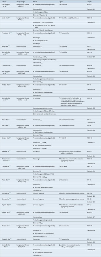
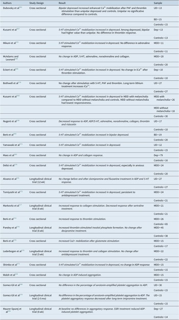
Otherwise, it should be briefly pointed out that several authors measured adrenoceptor-mediated platelet primary aggregation in depressed patients and healthy subjects. Both initial velocity and maximum amplitudes of platelet response to increasing concentrations of adrenaline were decreased in drug-free depressed, while suggesting a postsynaptic desensitization of platelet adrenoceptor function. 226–228 In a longitudinal clinical trial, the functional status of platelet α 2-adrenergic receptors in 30 MDD patients was assessed by simultaneously measuring both a biochemical mechanism of transduction of receptor activation (inhibition of adenylate cyclase activity) and a physiologic response of the receptor (induction of aggregation). The inhibitory effects, induced by adrenaline and [3H]-UK-14304 on adenylate cyclase activity, were unchanged, while, contrary to the previously mentioned studies, the aggregation responses induced by the same α 2-adrenergic receptor agonists were potentiated, which indicated receptor supersensitivity. Both functional responses are desensitized after long-term antidepressant treatment.Reference Garcia-Sevilla 229 A single cross sectional study, analyzing a sample of 20 MDD patients and 20 control subjects, revealed no significant intergroup differences in adrenaline and noradrenaline aggregatory responses.Reference McAdams and Leonard 230 Finally, Mikuni et alReference Mikuni 231 used noradrenaline and 5-HT to assess intracellular calcium mobilization in blood platelets from 11 depressed patients and 11 healthy control subjects. The 5-HT response resulted in a significant greater mobilization of intracellular calcium in depressed subjects than in control subjects, while noradrenaline provoked a higher, albeit not statistical intracellular calcium influx in the platelets of depressed patients (Table 6).
Studies on α2-Adrenergic Receptor Function

Abbreviations: Dep, depressive episode so it can be included in bipolar disorder, unipolar depression, and Schizoaffective disorder; MDD, major depressive disorder; UD, unipolar depression.
Most of the studies suggest that a noradrenergic receptor responsiveness may be abnormal in the affective disorders and may be altered by long-term antidepressant treatment. Several authors speculated on the possible dysfunctional noradrenergic states that may differ between patient subgroups. Specifically, depressed subjects would tend to denote a “hyper-reactive” adrenergic system, not only compared to healthy control subjects, but also to other psychiatric disorders. Unipolar depressed patients should be characterized by a condition of α2-adrenergic postsynaptic subsensitivity accompanied by episodic increase of central presynaptic noradrenergic availability. If the increased number of platelet α2-adrenergic receptors in patients with depressive disorder is relevant to the disease and reflects a state of supersensitivity of similar receptors in the brain, it follows that effective antidepressant treatment should induce receptor changes in the opposite direction. Different antidepressant treatments (SSRIs, TCIs, ECT, lithium salts, and mirtazapine) reduce the B max of high-affinity binding sites for many ligands ([3H]-clonidine, [3H]-yohimbine, [3H]-adrenaline, [3H]-para-aminoclonidine, [3H]-UK14304, and p125I-clonidine) on platelet membranes. However, this decrease in the number of α-adrenergic receptors was not associated with an increased affinity.
Nevertheless, due to the discrepancies attributable to several methodological variables, such as heterogeneous platelet or sample sizes and the choice of radioactive ligand and competitor (nonradioactive ligand) in the assay, it is not possible to draw definitive conclusions.
The strikingly greater variances in patients’ B max and K d values observed in these studies would be more consistent with the hypothesis that depressed patients might have an abnormal reactivity of the adrenergic system.
In any way, the role of the noradrenergic system in depression seems to be ancillary to that of other neurotransmitters, systems, and processes, and the impact of platelet studies’ results was limited and poorly explored in the last decades.
Platelet mono-amine oxidase activity
MAO is a mitochondrial outer membrane flavoenzyme catalyzing the oxidative deamination of neurotransmitters and exogenous arylalkylamines, widely expressed throughout central and peripheral tissues. Two MAO isoforms exist in mammals, namely MAO-A and MAO-B, which are products of distinct genes both located on the short arm of the X chromosome. MAO-A and MAO-B genes comprise 15 exons with identical intron-exon organization. Substrates for MAO include monoaminergic neurotransmitters, such as catecholamines (ie, dopamine, norepinephrine, and epinephrine) and 5-hydroxy-tryptamine (serotonin). MAO isoenzymes display different substrate specificity. However, in living organisms, the contribution of MAO-A and MAO-B in the deamination of substrates depends not only on their kinetic properties, but also on the relative distribution and abundance in the different organs and brain regions. Both MAOs are more distributed in neurons and in the astrocyte, but the presence outside the CNS is more differentiated: while MAO-A are also present in the liver, pancreas, spleen, gastrointestinal tract, and placenta, MAO-B predominates in skin and skeletal muscle, and is the sole form in platelets. Human brain MAO is 70 to 80% of type B.Reference Blaschko 149 , Reference Yeung 232 Platelets also possess MAO-B, generally considered an unspecific biological vulnerability marker toward some neuropsychiatric conditions (such as mood, psychotic eating, post-traumatic stress disorder, etc.), or related to personality traits. 233–243
Using the selection criteria indicated above, we included four longitudinal studies and one cross-sectional study in this review.
In one of the first studies, the platelet MAO activity was investigated in unipolar and bipolar depressed patients with the substrates 14C-tyramine and 14C-benzylamine. Sixty-two patients were selected from consecutive admissions over a period of 2 years. They were untreated with psychotropic drugs (other than benzodiazepines) for at least 2 weeks, or by ECT, when necessary. A group of 59 healthy subjects and 20 in-patients with neuroses and mild depression were chosen as control groups. A higher mean platelet MAO activity, assessed by the substrate tyramine, but not benzylamine, was found in depressed than in control subjects that also resulted positively associated with illness severity. In any case, the increase was mainly due to bipolar patients. The ECT provoked no changes upon the platelet MAO activity of 15 depressed patients. On the contrary, a lithium treatment for approximately 6 months produced a statistically significant rise in platelet MAO activity with the substrate benzylamine but not tyramine. In the opinion of the author, the differences in platelet MAO activity in depressed patients compared with controls are due to MAO structural differences. The form of the enzyme may be the mechanism through which genetic factors influence both the type and severity of the affective illnesses. Furthermore, the difference in MAO activity between unipolar and bipolar patients could suggest a genetic effect acting as a predisposing factor to illness awaiting an appropriate stress.Reference Mann 244
In support of this hypothesis, another longitudinal study, assessing the platelet MAO activity in 25 MDD patients and 25 depressed patients (unmedicated at baseline and after 3 weeks and 2 months of imipramine) using 5-HT as substrate, showed a higher activity of the enzyme that progressively decreased after the treatment and in parallel with the clinical improvement.Reference Quintana 245 In contrast, other studies reported no significant differences between depressed and control subjects. In one open study, no difference was found between 36 female depressed patients (21 treated with tianeptine and 15 treated with paroxetine) and 11 drug-free healthy women, using kynuramine as substrate, before and after treatment. Furthermore, there were no correlations between MAO activity, peripheral biomarkers, platelet and serum 5-HT concentrations, plasma levels of cortisol and prolactin (PRL) either before or after antidepressant treatment.Reference Muck-Seler 200 The MAO activity measured by spectrofluorometer at baseline was similar in 15 female non-suicidal, nonpsychotic MDD patients and 15 drug-free healthy women. Long-term sertraline treatment induced a statistically significant decrease in platelet MAO activity after 24 weeks, in parallel with symptom remission.Reference Pivac 197 Finally, another cross-sectional study detected no difference in platelet MAO activity, with [14C]-phenylethylamine as substrate, among 60 suicidal inpatients, 28 non-suicidal depressed inpatients, and healthy control subjects. A lower MAO-B activity was observed only in suicidal female patients. In this case, the authors used [14C]-phenylethylamine as substrate.
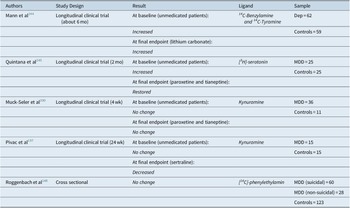
Several explanations might be put forward to this inconsistency, specifically, the use of heterogeneous biochemical methods and/or substrates, different diagnostic systems, or sample characteristics and eventual treatments that are all important sources of variation. Other sources of variation of the MAO-B activity are sex, age, medications, smoking habits, alcohol, or illicit drugs (Table 7).
Studies on Platelet MAO-Activity

Abbreviations: Dep, depressive episode so it can be included in bipolar disorder, unipolar depression, and Schizoaffective disorder; MDD, major depressive disorder; UD, unipolar depression.
In any case, the studies on platelet MAOs, although eliciting a great interest in the past decades, have progressively declined, especially in the field of pathophysiology of depression.
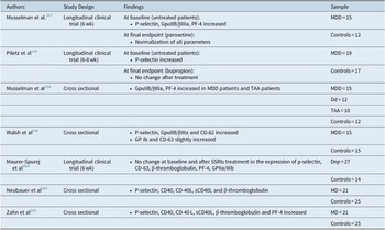
Biomarkers of platelet activation
As already mentioned in the introduction, platelets are now recognized as key players in inflammatory and innate immune responses, for their ability to interact with almost all immune cells. 52–62 Understanding platelet activation pathways and potential biomarkers might represent important clinical targets for several diseases, as well as new diagnostic and therapeutic possibilities in monitoring the disease activities and responses to treatment. Upon activation, platelet surface P-selectin is overexpressed, and platelets release their granule contents into circulation. Several markers of platelet activation, such as P-selectin, CD-40, CD-40L, sCD-40L, CD-63, β-thromboglobulin, PF4, and GPIIb/IIIa, have been identified to correlate with the presence of inflammation and atherosclerosis. 246–249 Platelet activation markers can be well studied by ELISA or western blot, but flow cytometer is currently the standardized method to study platelet function. 250 , Reference Lacroix 251
One of the first studies investigated the degree of platelet activation in a sample of 15 MDD patients and 12 age-matched healthy subjects.Reference Musselman, Marzec and Manatunga 252 At baseline, the depressed subjects exhibited greater procoagulant activity than controls, as detected by increased binding of the monoclonal antibodies anti-ligand-induced binding site (mAb anti-LIBS), which attaches to the fibrinogen binding site of the activated GP receptor, increased binding of the mAb GA6 to P-selectin, and increased plasma concentrations of the platelet-specific secretion protein PF4. The increased binding of the anti-LIBS reflects the conformational change in GP IIb/IIIa. After 6 weeks of open-label treatment with paroxetine, the depressed patients exhibited significant reductions in all three parameters with a normalization of the platelet activation. Although interesting, this study was carried out in a small sample with a lack of normal comparison subjects with other risk factors for ischemic heart disease (IHD). Given that the increased risk of IHD or cerebrovascular disease in MDD subjects might be due to an increase in platelet activity, on the basis of these findings, the authors speculated that a medium or long-term antidepressant treatment, by normalizing the degree of platelet activity, might reduce the cardio-cerebrovascular risk. Subsequently, the same authors provided further supporting evidence to their hypothesis. Markers of in vivo platelet stimulation and secretion were measured in 12 normal subjects and three patient groups: 15 MDD patients, 12 dialysis-dependent patients, and 10 patients with severe thoracic aortic atherosclerosis (TAA), transesophageal echocardiography (TEE)-documented. In comparison with the control subjects, depressed patients and patients with thoracic aortic atherosclerosis exhibited the greatest platelet stimulation, as detected by increased mAb anti-LIBS platelet binding. These findings extend previous observations of increased platelet activation not only in MDD patients but also in those with thoracic aortic atherosclerosis.Reference Musselman 253
Other authors used western blotting and revealed more P-selectin immunoreactivity in depressed patients with no cardiovascular history or other medical illness, compared to healthy individuals. Patients were assessed after 6 to 8 weeks of bupropion treatment, and it emerged that P-selectin remained high in depressed patient and was not related to HRSD score or with the post-treatment plasma concentrations of bupropion. In the opinion of the authors, the elevation of P-selectin could be considered a trait marker for depression.Reference Piletz 116
Supports to the presence of platelet hyperactivity in depression derived from a study reporting an increased expression of GPIb receptors, CD-62, and P-selectin, while GpαIIB/βIIIa and CD63, although also increased, did not reach the significant difference (MDD = 15; controls = 15). There was no difference between depressed patients and healthy volunteers in the expression in glycocalicin or vWF plasma concentration, or ADP-induced aggregation. Of note, the samples were not perfectly matched by age. The results of this study demonstrated that the number of GPIb receptors on platelets are increased in depression and would perhaps suggest a novel risk factor for thrombosis in MDD patients.Reference Walsh 254 In addition, in the same study, smoking has also been shown to increase CD62 on platelets, reflected significantly higher expression of CD62 observed among smokers versus nonsmokers in the control group. However, the causal relationship between smoking and depression is yet to be fully elucidated.Reference Walsh 254
More recently, another study, using whole blood aggregometry, flow cytometry, and ELISA, investigated the platelet expression of P-selectin, CD40-CD40L (atherosclerotic marker), PF-4, and β-thromboglobulin levels in 21 depressed patients and 25 healthy age- and sex-matched controls. All subjects were taking no antiplatelet medications within 7 days before sampling, and had no clinical evidence of peripheral artery disease, coronary or cerebral atherosclerotic, or inflammatory disease based on clinical history and examination. They also had no significant atherosclerotic risk profile except smoking. The results showed that P-selectin, CD40, CD-40 L, sCD40L, and β-thromboglobulin were significantly higher in depressed than in the control subjects, while CD41, CD42b, and PF-4, as well as the aggregometry results, did not differ between the two groups. The increase of these platelet markers in newly diagnosed patients would emphasize that depression is linked to a prothrombotic and proinflammatory state, possibly contributing to accelerate atherosclerosis.Reference Neubauer 255
One very interesting exploratory study examined cortisol awakening response (CAR), diurnal decrease in salivary cortisol concentrations (slope), and the expression of CD40, CD40L, sCD40L, p-selectin, β-thromboglobulin, and PF-4 in 20 MDD patients, and compared to 18 MDD patients with type II diabetes (T2DM) with MDD, and 24 healthy controls. Subgroups did not differ in 5-HT or cortisol slope, while T2DM patients without MDD showed significantly lower CAR than did healthy control subjects. Platelet markers were elevated in depressed and diabetics, while P-selectin and 5-HT levels were positively correlated in depressed patients only. The measurement of plasma cortisol on a single day and the relatively small sample represent the major limitations of this study; again, the cortisol parameters might reflect an actual state rather than chronic cortisol burden.Reference Zahn 203
A pilot study evaluated platelet 5-HT levels and biomarkers of platelet activation in platelets of untreated (n = 13) and SSRI-treated (14) depressed patients and normal subjects (n = 14). Depressed patients showed a significant reduction in platelet 5-HT, with no change after treatment. The expression of P-selectin, β-thromboglobulin, CD-63, and GpαIIB/βIII was similar in the different groups. It should be emphasized that the author did not provide information on the diagnostic assessment and did not show the values of the aforementioned proteinsReference Maurer-Spurej, Pittendreigh and Solomons 256 (Table 8).
Studies on Biomarkers of Platelet Activation

Abbreviations: Dd, dialysis-dependent patients; Dep, depressive episode so it can be included in bipolar disorder, unipolar depression, and Schizoaffective disorder; MDD, major depressive disorder; TAA, thoracic aortic atherosclerosis patients; UD, unipolar depression.
Most of the results emerging from the analyzed studies would seem to suggest that depressed subjects have an abnormal platelet activity. Based on the specific molecules and ligands secreted and expressed, platelets would seem to present a hyperactivation state. In other words, a depressive episode or a depressive disorder, through unidentified (neuro)-inflammatory mechanisms, would result in a pro-inflammatory and pro-thrombotic state. This condition, enhancing atherosclerosis processes, could possibly explain why depressed patients are more prone to develop IHD. This hypothesis would imply that an effective antidepressant treatment, managing the symptoms presented by the patients, could reduce the pro-inflammatory state, reducing the risk for IHD. At the same time, patients with a history of depressive disorder should seriously consider the introduction of drugs that reduce platelet activity, lowering the risk for IHD. Although there are still a few studies, SSRIs would appear the safest and the most efficient antidepressants to reduce platelet activation status in depression, although a debate is still open on their potentially increased risk for major bleeding (especially in combination with platelet inhibitor drugs or oral anticoagulants). 258–264
Platelet hyper-responsivity
Platelet hyper-reactivity is not merely an unavoidable adverse effect of conditions ranging from infections to diabetes, it is a strong factor exacerbating most aging-related human pathologies.Reference Podrez 265 Taken together, recent studies indicate that platelet reactivity is not just a biomarker of adverse effects, but it is by itself a condition that requires management, not limited to cardiovascular disease and stroke. The mechanisms, leading to platelet hyper-reactivity, include variations in platelet proteins, such as surface receptors or changes in the balance between procoagulant and anticoagulant factors in plasma. Several authors investigated platelet responsiveness to pro-activating agents, such as ADP, 5-HT, adrenaline, collagen, and thrombin in psychiatric patients, mainly in depressed patients, with somewhat controversial results. Some of them reported greater platelet reactivity with altered mobilization of intracellular calcium (Ca2+) deposits in response to stimulation with the aforementioned agents. In several studies, 5-HT stimulated Ca2+ mobilization was found increased in depressed patients, compared with the controls.Reference Mikuni 231 , 266–273 In one of these studies, the 5-HT stimulated Ca2+ response was significantly higher in the platelets of unmedicated depressive patients, with no intergroup difference in either basal Ca2+ concentration or thrombin-induced Ca2+ mobilization. In two different studies, Kusumi et alReference Kusumi, Koyama and Yamashita 267 found a significantly higher 5-HT-stimulated response in unmedicated patients with bipolar depression and melancholic MDD than in those with non-melancholic MDD and normal controls. The enhanced Ca2+ mobilization failed to correlate with severity of depressive symptoms. In patients with BD and melancholic MDD, a significant difference in 5 HT-stimulated Ca2+-response was noted between the unmedicated group and those in remission.Reference Kusumi, Koyama and Yamashita 266 , Reference Kusumi, Koyama and Yamashita 267 Interesting findings emerged from another study, in which not only there was a greater 5-HT response in 24 MDD patients than in 20 controls subjects, but, moreover, depressed patients with high anxiety showed significantly increased 5-HT responses compared to depressed patients with low anxiety. In addition, patients receiving SSRIs demonstrated lower 5-HT Ca2+ mobilization than those not taking drugs.Reference Delisi 270 One cross-sectional study investigated the intracellular free Ca2+ concentration in platelets of both untreated depressed patients and patients in remission treated with imipramine. The 5-HT-induced Ca2+ mobilization was significantly higher in both untreated patients and imipramine responders, compared with healthy controls. In addition, the responders showed a positive correlation between the duration of the remission and intracellular basal-free Ca2+ concentration, while suggesting that 5-HT-induced Ca2+ responses persisted after remission in depressed patients, and may perhaps be considered a trait marker.Reference Tomiyoshi 272
The increased mobilization of Ca2+ in depressed subjects resulted as a selective response to 5-HT, as other platelet activators, such as adrenaline,Reference Mikuni 231 thrombinReference Eckert 268 or ADP,Reference Shimbo 271 did not show an increased response.
Nevertheless, other authors described increased platelet secretion in response to some stimulants. One study detected a greater platelet secretion in depressed patients than in controls in response to collagen. Furthermore, patients with a family history of coronary disease had not significantly greater wound-induced fibrinogen receptor binding than the other groups. After treatment with sertraline, this higher responsiveness decreased.Reference Markovitz 117 Again, MDD patients showed a significantly higher platelet activation response to thrombin and/or collagen responsivity than patients with subsyndromal depression, schizophrenia, or controls.Reference Berk and Plein 274 , Reference Lederbogen 275 Antidepressants (SSRIs and TCIs) and ECT did not significantly change this apparent super-sensitivity.Reference Lederbogen 275 , Reference Pandey 276
One study examined the platelet glutamate receptor sensitivity using the platelet intracellular Ca2+ response to glutamate measured by spectrofluorometer. The results showed that unipolar depressed out- and inpatients showed a significantly greater platelet intracellular Ca2+ response to glutamate stimulation than the controls, in terms of both absolute values and percentage of response from baseline). In addition, depressed patients showed higher basal intracellular Ca2+ levels than controls, so that it is unclear whether the increased responsiveness to glutamate depends on a primary receptor super-sensitivity, or it is secondary to an alteration of the pathways of second messengers between the receptor activation and the mobilization of intracellular Ca2+ deposits.Reference Berk, Plein and Ferreira 277
Other authors detected no difference in platelet responsiveness to activating factors and matched controls, while showing an enhanced response to thrombin and PAF in bipolar.Reference Alvarez 141 , Reference McAdams and Leonard 230 , Reference Walsh 254 , Reference Maurer-Spurej, Pittendreigh and Solomons 256 , 278–282 Dubovsky et alReference Dubovsky 282 did not find any differences in basal intracellular Ca2+ between unipolar depressed, BD patients, and healthy controls. Bipolar patients showed an enhanced response to thrombin and PAF, compared to unipolar depressed and control subjects. In another study, intracellular Ca2+ was measured in the resting state and, after stimulation with thrombin, platelet-activating factor, and 5-HT in 27 unipolar depressed, 17 BD patients, and their matched control individuals. No intergroup differences were found in the resting state and after stimulation, with the exception of intracellular Ca2+ after stimulation with 5-HT that was significantly higher in the lithium-treated group, whereas antidepressants and antipsychotics did not seem to interfere with it.Reference Dubovsky 282
In two different studies, no baseline differences were found in depressed patients and healthy controls in the percentage of 5-HT-amplified platelet aggregation to AD.Reference Gomez-Gil 279 , Reference Gomez-Gil 280 The aggregatory response significantly decreased with remission after imipramine treatment, perhaps through a desensitization or downregulation of the 5-HT2A receptor.Reference Gomez-Gil 279 A 6-weeks clinical trial showed that SSRIs did not affect platelet aggregation and dense granule release in response to thrombin, but they significantly reduced ADP-induced platelet aggregation and dense granule release in both patient and normal control samples. According to the authors’ opinion, the active inhibition of platelet aggregation by SSRIs might explain their cardiovascular benefit.Reference Maurer-Spurej, Pittendreigh and Solomons 256
Finally, only one study identified a reduced platelet responsiveness to multiple activating factors (ADP, adrenaline, noradrenaline, 5-HT, collagen, thrombin, and ristocetin) in a small sample of 17 unipolar depressed patients, compared to 10 healthy controls, thus suggesting the presence of inhibitory factors in depressed patients, capable of reducing platelet aggregationReference Nugent, Dinan and Leonard 228 (Table 9).
Studies on Platelet Responsivity

Abbreviations: Dep, depressive episode so it can be included in bipolar disorder, unipolar depression, and Schizoaffective disorder; MDD, major depressive disorder; UD, unipolar depression.
All these findings suggest that the platelet response to activation in MDD patients is, in most of the case, supersensitive. This hyper-reactivity could lead to an increase in aggregability and consequently to an increased risk of thrombosis, representing a further confirmation of the close link between depression and cardiovascular disease, especially in untreated subjects. The positive effects of antidepressants on these parameters are noteworthy and deserve to be more thoroughly investigated in further studies.
Morphological changes
Five studies investigated eventual platelet morphological changes in depressed patients.
A first cross-sectional study conducted on a sample of 12 drug-free MDD patients compared to 22 healthy controls, reported a correlation between depression and discoid platelet form, and between HRSD score and platelet count.Reference Palmar, Marcano and Castejon 283 Furthermore, the patients showed dilated tubular and canalicular structures and an irregular and abundant glycocalyx on the outer membrane. The granules showed no changes in shape and size, although a denser structure was observed in the patients who also presented a greater amount of glycogen, or alterations of the mitochondria, which were smaller and more dilated than in healthy subjects.
Two studies deal with changes concerning the mean platelet volume (MPV). One of them reported elevated MPV in MDD patients (n = 15), as compared with controls (n = 17). Moreover, normalization of MPV levels and reduction of platelet count were observed after 8 weeks of treatment with escitalopram. However, this study is affected by the bias that the sample was relatively small and mainly composed of women in both patients (73.3%) and controls (64.7%). Moreover, the lack of control group with patients with other mental disorders or of patients treated with other types of antidepressant treatments (pharmacological and non-pharmacological) suggests the need of larger prospective trials to establish the clinical relevance of the described laboratory data.Reference Ataoglu and Canan 284
A cross-sectional study was carried out on a large sample of depressed patients (n = 287) compared with a control group of healthy subjects (n = 1999). The authors confirmed the increased MPV in depressed patients that was more pronounced among women. A significant negative correlation between MPV and platelet count was also found. However, despite the large sample size, the study suffers from some limitations: the preponderance of women in the whole sample, the lack of randomization of the selected subjects, the use of a list of questions following DSM-IV criteria, rather than a structured interview to make diagnosis, the lack of a distinction between bipolar and unipolar depressed patients, no assessment of illness severity and, finally, no consideration of other psychiatric or medical conditions that could potentially alter platelet functioning.Reference Canan 285
Two other studies detected no morphological changes of platelets in MDD. One of them investigated the effects on platelet shape induced by 5-HT in 29 MDD patients (12 treated with mianserin for 2-11 weeks) and 26 healthy controls, while reporting no intergroup difference. However, when the treated patients were compared with non-treated ones or healthy subjects, the first group showed an increased 5-HT-induced shape change. In the opinion of the authors, this would represent a sign of a marked influence on the intracellular signal transduction system due to the long-term treatment. However, it should be noted that the depressed patients were both bipolar and unipolar, were not sex-or age-matched with controls, and no information was provided regarding psychiatric and/or medical comorbidities that could potentially modify the results.Reference Kaneko 286
Finally, one study, by using phase-contrast microscopy, did not reveal any differences between unmedicated depressed and healthy controls in platelet morphology. These findings were confirmed by differential interference contrast micrographs. Platelets from control subjects and untreated depressed showed bright dots where the specific 5-HT antibody and the fluorescently labeled secondary antibody colocalize in dense granules. The platelets of the SSRI-treated patients showed differences in shape or in releasing dense granule content except for the lack of bright dots, while indicating very low levels of 5-HT in the dense granules. Generally, there was an inverse correlation between the SSRI dose and the number of 5-HT-labeled dense granulesReference Maurer-Spurej, Pittendreigh and Solomons 256 (Table 10).
Studies on Morphological Changes

Abbreviations: Dep, depressive episode so it can be included in bipolar disorder, unipolar depression, and Schizoaffective disorder; MDD, major depressive disorder; UD, unipolar depression.
In conclusion, the studies on morphological changes in platelets of depressed patients, although potentially interesting, are a few, heterogeneous, carried out with different techniques and, as such, not comparable.
Miscellany
Glycogen synthase kinase 3-beta (GSK3B) is a serine–threonine kinase with a key role in the regulation of the glycogen synthesis.Reference Embi, Rylatt and Cohen 287 GSK3B is also involved in the regulation of critical intracellular signaling pathways, including cell cycle, gene expression, and apoptosis.Reference Grimes and Jope 288 In neurons, GSK3B plays a major role in cytoskeleton organization and remodeling, synaptic plasticity, neurogenesis, and resilience to neuronal injury.Reference Bradley 289 , Reference Peineau 290 The studies highlighted the abnormal regulation of GSK3B in the pathophysiology of neuropsychiatric disorders and the impact of several psychotropic drugs (mood stabilizers, antidepressants, and atypical antipsychotics) on its activity. 291–297
In the present review, we reported a cross-sectional study on GSK3B activity in a group of 40 drug-free MDD elderly patients and 20 age-matched controls. The GSK3B activity was indirectly inferred by the GSK3B ratio, pGSK3B/tGSK3B that was determined by enzyme immunometric assays. The lower GSK-3B ratio would have indicated a higher proportion of the non-phosphorylated GSK-3B and, thus, a higher enzymatic activity. As compared with age and sex-matched control subjects, the depressed patients showed higher GSK3B activity than the control group. By subdividing the depressed group basing on severity and the presence of cognitive impairment, it emerged that both severity and cognitive decline positively correlated to GSK3B activity. These findings suggested additional evidence of the involvement of GSK3B in the pathophysiology of late-life MD. Abnormal GSK3B activity may reflect a severe disruption of neuronal homeostasis and neurodegeneration with potentially important implications to distinguish psychiatric and neurodegenerative disorders. In any case, this study is affected by a major bias, which is the different numerosity of the patient and control groups. Given the impact of cognitive impairment on GSK3B activity, it is difficult to ascertain if the statistical difference between the two groups was mainly due to patients with cognitive impairment.Reference Diniz 298
Although these data highlight the possible role of intracellular mechanisms in MDD, they are just a few and should be considered as mere suggestions to be explored in the future.
Brain-derived neurotrophic factor (BDNF) is a growth factor protein and member of the neurotrophin family. BDNF contributes to a variety of neural processes ranging from neurodevelopment to the survival and homeostatic maintenance of central and peripheral nervous system in adulthood and in aging. 299–301 During development, BDNF plays a key role in neurogenesis, differentiation, and maturation of neurotransmitter systems. 302–304 In adulthood, it is agreed that BDNF plays an important role in the modulation of synaptic plasticity and in the formation of memory and learning. 305–307 Animal models showed that BDNF is involved in the pathogenetic mechanism of MD, as well as postmortem studies showed an abnormal expression of BDNF in the hippocampus and prefrontal cortex of depressed patients. 308–312 The morphological changes in the brain, reported in depressed patients, seem to be reverted by the long-term administration of ADs that would induce an upregulation of BDNF. 313–316 BDNF is also present in large amounts in the blood where it is mostly stored in platelets (~99%), and only a small part of free BDNF is present in plasma. 317–319 Furthermore, BDNF is present in two distinct pools in platelets, in α-granules, and in the cytoplasm.Reference Serra-Millas 320 BDNF in the α-granules is released upon platelet activation, whereas the cytoplasmic BDNF is not.Reference Tamura 321 The maximum BDNF release is approximately 30% to 40% with stimulation, and the remaining 70% of BDNF is equivalent to that found in the cytoplasm, which is not released. 322 Thus, the plasma level of BDNF only partially reflects the amount effectively produced by platelets.
BDNF levels in the human brain are difficult to measure, so it is generally assessed in periphery. The relevance of this measure is supported by animal studies, showing that BDNF is able to cross the blood–brain barrier in both directions, and that peripheral and central BDNF levels are associated. 323–325 Since platelets cannot pass the blood–brain barrier, the BDNF level in the brain may not be reflected by the amount of BDNF associated with platelets, but rather by the amount of free BDNF in plasma.Reference Radka 318 Drug-free depressed patients and healthy subjects with depressive personality traits show decreased serum BDNF levels, which would increase after antidepressant treatment. 326–333 Some studies reported low BDNF levels in plasma of depressed subjects with gender differences, pointing out a specific role of gender in altering neurotrophin regulation.Reference Dwivedi 309 , Reference Karege 310 , Reference Gervasoni 329 , 334–340
One cross-sectional study revealed that platelet BDNF levels were significantly lower in 20 suicidal depressed patients and 20 nonrecent suicidal depressed patients than in healthy controls. As suggested by the authors themselves, this study has some limitations: the sample size was small and the platelet BDNF levels were within a relatively wide range; the depressed group was heterogeneous, being established by 15 men and 45 women and, as already noted,Reference Huang, Lee and Liu 335 the reduction of serum BDNF levels in MDD might be typical to women, not men; finally, they analyzed only platelet-rich plasma and platelet-poor plasma BDNF levels. Therefore, it is difficult to determine if the source of BDNF decline is brain or other peripheral cells.Reference Lee and Kim 341 Although several research studies described that changes in circulating BDNF may be used as a proxy for changes occurring in the brain,Reference Radka 318 , Reference Lommatzsch 342 , Reference Karege 343 it is uncertain how precisely BDNF levels measured in venous blood samples reflect circulating BDNF levels in vivo, and how different methodological approaches may affect the results.
To summarize, on the basis of the available information, the importance of assessing platelet BDNF appears limited both for the controversial results emerging from the studies and because there is no direct correspondence between BDNF produced by platelets and that released in plasma under stimulation.
Finally, two studies that analyzed endothelial nitric oxide synthase (eNOS) activity and plasma nitric oxide metabolites (NOx) levels in MDD patients were included in this review. Major depression has been associated with increased cardiovascular mortality in patients with coronary heart disease (CHD), and it has been described as an independent risk factor for the development of CHD in healthy subjects; however, the underlying mechanism remains unclear. It is known that nitric oxide plays a major role in cardiovascular regulation and its decreased production has been associated with several cardiovascular risk factors. In a cross-sectional study, Chrapko et alReference Chrapko 344 investigated the correlation between platelet endothelial nitric oxide synthase (eNOS) activity and plasma nitric oxide (NOx) levels. By comparing a sample of 15 depressed patients with healthy controls, NOx and eNOS resulted statistically significant decreased in the patient group. No correlation was found between the illness severity and eNOs activity or plasma NOx levels, or between platelet eNOS and NOx. In addition, by analyzing MDD patients with and without comorbid anxiety disorders, no difference was found. This would suggest a lack of a major confounding effect of comorbid anxiety disorder in these results, even if, due to the limited representation of each anxiety disorder in the sample, it cannot be excluded that certain anxiety disorders might be associated with specific dysregulation of endothelial NO production and platelet eNOS activity.Reference Chrapko 344
In a separate study with a different sample, the same research group confirmed the previous findings, and furthermore analyzed the effect of 8-weeks paroxetine treatment on NOx and eNOS in both depressed (n = 17) and controls (n = 12). After paroxetine treatment, plasma NOx levels increased in both samples. On the other hand, platelet eNOS activity was reduced in the control group, while no change was detected in MDD patients. According to the authors, this reduced production of NO by vascular endothelium and platelets could contribute to an increased risk of developing cardiovascular disease in depressed subjects. The impact of this risk factor could be reduced by the administration of paroxetine. It should be highlighted, however, that the small sample size and the choice of including in the depressed group people with other psychiatric disorders in comorbidity could be considered as limitsReference Chrapko 345 (Table 11).
Studies on Different Platelet Molecular Pathway

Abbreviations: Dep, depressive episode so it can be included in bipolar disorder, unipolar depression, and Schizoaffective disorder; MDD, major depressive disorder; UD, unipolar depression.
These studies are potentially interesting but deserve to be replicated in larger samples.
Conclusions
The studies reviewed in the present article reported conflicting results. As already explained in each chapter, the majority of these discrepancies may derive from differences in laboratory methodologies, in the selection of the samples, in the diagnostic criteria employed throughout the decades, and in the eventual presence of cofactors that can potentially influence the platelet activity (drugs, comorbidity, etc.).
However, the findings of several studies on structural and metabolic alterations, modifications in the expression of specific proteins, changes in the aggregability, or in the reactivity to different pro-activating stimuli, may be suggestive of potential platelet dysfunctions in depressed subjects. What is less understood is the etiology of this altered platelet function in depressed subjects. The possible pro-inflammatory state in relation to states of chronic stress with related dysfunction of the HPA axis system and alterations of the monoaminergic pathways could be responsible for these platelet changes in depressed patients, causing a state of hyperreactivity.
This implies that platelets of MDD patients are more likely to cause the onset and progression of atherosclerotic lesions and to lead to cardiovascular disease, potentially explaining the increased morbidity and mortality rate in these individuals.Reference Vieweg 346 , Reference Wang, Hoffman and Blumenthal 347 Furthermore, these conditions can share a common pathophysiology, since dysregulated serotonergic and adrenergic signaling might be the basis of each of these diseases.
At the same time, accumulated evidence would indicate that SSRIs might show antiplatelet effects in vivo and protect against cardiovascular events.Reference Kubera 348 However, given the paucity of available studies, it is currently impossible to establish with certainty which antidepressant treatment may have the best tolerability profile and greater efficacy in reducing cardiovascular risk. In addition, in the future, it would be interesting to also compare the impact of antidepressants with that of non-pharmacological treatments, such as ECT, cognitive-behavioral therapy (CBT), and transcranial magnetic stimulation (TMS). We hope that, in the future, further studies will be able to explain the pathophysiological bases of this cascade of events in MDD patients, determine whether specific treatment can be an effective tool to improve depressive symptoms, and, at the same time, reduce the incidence of cardiovascular events.
Disclosures
Claudia Tagliarini, Manuel Glauco Carbone, Giovanni Pagni, Donatella Marazziti, and Nunzio Pomara do not have an affiliation with or financial interest in any organization that might pose a conflict of interest.