Introduction
Entomopathogenic nematodes (EPNs) of the families Steinernematidae and Heterorhabditidae are important biocontrol agents of a broad range of insects that damage agricultural crops throughout the world (Shapiro-Ilan et al., Reference Shapiro-Ilan, Han, Qiu, Morales-Ramos, Rojas and Shapiro-Ilan2014; Lacey et al., Reference Lacey, Grzywacz, Shapiro-Ilan, Frutos, Brownbridge and Goettel2015; Kergunteuil et al., Reference Kergunteuil, Bakhtiari, Formenti, Xiao, Defossez and Rasmann2016). They are used as an alternative for insect control due to their ability to locate host insects, great reproductive capacity and virulence, and association with bacteria of the genera Xenorhabdus spp. (Steinernematidae) and Photorhabdus spp. (Heterorhabditidae) (Gaugler & Kaya, Reference Gaugler and Kaya1990; Kaya & Gaugler, Reference Kaya and Gaugler1993; Burnell & Stock, Reference Burnell and Stock2000; Kaya et al., Reference Kaya, Aguillera, Alumai, Choo, De la Torre, Fodor, Ganguly, Hazir, Lakatos and Pye2006). Steinernematids are cosmopolitan and, to date, 95 species (Hunt & Nguyen, Reference Hunt and Nguyen2016) have been described in all the continents except Antarctica, and this number is growing every year.
In India, little EPN diversity has been found during surveys of agricultural soils. Currently, a total of nine EPN species (seven steinernematids and two heterorhabditids) have been reported from India, of which only one new species (Heterorhabditis indica Poinar, Karunakar and David, 1992) has been described from the country. Among steinernematids, no new species have been reported to date, but two previously described new species have been synonymized with existing ones, namely Steinernema thermophilum (Gaungly & Singh, 2000) was synonymized with S. abbasi (Hunt, Reference Hunt, Nguyen and Hunt2007) and Steinernema dharanai (Kulkarni, Rizvi, Kumar, Paunikar & Mishra, 2012) was recognized as a junior synonym of S. hermaphroditum (Stock, Griffin & Chaerani, 2004).
For the commercial and successful establishment of EPNs as biological control agents, it is important to isolate and identify the effective EPN strain. In the search for EPN populations, a survey was conducted in the agricultural fields of the Meerut District of Western Uttar Pradesh, India during the month of June, when temperatures can reach up to 40°C, to assess their presence during such high temperatures. Two nematode populations belonging to the genus Steinernema Travassos, 1927, were recovered from soil samples of pepper (Capsicum annuum L. (Solanales: Solanaceae)) and pearl millet (Pennisetum glaucum L. (Poales: Poaceae)) fields. The third-stage infective juveniles display two horn-like structures on the labial region, and thus belong to the ‘bicornutum’ group. The morphology, morphometry and molecular characterization of these isolates revealed that they are conspecific to S. pakistanense Shahina, Anis, Reid, Rowe and Maqbool, 2001; hence it is the first report of S. pakistanense from the Indian subcontinent.
For many steinernematids, the identity of the bacterial symbiont is unknown, even though such information is necessary for our better understanding of the diversity of these nematode–bacteria complexes. In the only study focused on the symbiont of S. pakistanense, Shahina et al. (Reference Shahina, Manzar and Tabassum2004) reported this nematode to be associated with Xenorhabdus nematophila. This bacterium is known to be associated with Steinernema carpocapsae, and its presence in S. pakistanense, a nematode from a different phylogenetic clade, seems surprising, although host switches in the Steinernema–Xenorhabdus complex have been reported to be frequent (Lee & Stock, Reference Lee and Stock2010). However, in their study, Shahina et al. (Reference Shahina, Manzar and Tabassum2004) identified the bacterium based solely on phenotypic data, which are now considered insufficient for the precise identification of bacteria. Thus we have performed the first molecular characterization of the bacterium associated with S. pakistanense.
Because the isolated bacterium was found to be related to the symbionts reported from other related nematode species, suggesting a possible co-evolutionary history for the nematodes and bacteria, we have also performed a co-phylogenetic analysis, to test the degree of nematode–bacteria co-speciation.
Finally, the isolates were tested in laboratory bioassays for pathogenicity against Spodoptera litura (Fabricius, 1775) (Lepidoptera: Noctuidae) and Helicoverpa armigera (Hübner, 1808) (Lepidoptera: Noctuidae).
Materials and methods
Nematode origin and culture
A total of 25 soil samples were collected by taking five random sub-samples at a depth of 0–20 cm from different agricultural fields of the Meerut district (28°59′N, 77°42′E and 225 m above sea level) of Western Uttar Pradesh. EPNs were recovered from only two soil samples using the insect-baiting technique (Bedding & Akhurst, Reference Bedding and Akhurst1974) with the last instars of Galleria mellonella (L.) (Lepidoptera: Pyralidae), and the remaining soil samples were found to be negative for EPN infection. Infective juveniles (IJs) were maintained by recycling through G. mellonella larvae, washed properly with double-distilled water (DDW), disinfected with 0.1% sodium hypochlorite and stored in approximately 150 ml of sterilized distilled water in 500-ml vented tissue-culture flasks at 15°C for subsequent identification and the establishment of stock cultures.
Morphological and morphometric characterization of the nematodes
For taxonomic studies, first- and second-generation adults were obtained by dissecting the dead G. mellonella cadavers in Ringer's solution on the third and fifth days after infection, respectively, while fresh IJs were collected from a White trap (White, Reference White1927). They were killed in hot water (60°C), fixed in TAF (7 ml formalin, 2 ml triethanolamine, 91 ml distilled water) (Courtney et al., Reference Courtney, Polley and Miller1955), processed to glycerin (Seinhorst, Reference Seinhorst1959) and mounted in a small drop of glycerin on a glass slide. The cover slip was placed on to the glycerin drop with some extra paraffin wax to prevent the flattening of the nematodes. Morphological observations were made using a light compound microscope (Magnus MLX; Olympus, New Delhi, India) and phase-contrast microscope (Nikon Eclipse 50i; Nikon, Tolyo, Japan). Morphometric details were gathered with the help of the inbuilt software of a phase-contrast microscope (Nikon DS-L1).
DNA extraction, amplification and sequencing of the nematodes
The total genomic DNA was extracted from the pool of infective juveniles using a Quiagen DNA Blood and Tissue Kit (Qiagen, Hilden, Germany) (Bhat et al., Reference Bhat, Istkhar, Chaubey, Půža and San-Blas2017) followed by agarose gel electrophoresis (AGE) for the detection of DNA in the collected eluate. Molecular characterizations were based on internal transcribed spacer (ITS) regions of rDNA and a partial sequence of 28S rDNA (D2–D3 domain). These regions were amplified using the primers suggested by Vrain et al. (Reference Vrain, Wakarchuk, Levesque and Hamilton1992) and Joyce et al. (Reference Joyce, Burnell and Powers1994), respectively. The amplifications of the ITS and D2–D3 regions of the rRNA were performed according to Bhat et al. (Reference Bhat, Istkhar, Chaubey, Půža and San-Blas2017). The products were analysed on 1% agarose gels with TAE (Tris–acetic acid–EDTA) buffer. The amplified polymerase chain reaction (PCR) products were purified and sequenced in both directions by Bioserve Pvt. Ltd (Hyderabad, India). Amplified regions were annotated and submitted to the National Center for Biotechnology Information (NCBI) under accession numbers MF289980 and MF289982 for ITS and D2–D3 genes, respectively, for CS31; and for CS32, MF289981 and MF289983 for the same respective regions.
Isolation and molecular characterization of entomopathogenic bacteria
The endosymbiont bacterium associated with S. pakistanense CS31 was obtained from the haemolymph of G. mellonella 1 day after infection with CS31, by adopting the method of Akhurst (Reference Akhurst1980). The haemolymph was streaked on nutrient agar supplemented with 0.004% (w/v) triphenyltetrazolium chloride and 0.0025% (w/v) bromothymol blue (NBTA medium) and left overnight at 28°C (Akhurst, Reference Akhurst1980). Single colonies were transferred with a sterile toothpick to YS broth (Akhurst, Reference Akhurst1980) and cultivated on an orbital shaker (180 rpm) at 25°C.
Bacterial DNA was extracted from a 2-day-old culture using a DNeasy Blood and Tissue Kit (Qiagen) according to the manufacturer's instructions. The 16S RNA was amplified using primers 10 F: 59-AGTTTGATCATGGCTCAGATTG-39 (forward) and 1507R: 59-TACCTTGTTACGACTTCACCCCAG-39 (reverse) (Sandstrom et al., Reference Sandstrom, Russel, White and Moran2001). The recombinase A gene (recA) was amplified using primers RecA1 F: 59-GCTATTGATGAAAATAAACA-39 (forward) and RecA2R: 59-RATTTTRTCWCCRTTRTAGCT-39 (reverse) (Tailliez et al., Reference Tailliez, Laroui, Ginibre, Paule, Pages and Boemare2010). The gyrase B gene (gyrB) was amplified using primers 8SF gyrB: 59-TACACGAAGAAGAAGGTGTTTCAG-39 (forward) and 9Rev gyrB: 59-TACTCATCCATTGCTTCATCATCT-39 (reverse) (Tailliez et al., Reference Tailliez, Laroui, Ginibre, Paule, Pages and Boemare2010). The PCR master mix consisted of 7.2 ml ddH2O, 0.5 ml bovine serum albumin (BSA), 1.25 ml 103 PCR buffer, 1 ml deoxyribonucleoside triphosphates (dNTPs), 0.75 ml of each forward and reverse primer, 0.1 ml polymerase and 1 ml of DNA extract. The PCR profiles were used as follows. For 16S: one cycle at 94°C for 1 min; followed by 33 cycles at 94°C for 60 s, 55°C for 60 s and 72°C for 2 min; and a final extension at 72°C for 3 min. For recA: one cycle at 94°C for 2 min; followed by 35 cycles at 94°C for 30 s, 49.5°C for 35 s and 72°C for 60 s; and a final extension at 72°C for 2 min. For gyrB: one cycle at 94°C for 2 min; followed by 35 cycles at 94°C for 30 s, 66°C for 35 s and 72°C for 60 s; and a final extension at 72°C for 2 min. All PCR products were sequenced and deposited in GenBank under the following accession numbers: MF592490 (recA sequence) and MF592491 (gyrB sequence).
Phenotypic and biochemical characterization of symbiotic bacteria
Phenotypic variations were observed in symbiotic bacteria on the basis of adsorption properties towards bromothymol blue (BTB) and neutral red. The adsorption of BTB was examined on NBTA agar (Akhust, Reference Akhurst1980). Bacteria were present in two phases, namely phase I and phase II. Phase II bacteria reduced tetrazolium chloride (TTC) to formazan and produced red-coloured colonies. In the case of phase I bacteria, the reduction of TTC was hidden as they adsorbed BTB and produced blue-green colonies. The production of a blue-green or red colour was taken as the characteristic mark of phase I or phase II bacterial colonies.
For neutral red adsorption, the bacteria were grown by streaking on MacConkey agar and were incubated for 24–48 h. Phase I variants of bacteria showed adsorption of neutral red and appeared as red/reddish-brown while the phase II variants appeared as light yellow/off-white.
The biochemical characterization was examined using a KB003 Hi25 Enterobacteriaceae Identification Kit from Hi-media (Mumbia, India), designed for the identification of Gram-negative Enterobacteriaceae species. A total of 13 conventional biochemical tests and 11 carbohydrate utilization tests were performed using this kit. For biochemical characterization, bacteria were cultured on NBTA media and blue-green colonies were shifted into 5 ml heart infusion broth (Hi-media). The culture was grown overnight and 50-μl aliquots were then inoculated into each of 24 wells of the kit. The kit was incubated according to the manufacturer's instructions and changes in the colour of media were recorded as ‘plus’ and ‘minus’.
Sequence alignment and phylogenetic analyses
The sequences were edited and compared with those present in GenBank by means of a Basic Local Alignment Search Tool (BLAST) (Altschul et al., Reference Altschul, Gish, Miller, Myers and Lipman1990) of the National Center for Biotechnology Information. An alignment of nematode samples together with sequences of related steinernematid species was produced for the ITS and 28S regions using default Clustal W parameters in MEGA 6.0 (Tamura et al., Reference Tamura, Stecher, Peterson, Filipski and Kumar2013) and optimized manually in BioEdit (Hall, Reference Hall1999). Pairwise distances were computed using MEGA 6.0 (Tamura et al., Reference Tamura, Stecher, Peterson, Filipski and Kumar2013). The phylogenetic trees of the ITS and 28S genes were obtained by the Minimum Evolution method (Rzhetsky & Nei, Reference Rzhetsky and Nei1992) in MEGA 6.0 (Tamura et al., Reference Tamura, Stecher, Peterson, Filipski and Kumar2013). Steinernema nepalense and Steinernema scapterisci were used as outgroup taxa. The Minimum Evolution tree was searched using the Close-Neighbour-Interchange (CNI) algorithm (Nei & Kumar, Reference Nei and Kumar2000). The neighbour joining algorithm (Saitou & Nei, Reference Saitou and Nei1987) was used to generate the initial tree. The evolutionary distances were computed using the p-distance method (Nei & Kumar, Reference Nei and Kumar2000) and are expressed as the number of base differences per site.
Alignments of the bacterial recA and gyrB genes were concatenated into one dataset. The tree was constructed in the same way as the nematode trees and Xenorhabdus bovienii was used as the outgroup taxon.
Co-phylogenetic analysis
The bacteria from the monophyletic clade of X. indica, Xenorhabdus stockiae and Xenorhabdus innexi, and several unidentified bacterial strains, together with their nematode hosts were used in the co-phylogenetic analysis. For the analysis concatenated datasets of the ITS and D2–D3 regions, and recA and gyrB genes, were used to infer the nematode and bacterial trees, respectively. The trees were constructed using the Minimum Evolution algorithm, as described above. A comparison of the nematode and bacterial phylogenies was made and the significance of the observed co-divergence was assessed using TreeMap version 3.0 (Charleston & Page, Reference Charleston and Page2002). The randomization tests were used to test the null hypothesis that the topological similarities could be accounted for by chance.
Virulence tests on Spodoptera litura and Helicoverpa armigera
Mortality bioassays of both the Steinernema isolates, CS31 and CS32, were performed against two pests, S. litura and H. armigera. Six-well plates (well size 3.5 cm) lined with a double layer of Whatman Filter Paper No. 1 were used for bioassay experimentation using 1-week-old IJs. Concentrations of 0, 25, 50, 100 and 200 IJs in 450 μl distilled water were placed over the filter paper. For each concentration, ten insect larvae of the same size and weight were used and a single larva was set per well. The plates were incubated at 28 ± 2°C. Mortality was recorded at 12-h intervals until 100% mortality was achieved. Larvae infected with 100 IJs/larva (preferred optimum dose for counting progeny) were transferred after 7 days to a modified White trap (White, Reference White1927) to observe the persistence of infection and emergence of IJs (18–20 days). Each bioassay was placed separately and all experiments repeated twice.
The insect larval mortality assay was analysed statistically through probit analysis, and median lethal time (LT50) values were calculated at a 95% confidence limit. Differences between percentages of mortality depending on different isolates were assessed using Wald statistics in the STATISTICA program v. 10 (StatSoft Inc., Tulsa, Oklahoma, USA). Data were presented as a percentage ± SD. Nematode progeny production was analysed by analysis of variance in the same program, and presented in the form of mean ± SD.
Results and discussion
The morphological, morphometric and molecular analysis identified both of the isolated nematodes, CS31 and CS32, as S. pakistanense (Shahina et al., Reference Shahina, Anis, Reid, Rowe and Maqbool2001). This is the first report of this species from India, and three slides of IJs, each bearing seven specimens, three slides of first-generation females bearing one female on each slide, two slides of first-generation males bearing three males on each slide, two slides each of second-generation females and males bearing two and three specimens, respectively, on each slide were deposited in the National Nematode Collection of India, IARI, New Delhi, India. Steinernema pakistanense has been reported from Karachi, Sindh, and near Sonmiani beach and Mubarak village, Balochistan, Pakistan. It has also been reported from a horticultural farm at Hisar, India and several other places but only on a morphological and morphometric basis, which is not valid proof of its existence in India.
Morphology and morphometry
Steinernema isolates CS31 and CS32, obtained during the present investigation, were identified as S. pakistanense; however, some disparity with the original report may be noted. The mucron was found to be absent in all generations except the second-generation females, while in the original description it was observed in second-generation females and 10% of first-generation males. Epiptygmata were absent in the females of the present specimens while weakly developed, double-flapped epiptygmata were reported originally. A post-anal swelling was visible in second-generation females but this was not the case in the original description.
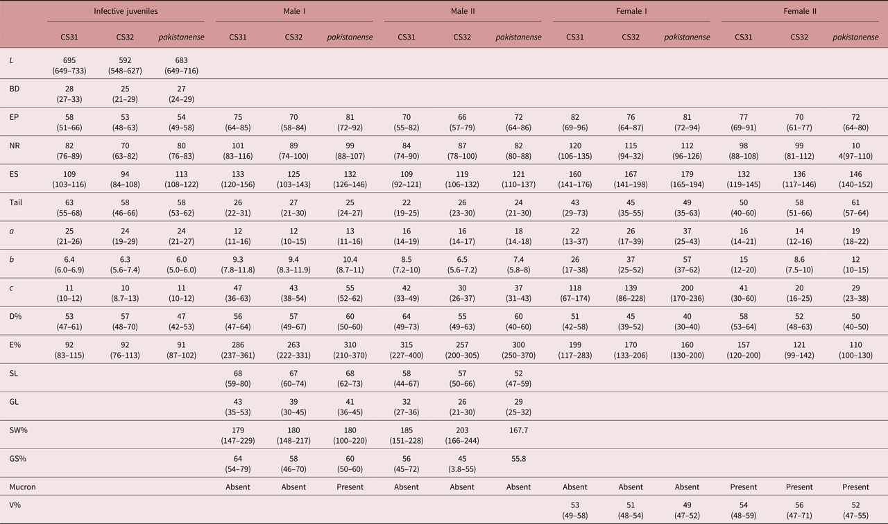
Morphometrical measurements of all the generations of isolates CS31 and CS32 were similar to the topotype population of S. pakistanense (Shahina et al., Reference Shahina, Anis, Reid, Rowe and Maqbool2001) but little divergence is seen when they are compared with each other or with the original description. A comparison in morphometrical parameters in all generations is given in table 1.
Comparative morphometrics of all generations of CS31 and CS32 with the original description of Steinernema pakistanense. All measurements are in micrometres (except percentage) and in the form of mean (range).

L, Total body length; BD, max. body diameter; EP, anterior to excretory pore; NR, anterior to nerve ring; ES, pharynx length; a, total body length/maximum body diameter (L/BD); b, total body length/pharynx length (L/ES); c, total body length/tail length (L/T); D%, excretory pore position/ pharynx length*100 (EP/ES*100); E%, excretory pore position/tail length (EP/T*100); GL, gubernaculum length; SW%, spicule length/ anal or cloacal body width*100 (SL/ABD*100); GS%, gubernaculum length/ spicule length (GL/SL*100); V%, vulva position from anterior end expressed as percentage of L (AV/L*100).
Molecular characterization
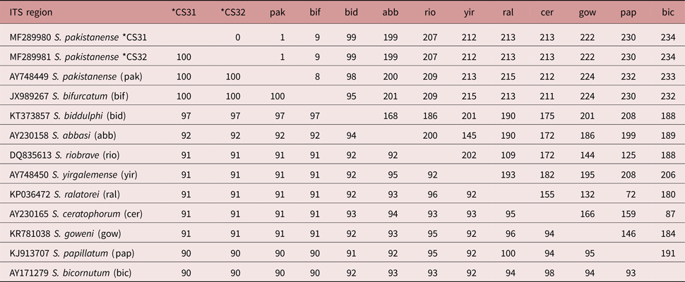
ITS sequences of S. pakistanense of both the Indian strains (CS31 and CS32) differ from the original ITS sequences by 1 bp at position 268, which is T in the present specimens and C in the type population, while they show no nucleotide differences between each other. The ITS sequence of CS31 and CS32 is separated from those of other related species by 9–234 bp (table 2). No sign of intra-individual variability in the ITS sequence was observed.
Pairwise distances of the ITS region between Steinernema species from the ‘bicornutum’ group.

Below diagonal, percentage similarity; above diagonal, total character differences.
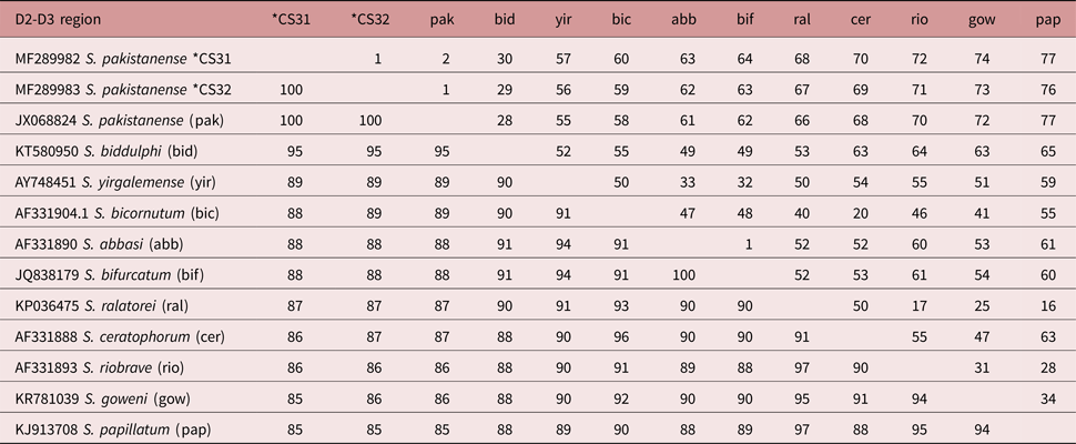
No variation in the D2–D3 sequence was found between the CS31 and CS32 strains, but a variation was seen within S. pakistanense. The D2 and D3 expansion fragments of the 28S rRNA gene of both the Indian isolates were separated by 30–77 bp from other related species (table 3).
Pairwise distances of the D2–D3 regions between Steinernema species of the ‘bicornutum’ group.

Below diagonal, percentage similarity; above diagonal, total character differences.
Phylogenetic analysis
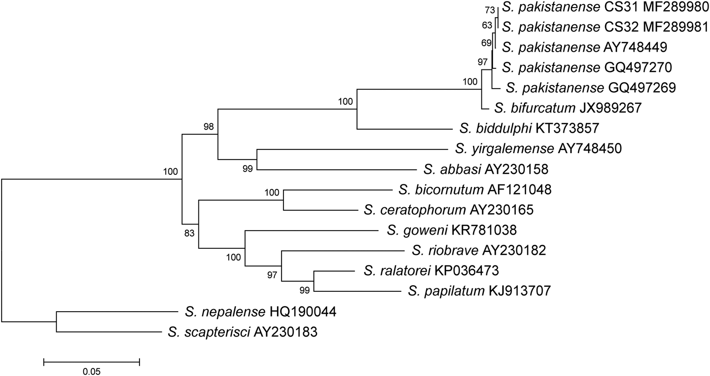
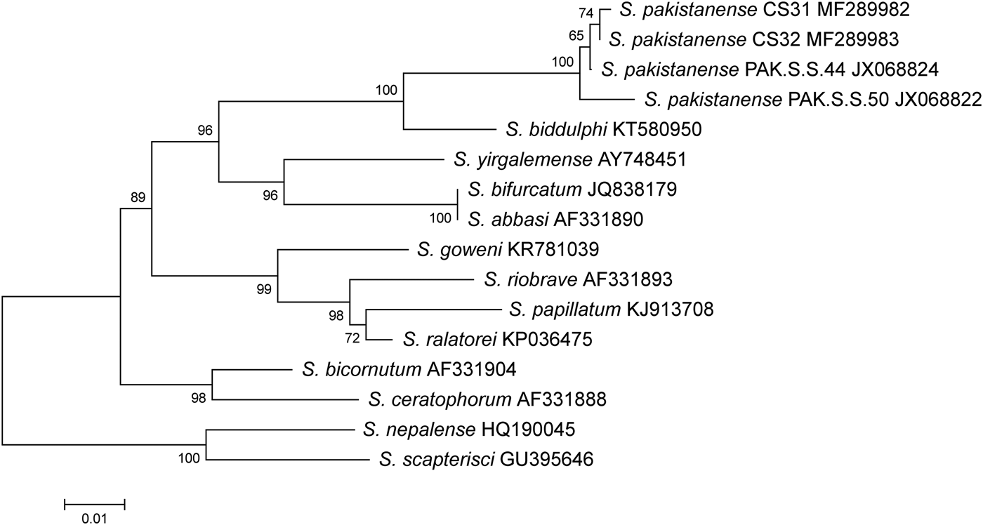
The phylogenetic analyses of the ‘bicornutum’ group based on ITS regions and the 28S rRNA gene showed the apparent monophyly of the group formed by the isolates of CS31 and CS32 and the original S. pakistanense, thus confirming their identification (figs 1 and 2). Both phylogenetic analyses have shown that S. pakistanense is a sister species to the well-supported monophyletic clade containing S. biddulphi, S. bifurcatum, S. abbasi and S. yirgalemense. Within this group, S. pakistanense clusters with another Asian species, S. bifurcatum (fig. 1) and the African species, S. biddulphi (figs 1 and 2). However, our results suggest that, based on molecular data, the status of S. bifurcatum is problematic. The ITS sequence of this species shows only 12 bp differences from S. pakistanense, corresponding to a 98.5% similarity. Such a similarity, however, falls well within the range for intraspecific variation in steinernematids (Půža et al., Reference Půža, Chundelová, Nermuť, Žurovcová and Mráček2015). On the other hand, the analysis based on the D2–D3 sequence places S. bifurcatum as a sister species to S. abbasi. In the analysis of pairwise distances, the sequences of these two species differ by 37 bp; however, the differences occur only in the first c. 150 bp, while in the rest of the alignment, the sequence of S. bifurcatum is identical to that of S. abbasi. It thus seems that the differences were created by an incorrect editing of the JQ838179 sequence. Our results are in agreement with the work of Cimen et al. (Reference Cimen, Půža, Nermuť, Hatting, Ramakuwela and Hazir2016), who state that the 28S rDNA sequence attributed to S. bifurcatum (JQ838179) obviously belongs to S. abbasi.
Phylogenetic relationships in the ‘bicornutum’ group of Steinernema, based on analysis of ITS rDNA regions. Steinernema scapterisci and S. nepalense were used as outgroup taxa. The percentage of replicate trees in which the associated taxa clustered together in the bootstrap test (10,000 replicates) is shown next to the branches. Branch lengths indicate evolutionary distances and are expressed in units of number of base differences per site.

Phylogenetic relationships in the ‘bicornutum’ group of Steinernema, based on the analysis of D2–D3 regions of the 28S rDNA. Steinernema scapterisci and S. nepalense were used as outgroup taxa. The percentage of replicate trees in which the associated taxa clustered together in the bootstrap test (10,000 replicates) is shown next to the branches. Branch lengths indicate evolutionary distances and are expressed in units of number of base differences per site.

Endosymbiont bacteria: molecular, phenotypical and biochemical diagnosis
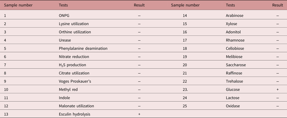
Bacteria isolated from S. pakistanense CS31were grown on NBTA media, producing both phase I and II variants, confirmed by their dye-absorbance ability. Gram staining differentiation showed that the bacterial isolates were Gram-negative rods. The culture grown overnight on heart infusion broth, when transferred and incubated on the KB003 Hi25 Enterobacteriaceae Identification Kit of Hi-media, produced mostly negative reactions (table 4). On nutrient agar, colonies have a brownish pigmented centre, appear shiny and opaque, and are circular to irregular and convex. Phase I colonies adsorb neutral red, forming red colonies on MacConkey agar, and adsorb bromothymol blue forming blue colonies on NBTA. Esculin was weakly hydrolysed. Acids were weakly assimilated (table 4). Most of the phenotypic and biochemical results of symbiotic bacteria reported by Shahina et al. (Reference Shahina, Manzar and Tabassum2004) were identical to the results found here. Only one difference was noted in that phenylalanine deamination was found to be negative in the present investigations, while Shahina et al. (Reference Shahina, Manzar and Tabassum2004) reported it as 26–75% positive. The secondary forms of Xenorhabdus species were reported as stable colony variants that have lost their ability to produce pigment, antimicrobial agents and secondary metabolites, are not able to take up dye, and have sometimes lost their luminescent properties, as in the case of Photorhabdus luminescens (Boemare et al., Reference Boemare, Akhurst and Mourant1993).
Biochemical characteristics of X. indica CS31 isolates confirmed by the Hi-media Enterobacteriaceae Identification Kit.

ONPG, O-nitrophenyl-beta-d-galactopyranoside; −, negative; +, positive.
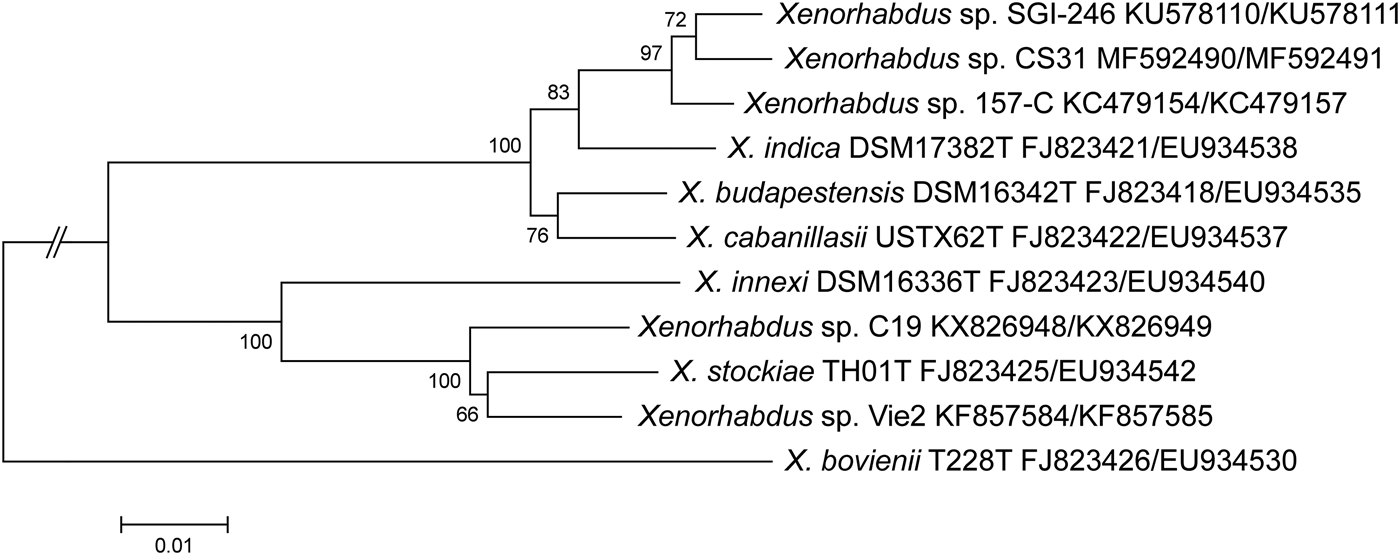
The sequences of the recA and gyrB genes of Xenorhabdus sp. CS31 exhibited the highest similarity to the sequences of X. indica 157-C (98.4 and 99.1%, respectively) and Xenorhabdus sp. SGI-246 (98.1 and 99.1%, respectively) (fig. 3). Thus Xenorhabdus sp. CS31 probably represents another strain of X. indica. This result is in contradiction to those of Shahina et al. (Reference Shahina, Manzar and Tabassum2004), who identified the symbiont as X. nematophila, but only based on the phenotypic characters. In the light of our results, their identification can be regarded as incorrect.
Phylogenetic relationships of Xenorhabdus sp. isolated from Steinernema pakistanense (CS31) with other closely related species of Xenorhabdus, based on analysis of recA and gyrB gene sequences. The percentages of replicate trees in which the associated taxa clustered together in the bootstrap test (1000 replicates) are shown next to the branches. Branch lengths indicate evolutionary distances and are expressed in units of number of base differences per site.

Interestingly, X. indica 157-C and Xenorhabdus sp. SGI-246 are associated symbiotically with S. yirgalemense (Ferreira et al., Reference Ferreira, van Reenen, Tailliez, Pagès, Malan and Dicks2014) and S. biddulphi (Cimen et al., Reference Cimen, Půža, Nermuť, Hatting, Ramakuwela and Hazir2016) and both these nematode species are closely related to S. pakistanense.
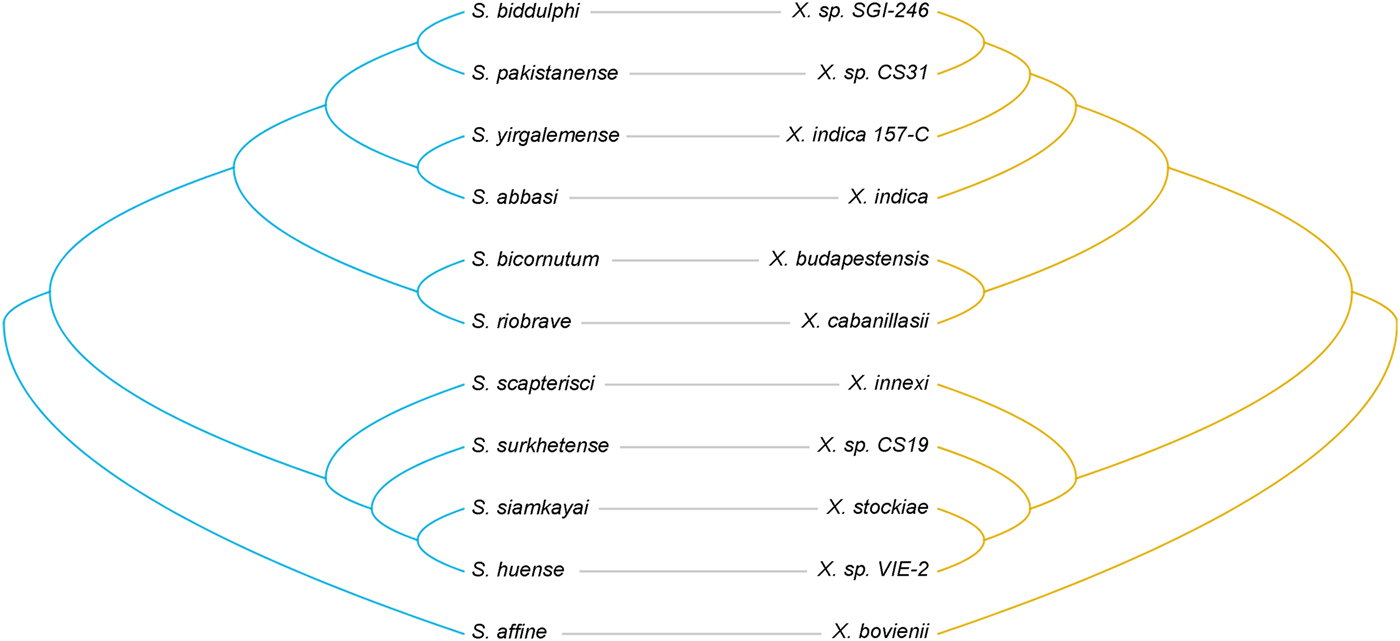
The co-phylogenetic analysis suggests that there are significantly more co-divergence events between the nematodes and bacteria (P = 0.0001) than would be expected by chance if their phylogenies were independent (fig. 4). Reconciling the nematode and bacteria phylogenies indicates remarkable topological congruence, with 18 co-divergence events out of a possible 23, with two duplications, two losses and no host switch.
Tanglegram showing correspondence between Xenorhabdus sp. CS31 and related Xenorhabdus spp. (right) and their nematode partners (left). Associated pairs are linked by a grey line.

In the Steinernema and Xenorhabdus complex, the only study addressing co-speciation (Lee & Stock, Reference Lee and Stock2010) found no evidence for co-speciation and suggested that there have been at least 17 host switches of strains of Xenorhabdus spp. within clades and even between clades. Recent reports of switches of symbionts between nematodes of distantly related clades (Cimen et al., Reference Cimen, Půža, Nermuť, Hatting, Ramakuwela and Hazir2016; Dreyer et al., Reference Dreyer, Malan and Dicks2017) suggest that switches can be frequent in the Steinernema and Xenorhabdus complex. In the light of these facts, our results are surprising. Our analyses suggest that in some steinernematid lineages the nematodes have undergone co-speciation with their bacterial symbionts. This fact suggests that the specificity of the Steinernema spp. / Xenorhabdus spp. pairs might differ in different lineages. On the other hand, our analysis did not include S. carpocapsae, which is a member of the ‘carpocapsae’ clade, but is associated with the symbiont X. nematophila, belonging to a different clade within the Xenorhabdus phylogeny. The switch of X. nematophila to S. carpocapsae was documented by Stock (Reference Stock and Campos-Herrera2015).
Virulence tests
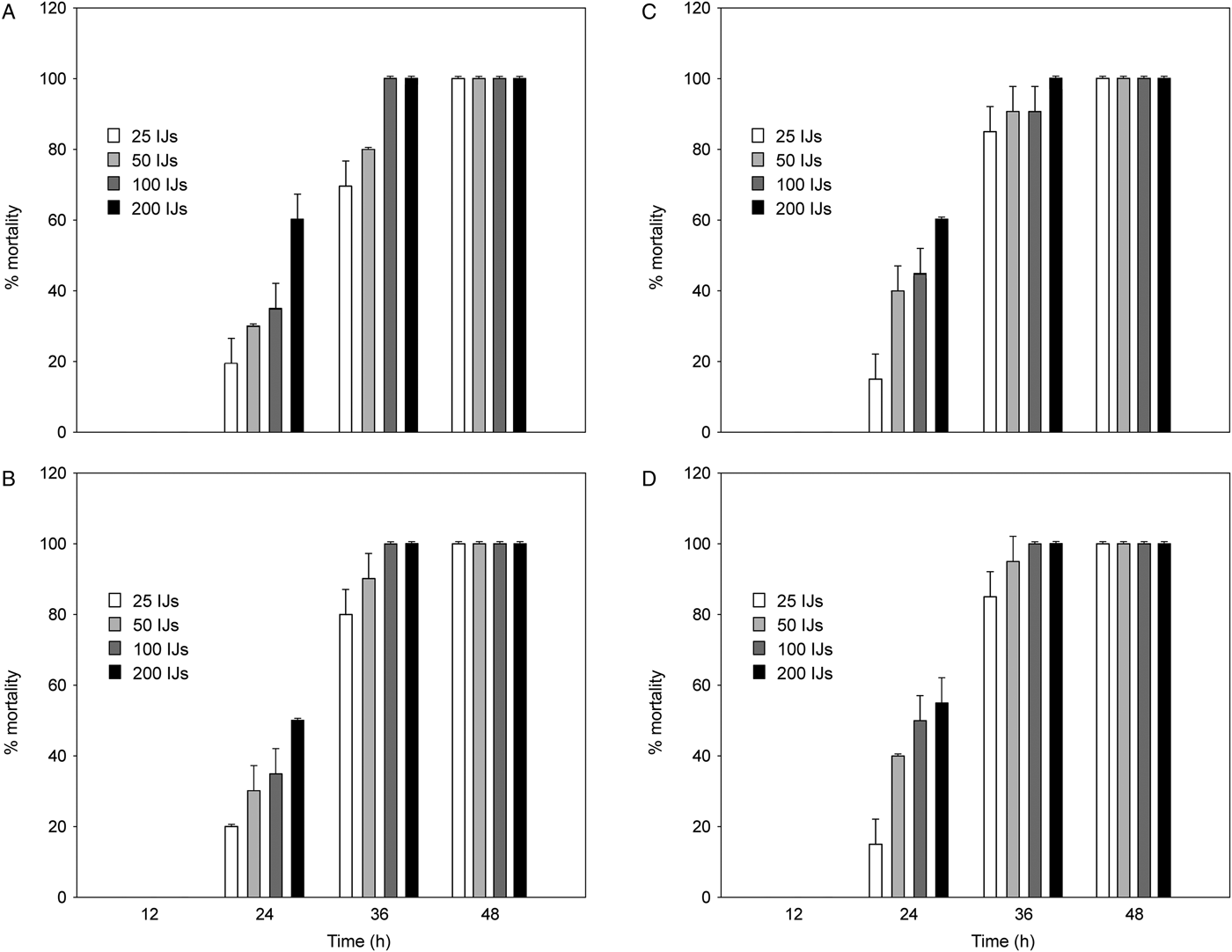
Both strains of S. pakistanense were able to kill and reproduce in both of the insects tested. The mortality of S. litura and H. armigera caused by strains CS31 and CS32 did not differ (Wald stat. = 0.0339, P = 0.854 and Wald stat. = 0.086, P = 0.768, respectively) and reached 100% after 48 h at all doses (fig. 5). The median lethal time (LT50) for the lowest dose of 25 IJs/larva was c. 30 h in both strains and both hosts. The onset of mortality is very rapid. With the same insects, and with comparable doses of S. abbasi, Kalia et al. (Reference Kalia, Sharma, Shapiro-Ilan and Ganguly2014) observed that 100% mortality occurred no earlier than 96 h in H. armigera and 192 h in S. litura.
Percentage mortality (mean and SD) of Spodoptera littura (A and B) and Helicoverpa armigera (C and D) larvae with different doses of Steinernema pakistanense strains CS31 (A and C) and CS32 (B and D).

The production of progeny in both of the tested hosts did not differ significantly between strains (F = 1.5, P = 0.23), and the mean production of progeny by the strains CS31 and CS32 in S. litura was 54,980 ± 22,245 IJs and 60,680 ± 20,826 IJs, respectively. In H. armigera, the production of nematode progeny was significantly lower (F = 7.63, P = 0.008) and reached 39,240 ± 5598 IJs (strain CS31) and 46,780 ± 13,855 IJs (strain CS32). In conclusion, our data show that S. pakistanense is very virulent towards the insect pests tested, and thus this species could be considered for use in the biocontrol of these organisms in India.
Acknowledgement
We thank Mr Christopher Steer, University of South Bohemia, Czech Republic, for language correction.
Financial support
The authors thank the Department of Science and Technology (DST), New Delhi, India, for providing financial assistance through DST INSPIRE Fellowship/2014/76.
Conflict of interest
None.