In his seminal work describing the endocranial anatomy of Ectosteorhachis nitidus, Romer (Reference Romer1937) stated that “rhipidistian crossopterygians are of great phylogenetic importance, since they are unquestionably the closest known relatives of the ancestors of land vertebrates”. This apt observation has since been echoed in modern cladistic analyses, with tetrapodomorph fishes such as Ectosteorhachis positioned basal to early tetrapods within the Tetrapodomorpha (Johanson & Ahlberg Reference Johanson and Ahlberg2001). Sequential changes in anatomy between tetrapodomorph fishes and tetrapods have been noted in bones of the skull roof (e.g. Schultze & Arsenault Reference Schultze and Arsenault1985, fig. 1) and pectoral fin/limb (e.g. Long et al. Reference Long, Young, Holland, Senden and Fitzgerald2006, fig. 3; Shubin et al. Reference Shubin, Daeschler and Jenkins2006, fig. 4). However, the early evolution of internal braincase anatomy amongst the Tetrapodomorpha is comparatively poorly known, with the majority of descriptions from ‘osteolepiform’ and ‘elpisostegid’ taxa restricted to provisional details of internal snout morphology. Such work includes studies on the nasal capsules and olfactory nerve canals of Thursius estonicus (Worobjewa Reference Worobjewa1975), Gyroptychius sp. (Thomson Reference Thomson1964a), G. elgae Vorobyeva, Reference Vorobyeva1977, Megistolepis klementzi (Vorobyeva Reference Vorobyeva1977), Shirolepis ananjevi Vorobyeva, Reference Vorobyeva1977, “osteolepid A” (Janvier Reference Janvier and Panchen1980); the megalichthyid Megalichthys cf. hibberti (Schultze Reference Schultze1974), Jarvikina wenjukowi (Jarvik Reference Jarvik1937; Vorobyeva Reference Vorobyeva1962, Reference Vorobyeva1977), Platycephalichthys Vorobyeva, Reference Vorobyeva1959 (Vorobyeva Reference Vorobyeva1962; Worobjewa Reference Worobjewa1975) and Panderichthys stolbovi Vorobyeva, Reference Vorobyeva1960 (Vorobyeva Reference Vorobyeva1962, Reference Vorobyeva1971). Complete endocranial material has been described from relatively few tetrapodomorph fish taxa. The first such description was on eroded natural casts preserving the internal ear region of the ‘osteolepidid’ Osteolepis macrolepidotus (Watson Reference Watson1925a, Reference Watsonb) from the Middle Old Red Sandstone of Scotland. This work was followed by a complete description of the endocranial morphology of Osteolepis by Thomson (Reference Thomson1965). The only other internal ethmosphenoid or otico-occipital material described from cosmine-covered ‘osteolepiform’ fishes includes poorly preserved partial internal moulds of Sengoerichthys ottoman Janvier et al. Reference Janvier, Clément and Cloutier2007, several broken specimens of Cladarosymblema narrienense Fox et al., Reference Fox, Campbell, Barwick and Long1995, a cross section through the otico-occipital of Medoevia lata Lebedev, Reference Lebedev1995a, radiographs of Gogonasus andrewsae Long, Reference Long1985 (Long et al. Reference Long, Barwick and Campbell1997), and several complete braincase units of Ectosteorhachis (Romer Reference Romer1937) from the Cisuralian Red Beds, of Texas, USA. This latter material was sectioned by Romer (Reference Romer1937) using the “peel” method, resulting in detailed reconstructions of the brain endocast, cranial nerves and blood vessels, with subsequent investigations focusing on the nasal capsule (Thomson Reference Thomson1964b) and semicircular canals (Thomson Reference Thomson1966). Similar studies were later undertaken by Stensiö (Reference Stensiö1963) and Jarvik (Reference Jarvik1942, Reference Jarvik1954, Reference Jarvik1966, Reference Jarvik1980, Reference Jarvik, Schultze and Cloutier1996) on the endocranium of the tristichopterid Eusthenopteron foordi from the Frasnian of Miguasha, Quebec, Canada. Jarvik's work on the internal structure of the ethmosphenoid (Reference Jarvik1942, Reference Jarvik1966, Reference Jarvik1980) and otico-occipital (1980, 1996) was based on drawings and wax models produced using the Sollas grinding method by Erik Stensiö (Jarvik Reference Jarvik1980). Although this technique and the “peel” method used by Romer (Reference Romer1937) ultimately destroyed the endocrania under study, they allowed important comparisons to the pertinent anatomy of other gnathostome groups, including tetrapods, dipnoans, actinopterygians and chondrichthyans (Romer Reference Romer1937; Jarvik Reference Jarvik1980).
Recently, x-ray computed tomography has been used on tetrapodomorph fish endocrania to reveal internal details without destroying material. Preliminary work using this technique includes study of the braincase of Eusthenopteron (Ahlberg et al. Reference Ahlberg, Blom, Brazeau, Clément, Snitting, Ivanov and Young2005; Ahlberg Reference Ahlberg2006) and the ethmosphenoid and canals feeding the vagus nerve and occipital arteries of the otico-occipital of Spodichthys buetleri (Snitting Reference Snitting2008). More rigorous studies using micro-tomography include the ethmosphenoid of the most basal tetrapodomorph Tungsenia Lu et al., Reference Lu, Zhu, Long, Zhao, Senden, Jia and Qiao2012, from the Pragian Posongchong Formation of Yunnan Province, southwestern China.
This paper presents the first complete description of internal braincase anatomy in a tetrapodomorph fish based on computed tomography. The material under study, the first complete specimen of Gogonasus andrewsae (NMV P221807) is preserved in uncrushed, 3D condition. Small parts of the internal otico-occipital unit were scanned using micro-computed tomography by Long et al. (Reference Long, Young, Holland, Senden and Fitzgerald2006, suppl. info), but were not thoroughly described. First collected in 1967 from the Frasnian Gogo Formation, 100 km East of Fitzroy Crossing, Kimberley Region, Western Australia, the initial description of Gogonasus was of a single ethmosphenoid unit complex (Long Reference Long1985). Two additional partially complete cranial unit complexes, clavicles, along with associated centra and scales were described from the same general locality (Long et al. Reference Long, Barwick and Campbell1997). Other remains, possibly referable to Gogonasus, include three isolated jaws, two ethmoid ossifications, isolated dermal bones and scales from the Early Frasnian Gneudna Formation of the Carnarvon Basin, Western Australia (Long & Trinajstic Reference Long and Trinajstic2000; Trinajstic & George Reference Trinajstic and George2009). These specimens are significantly larger (20%) than comparable Gogonasus specimens from the Gogo Formation, leading Long & Trinajstic (Reference Long and Trinajstic2000) to suggest two separate species. Discovered in 2005, the specimen of Gogonasus NMV P221807 was initially described as possessing several derived features of the spiracular chamber and pectoral fin endoskeleton. This resulted in the proposing of a novel phylogenetic position of Gogonasus, crownward of the Tristichopteridae, as the sister taxon to ‘elpistostegid’ fishes and tetrapods (Long et al. Reference Long, Young, Holland, Senden and Fitzgerald2006). This hypothesis was rejected by Friedman et al. (Reference Friedman, Coates and Anderson2007), who posited a more stemward relationship for Gogonasus, as a member of the ‘Osteolepididae’. Later work by Holland & Long (Reference Holland and Long2009) placed Gogonasus stemward of the Tristichopteridae, yet crownward of other cosmine-covered tetrapodomorph fishes. Recent work by Swartz (Reference Swartz2012) has positioned Gogonasus basal to Osteolepis, Gyroptychius, Medoevia and members of the Megalichthyidae within the Megalichthyiformes Coates & Friedman, Reference Coates, Friedman, Elliott, Maisey, Yu and Miao2010. While rigorous in nature, such phylogenetic studies involving tetrapodomorph fishes are yet to include extensive internal braincase characters, owing to a dearth of data. As such, the following description of the internal braincase of Gogonasus represents a vital opportunity to examine previously unknown morphology from a cosmine-covered ‘osteolepidid’ megalichthyiform fish.
1. Materials and methods
1.1. Specimen preparation and study
Specimen NMV P221807 was collected in a limestone nodule by Professor Tim Senden in 2005 in the Frasnian, Gogo Formation of Gogo Station, Western Australia. It was prepared by Dr John Long through immersion in 10% acetic acid, for two-day intervals to dissolve surrounding matrix. Bone material was then washed in water and air-dried following every acid treatment, thereupon being strengthened using Mowital B30 (Clariant Chemicals www.clariant.com.au) diluted in pure ethanol. Photographs of the specimen were taken with a Leica DFC500 camera using a Leica M205C microscope.
Microtomography was undertaken by Professor Tim Senden at the Department of Applied Mathematics, Research School of Physical Sciences and Engineering, Australian National University. The specimen was mounted on a Newport RV120PP rotation stage and fired upon by a polychromatic X-ray beam via bremsstrahlung (30–225 kV=1 mA with 2–5 µm spot size) from an X-Tek RTR-UF225 X-ray source. Spatial resolution was set to 13 µm, and a Roper PI-SCX100:2048 X-ray Camera was used to record radiographs at an active area of 70×70 mm2 with 2048×2048 16-bit pixels and a download time of one megapixel/second (Sakellariou et al. Reference Sakellariou, Sawkins, Limaye and Senden2004). The freeware program Dristhi was used for volume rendering and screen captures (Limaye Reference Limaye2006).
With regard to the planes of section used in the figures, different slices are defined as follows: sagittal section, pertaining to a vertical slice through the longitudinal axis; transverse section, as a vertical slice perpendicular to the longitudinal axis; and frontal section, as a horizontal slice through the longitudinal axis.
Anatomy described here will generally be limited to internal anatomy, as external braincase features have been thoroughly described by Long (Reference Long1985) and Long et al. (Reference Long, Barwick and Campbell1997).
Institutional abbreviations. AMNH, American Museum of Natural History, New York, USA; ANU, Australian National University, Canberra, Australia; NMS G/HM, National Museum of Scotland, Edinburgh, Scotland; NMV P, Museum Victoria, Melbourne, Australia; WAM, Western Australian Museum.
2. Systematic palaeontology
Osteichthyes Huxley, Reference Huxley1880
Sarcopterygii Romer, Reference Romer1955
Tetrapodomorpha Ahlberg, Reference Ahlberg1991
Megalichthyiformes Coates & Friedman, Reference Coates, Friedman, Elliott, Maisey, Yu and Miao2010
Gogonasus Long, Reference Long1985
Gogonasus andrewsae Long, Reference Long1985
For synonymy list, see Long et al. (Reference Long, Barwick and Campbell1997).
Etymology. Snout from Gogo. Species name in honour of the late Mahala Andrews.
Type material. ANU 21885, ethmosphenoid portion of the braincase.
Additional material. ANU 49259, complete skull and branchial skeleton; WAM 86.9.661 complete skull, clavicles, and scales; NMV P221807, complete skull, branchial skeleton, pectoral girdle and fins, articulated body, vertebrae and scales.
Locality and horizon. Specimens NMV P221807, ANU 21885, ANU 49259 and WAM 86.9.661 were recovered from Early Frasnian limestone nodules constituting the basal facies of the Gogo Formation, Paddy's Valley, Mt. Pierre Station, approximately 100 km east of Fitzroy Crossing, Western Australia (Long et al. Reference Long, Barwick and Campbell1997).
Remarks. An emended diagnosis for Gogonasus andrewsae detailed in Long et al. (Reference Long, Barwick and Campbell1997, p. 5) is primarily based on external anatomy. While NMV P221807 exhibits the diagnostic features of the taxon, further work is needed to review the distribution of relevant characters throughout all specimens of Gogonasus and amongst other ‘osteolepidid’ tetrapodomorphs. Several possible autapomorphies relating to pectoral girdle morphology are listed in Holland (Reference Holland2013, p. 162). As details pertaining to internal braincase morphology are sparsely-known from other tetrapodmorph fishes, such features from NMV P221807 are not included here within an emended diagnosis. Incomplete Gogonasus cranial elements from the Gneudna Formation display comparable character combinations to specimens described from the Gogo Formation.
3. Description
3.1. Ethmosphenoid division
3.1.1. Nasal capsule and associated anatomy
Situated inside the anterior of the ethmosphenoid unit (Fig. 1), the nasal capsule (Figs 2a, 3a, 5a, 6a, 7a, 8a, 9a, b; nas.cap.) is a roughly spherical casing of thin periosteal bone, measuring approximately one quarter of the breadth of the snout. Both left and right capsules are extensively separated by a relatively broad nasal septum (Fig. 2a; nas.sep.). Broadly separated nasal capsules also occur in porolepiforms (e.g. Jarvik Reference Jarvik1972) and other cosmine-covered tetrapodomorph fishes (e.g. Romer Reference Romer1937; Thomson Reference Thomson1965). The anterior surface or prenasal wall of the nasal capsule is concave in shape (Fig. 3a), being buttressed by a thick layer of periosteal bone emanating from the premaxilla and an inner layer of trabecular bone.
The ethmosphenoid unit of Gogonasus andrewsae, NMV P221807: (a) right lateral view; (b) ventral view; (c) dorsal view. Scale bar=2 mm. Abbreviations: ant.aut. art=anterior autopalatine articulation site; bpt.pr=basipterygoid process; buc.for=buccohypophysial foramen; c.int.cor=canal for the internal carotid; c.naso=nasobasal canal; c.ocu=canal for oculomotor nerve; c.oph.mag=canal for the ophthalmica magna; c.opt=canal for optic nerve; c.pin=canal for pineal organ; c.pit.v=canal for the pituitary vein; cri.sus=crista suspendens; f.endcho=fenestra endochoanalis; f.exo.ant=fenestra exonarina anterior; f.vom=vomerine fang; ioc=infraorbital sensory line canal; it=intertemporal; med.po.ro=median post rostral; na=nasal; not.pit=notochordal pit; pa=parietal; pit=pit-line canal; pmx.teeth=premaxillary teeth; pos.so=posterior supraorbital; pre.fos=prenasal fossa; pr.con=processus connectens; psp=parasphenoid; vom=vomer.

Frontal section through the nasal septum and nasal capsules of Gogonasus andrewsae, NMV P221807: (a) CT slice of nasal septum in dorsal view; (b) endocast of the ethmosphenoid unit in right lateral view, with dark shading representing the section in (a); (c) endocast of the ethmosphenoid unit in dorsal view, with dark shading representing the section in (a). Scale bars=2 mm. Abbreviations: c.cut.v=canal for cutaneous blood vessel; c.ext=canal for r. externus narium; c.nva=anterior canal for nerve or vessel; c.olf=canal for olfactory nerve; c.pr=canal for profundus nerve; div.nas.com=division nasalis communis; div.pre.com=division prenasalis communis; f.endcho=fenestra endochoanalis; f.exo.ant=fenestra exonarina anterior; nas.cap=nasal capsule; nas.sep=nasal septum; pr.der=processus dermintermedius; trab=trabecular bone; tub=tubuli; vm.rec= ventromedial recess possibly housing Jacobson's organ.

Transverse section through the nasal capsule and nasal septum of Gogonasus andrewsae, NMV P221807: (a) CT slice through right nasal capsule showing the prenasal wall in posterior view; (b) CT slice through right side of nasal septum in anterior view; (c) endocast of the ethmosphenoid unit in left lateral view, with dark shading representing CT sections in (a)–(b); (d) endocast of the ethmosphenoid unit in dorsal view, with dark shading representing CT sections in (a)–(b). Scale bars=2 mm. Abbreviations: c.cut.v=canal for cutaneous blood vessel; c.ext=canal for r. externus narium; c.naso=nasobasal canal; div.nas.com=division nasalis communis; div.pre.com=division prenasalis communis; f.endcho=fenestra endochoanalis; f.exo.ant= fenestra exonarina anterior; f.vom=vomerine fang; ioc=infraorbital sensory line canal; lam.nar=lamina nariochoanalis; nas.cap=nasal capsule; pr.der=processus dermintermedius; trab=trabecular bone; tub=tubuli; vm.rec=vetromedial recess possibly housing Jacobson's organ; vom=vomer.

A small opening, the nasobasal canal (Figs 3a, 6a; c.naso), incises the ventrolateral region of the prenasal wall, passing a short distance through the division prenasalis communis (Figs 2a, 3b; div.pre.com) to exit in the prenasal fossa (=palatal lamina (Long et al. Reference Long, Barwick and Campbell1997)) (Fig. 1b; pre.fos) of the ethmosphenoid. It differs from that present in Eusthenopteron, which splits into three separate branches (Jarvik Reference Jarvik1942). Two small openings on the dorsomedial portion of the prenasal wall are the outlet of the r. externus narium (Figs 2a, 3a, 5a, 6a, 7b; c.ext). These passageways continue medially within the anterior part of the nasal septum, each giving off at least two branches each, directed anteriorly towards the infraorbital sensory line canal, and medially to join a complex network of tubuli. Lateral to these foramina is a single anterior canal (Figs 2a, 5a; c.nva) close to the fenestra exonarina anterior (Figs 1a, c, 2a. 3a, 4a; f.exo.ant). Similar canals for either nerves or blood vessels occur in Eusthenopteron (Jarvik Reference Jarvik1942, fig. 57). The ventromedial wall of the nasal capsule in Gogonasus shows a large depression (Figs 2a, 3a, 6a; vm.rec), corresponding to the recess suggested to house Jacobson's organ in Ectosteorhachis (Romer Reference Romer1937, p. 22) and Eusthenopteron (Jarvik Reference Jarvik1980, fig. 81c). Posterior to this, the medial wall of the nasal capsule is pierced by a small foramen, which opens abruptly above the corresponding vomer (Fig. 6a; c.nvv).
The processus dermintermedius and associated structures of the lateral surface of the nasal capsule in Gogonasus andrewsae, NMV P221807: (a) CT image of processus dermintermedius from inside the right nasal capsule in medial view; (b) transverse CT section through lateral wall of the right nasal capsule in posterior view; (c) transverse CT section through the right side of the prenasal wall, showing the prenasal cavity in posterior view; (d) endocast of the ethmosphenoid unit in left lateral view, with dark shading representing CT sections in (a)–(c); (e) endocast of the ethmosphenoid unit in dorsal view, with dark shading representing CT sections in (a)–(c). Scale bars=2 mm. Abbreviations: c.lam.nar=lamina nariochoanalis canal; c.pn.pal=canal emanating from prenasal cavity entering the palate; dent=denticulate surface; f.exo.ant=fenestra exonarina anterior; f.lam.nar=foramen perforating lamina nariochoanalis; ioc=infraorbital sensory line canal; lam.nar=lamina nariochoanalis; nas.cap=nasal capsule; pn.cav=prenasal cavity; pr.der=processus dermintermedius.

The lateral wall of the nasal capsule is perforated by the fenestra exonarina anterior (Fig. 4a; f.exo.ant), which occupies approximately half the length of the nasal cavity. This fenestra is bordered ventrally by the lamina nariochoanalis (Figs 3a, 4a, b; lam.nar), which is laterally supported by the lateral rostral. The lamina nariochoanalis underlies the processus dermintermedius (Figs 2a, Reference Ahlberg, Blom, Brazeau, Clément, Snitting, Ivanov and Young3b, 4a, b, 5a; pr.der), which extends medially within the nasal capsule. It comprises a vertical anterior region, which abuts the lateral side of the prenasal wall, and a horizontal/transverse posterior region. A dense, denticulate field (Fig. 4a, b; dent) covers the surface of the processus dermintermedius, which appears continuous with the smooth periosteal lining of the rest of the nasal capsule. This appears distinct from the processus dermintermedius of Eusthenopteron, which supposedly originates from the lateral rostral, and is not continuous with the lining of the nasal capsule (Jarvik Reference Jarvik1980, fig. 81c). Ventral to the lamina nariochoanalis are three small foramina (Fig. 4a, b; f.lam.nar), which open into a canal (Fig. 4b, c; c.lam.nar) in the lateral wall of the nasal capsule. This canal passes through the lamina nariochoanalis to run parallel to the larger infraorbital sensory line canal (Fig. 4b, c; ioc). Although it terminates posteriorly, the lamina nariochoanalis canal connects to a large anterior cavity situated inside the prenasal wall (Figs 4c, 6a; pn.cav), positioned laterally to the nasobasal canal and sagittally in line with the processus dermintermedius. It is connected ventrally to the infraorbital sensory line canal, while sending off a small medially descending canal which opens up inside the palate (Fig. 4c, c.pn.pal).
The olfactory tract and associated structures of the ethmosphenoid unit in Gogonasus andrewsae, NMV P221807: (a) frontal CT section of the ethmosphenoid unit in dorsal view, showing the nasal capsules and olfactory canals; (b) frontal CT section through the right olfactory canal, showing the internal structure in ventral view; (c) CT image of the medial surface of the right olfactory canal in medial view; (d) endocast of the ethmosphenoid unit in left lateral view, with dark shading representing CT sections in (a)–(c); (e) endocast of the ethmosphenoid unit in dorsal view, with dark shading representing CT sections in (a)–(c). Scale bars=2 mm. Abbreviations: c.cut.v=canal for cutaneous blood vessel; c.ext=canal for r. externus narium; c.nva=anterior canal for nerve or vessel; c.olf=canal for olfactory nerve; c.pr=canal for profundus nerve; hyp.fo= hypophysial fossa; nas.cap=nasal capsule; pr.der=processus derintermedius; ros.cav=rostral cavity; trab=trabecular bone; tub=tubuli.

The medial wall of the nasal capsule and the dorsal region of the nasal septum of Gogonasus andrewsae, NMV P221807: (a) sagittal CT section through the right nasal capsule, showing the medial wall in lateral view; (b) frontal CT section through the nasal septum, showing the region dorsal to the nasal capsules in dorsal view; (c) endocast of the ethmosphenoid unit in left lateral view, with dark shading representing CT sections in (a)–(b); (d) endocast of the ethmosphenoid unit in dorsal view, with dark shading representing CT sections in (a)–(b). Scale bars=2 mm. Abbreviations: c.ant.cer.v=canal for anterior cerebral vein; c.cut.v=canal for cutaneous blood vessel; c.ext=canal for r. externus narium; c.naso=nasobasal canal; c.nvv=ventral canal for nerve or vessel; c.olf=canal for olfactory nerve; c.sup.oph=canal for superficial ophthalmic nerve; nas.cap=nasal capsule; pn.cav=prenasal cavity; ros.cav=rostral cavity; soc=supraorbital sensory line canal; trab=trabecular bone; tub=tubuli; vm.rec=vetromedial recess possibly housing Jacobson's organ.

3.1.2. Olfactory canal and nasal septum
The concave surface of the postnasal wall (Fig. 8a) is marked with a large medially directed canal for the olfactory nerve. The olfactory canal (Figs 2a, 5a, 6b, 8a, b, 10a–c; c.olf) stretches over half the entire length of the ethmosphenoid unit, upon emerging in the cavity for the diencephalon. Elongate olfactory canals also occur in megalichthyids (e.g. Romer Reference Romer1937, figs 8, 9; Fox et al. Reference Fox, Campbell, Barwick and Long1995, fig. 24a), Shirolepis (Vorobyeva Reference Vorobyeva1977, fig. 26), Gyroptychius (Thomson Reference Thomson1964a, fig. 3) and Spodicthys (Snitting Reference Snitting2008, fig. 5a–c), but are relatively shorter in Tungsenia (Lu et al. Reference Lu, Zhu, Long, Zhao, Senden, Jia and Qiao2012), Osteolepis (Thomson Reference Thomson1965, fig. 2) and Eusthenopteron (Jarvik Reference Jarvik1980, fig. 89). The dorsal (Fig. 5b) and medial walls (Figs 5c, 6a, 8b) of the olfactory canal show more than a dozen openings for cutaneous blood vessel canals (Figs 2a, 5b, c, 6a, 8b; c.cut.v). This differs from the 1–3 main canals for cutaneous blood vessels restored in other tetrapodomorph fishes (Romer Reference Romer1937, fig. 8; Thomson Reference Thomson1967, fig. 1c; Jarvik Reference Jarvik1942, fig. 57). In Gogonasus, these canals are supported by fine webs of trabecular bone (Figs 2a, 3a, b, 5a, c, 6b, 8b, 9b; trab), which also support a complex network of tubuli (Figs 2a, 3b, 5a, 6b, 7a, b, 9b; tub) throughout the entire nasal septum, similar to the rostral tubuli of Devonian lungfishes (Cheng Reference Cheng1989), Youngolepis (Chang Reference Chang1982), Powichthys (Jessen Reference Jessen and Lehman1975), Porolepis (D. Goujet pers. comm. 2009) and Cladarosymblema (Fox et al. Reference Fox, Campbell, Barwick and Long1995). These vessels extend from the medial wall of the nasal capsule to halfway along the olfactory canal. Inside the nasal septum, the arrangement of tubuli is somewhat erratic, as in Cladarosymblema, lacking the discernable longitudinal arrangement of Devonian lungfish (Fox et al. Reference Fox, Campbell, Barwick and Long1995). Nested amongst this dense mesh in the center of the nasal septum is a distinct, irregularly shaped capsule (Figs 5a, 6a, 7a, b; ros.cav), not recorded definitively in any other tetrapodomorph fishes. A similarly placed structure is visible in radiographs of Gogonasus specimen WAM 86.9.661, although it appears slightly larger (Long et al. Reference Long, Barwick and Campbell1997, fig. 9). This structure is positioned directly underneath the median postrostral bones (Fig. 1c; med.po.ro) in the ethmoid shield, and is approximately the same length as the olfactory caspsule, although half the width. Several vessels emanating from the olfactory tract converge with the ventral surface, while numerous anterior branches of the superficial ophthalmic nerve reach the uneven lateral extremities. Its position inside the snout and relation to the superficial ophthalmic nerve is remarkably similar to the space for the electoreceptive rostral organ of actinistians (Bemis & Hetherington Reference Bemis and Hetherington1982). Several raised knob-like undulations from the capsule contact the base of the rostral and postrostral, but do not perforate the snout, as opposed to the three large canals associated with the actinistian rostral organ (Bemis & Hetherington Reference Bemis and Hetherington1982).
The rostral cavity of the nasal septum in Gogonasus andrewsae, NMV P221807: (a) frontal CT section through the nasal septum, showing the bottom of the rostral cavity in ventral view; (b) sagittal CT section through right side of the rostral cavity in lateral view; (c) endocast of the ethmosphenoid unit in left lateral view, with dark shading representing CT sections in (a)–(b); (d) endocast of the ethmosphenoid unit in dorsal view, with dark shading representing CT sections in (a)–(b). Scale bars=2 mm. Abbreviations: c.ext=canal for r. externus narium; c.nvd=canal for nerve of vessel perforating the roof of the nasal capsule; nas.cap=nasal capsule; ros.cav=rostral cavity; tub=tubuli.

3.1.3. Postnasal wall and associated anatomy
Dorsolateral to the opening for the olfactory canal, inside the nasal capsule, is a smaller foramen for the profundus nerve (Figs 2a, 5a, 8a; c.pr). This foramen is approximately half the diameter of the olfactory canal and is relatively short, opening externally through the medial side of the postnasal wall (Fig. 8b). Lateral to this in the nasal capsule are two smaller foramina (Fig. 8a, b; c.pr.sub), similar in position to the numerous openings for subdivisions of the profundus nerve in Powichthys and Porolepis (Clément & Janvier Reference Clément and Janvier2004). The ventrolateral region of the postnasal wall is wide and lacks any prominent fenestra, as in Cladarosymblema (Fox et al. Reference Fox, Campbell, Barwick and Long1995, fig. 21b). This is distinct from the nasal capsule in Eusthenopteron (Jarvik Reference Jarvik1980, fig. 81b), which is perforated by the fenestra endonarina posterior, and that of Ectosteorhachis, which displays a similar opening (=posteroventral fenestra; Thomson Reference Thomson1964b, fig. 5). Instead, a notch is present ventral to this area in the bottom of the postnasal wall (Figs 8a, b, 9a; pos.not), as in Medoevia (Lebedev Reference Lebedev1995a, p. 303) and other specimens of Gogonasus (pers. obs.). Curiously, the external surface of the postnasal wall is marked with a strong indentation in this area (Fig. 8b; pos.ind), similar in position to the fenestra endonarina posterior in Eusthenopteron (Jarvik Reference Jarvik1980). However, this embayment in Gogonasus does not enter the nasal capsule, and may possibly represent a myodome. The tectum nasi (dorsal roof of the nasal capsule) (Fig. 9a) is perforated by several small canals (Figs 7a, 9a; c.nvd) which appear to connect with the canal for the superficial ophthalmic nerve (Figs 6b, 8b, 9b; c.sup.oph), which passes dorsal to the nasal capsule. This position appears similar to the state in Eusthenopteron (Jarvik Reference Jarvik1980, fig. 81b; =canal for r. ophthalmicus lateralis) and some specimens of Ectosteorhachis (Thomson Reference Thomson1967, p. 663), but differs from previous restorations of Osteolepis (Thomson Reference Thomson1967, fig. 1b) and Gyroptychius (Thomson Reference Thomson1967, fig. 1d), in which the superficial ophthalmic nerve was shown to pass through the nasal capsule. The canal for the superficial ophthalmic nerve enters the ethmosphenoid unit externally via an opening dorsal to that for the profundus nerve (Fig. 8b), and connects to several laterally positioned tracts for cutaneous blood vessels and branches from the median rostral capsule. It provides several branches to the larger, dorsolaterally positioned sensory line canal system spanning the anterolateral portion of the ethmoid shield, upon extending anteriorly and sending branches towards the canals for the r. externus narium.
The postnasal wall of Gogonasus andrewsae, NMV P221807: (a) transverse CT section of the right nasal capsule showing the postnasal wall in anterior view; (b) transverse CT section of the right side of the ethmosphenoid unit, showing the postnasal wall in posterior view; (c) endocast of the ethmosphenoid unit in left lateral view, with dark shading representing CT sections in (a)–(b); (d) endocast of ethmosphenoid unit in dorsal view, with dark shading representing CT sections in (a)–(b). Scale bars=2 mm. Abbreviations: c.cut.v=canal for cutaneous blood vessel; c.olf=canal for olfactory nerve; c.pr=canal for profundus nerve; c.pr.sub=canal for subdivisions of the profundus nerve; c.sup.oph=canal for superficial ophthalmic nerve; f.vom=vomerine fang; nas.cap=nasal capsule; pos.ind=postnasal indentation; pos.not=postnasal notch; soc=supraorbital sensory line canal; trab=trabecular bone; vom=vomer.

The roof of the nasal capsule in Gogonasus andrewsae, NMV P221807: (a) frontal CT section through the right nasal capsule, showing the tectum nasi in ventral view; (b) sagittal CT section through the roof of the left nasal capsule and the canal for the superficial ophthalmic nerve (showing medial surface in lateral view); (c) endocast of the ethmosphenoid unit in left lateral view, with dark shading representing CT sections in (a)–(b); (d) endocast of the ethmosphenoid unit in dorsal view, with dark shading representing CT sections in (a)–(b). Scale bars=2 mm. Abbreviations: c.nvd=canal for nerve of vessel perforating the roof of the nasal capsule; c.sup.oph=canal for superficial ophthalmic nerve; nas.cap=nasal capsule; pos.not=postnasal notch; trab=trabecular bone; tub=tubuli.

3.1.4. Posterior portion of the ethmosphenoid unit
Posterior to the origin of the olfactory canal is a relatively short bulbar portion of the cranial cavity, housing the telencephalon bulb (Fig. 10b; c, bulb). It is incised medially with a single anterodorsally directed canal for the anterior cerebral vein (Figs 6b, 10a, b; c.ant.cer.v), which is approximately two thirds of the width of the olfactory canal. Similar morphology has been described from the telencephalon region of the braincase in Spodichthys (Snitting Reference Snitting2008, fig. 5a, c), but appears distinct from that of Tungsenia (Lu et al. Reference Lu, Zhu, Long, Zhao, Senden, Jia and Qiao2012), in which the narrow canal for the anterior cerebral vein is labially directed, and from Eusthenopteron (Jarvik Reference Jarvik1980, fig. 89a), Osteolepis (Thomson Reference Thomson1965, fig. 2a) and Youngolepis (Chang Reference Chang1982, fig. 17), which have paired canals in this region. Towards its distal end, the canal for the anterior cerebral vein veers to the left lateral side of the ethmosphenoid unit.
The posterior half of the ethmosphenoid cranial cavity in Gogonasus andrewsae, NMV P221807: (a) transverse CT section through the optic canal showing the cranial cavity in posterior view; (b) sagittal CT section through cranial cavity, showing the internal structures of the right lateral surface in medial view; (c) frontal CT section through the cranial cavity underneath the anterior cerebral vein in dorsal view; (d) endocast of the ethmosphenoid unit in left lateral view, with dark shading representing CT sections in (a)–(c); (e) endocast of the ethmosphenoid unit in dorsal view, with dark shading representing CT sections in (a)–(c). Scale bars=2 mm. Abbreviations: ant.ven.cav=anteroventral cavity; buc.duct=buccohypophysial duct; bulb=bulbar portion of the braincase; c.ant.cer.v=canal for anterior cerebral vein; c.int.cor=canal for the internal carotid; c.ocu=canal for oculomotor nerve; c.olf=canal for olfactory nerve; c.oph.mag=canal for the ophthalmica magna; c.opt=canal for optic nerve; c.ot.art=canal for the otic artery; c.para.pin=canal for parapineal organ; c.pin=canal for pineal organ; c.pit.v=canal for the pituitary vein; c.troc=canal for troclear nerve; die.cav=cavity for the diencephalon; for.bulb=foramina through the bulbar portion of the braincase; hyp.fo=hypophysial fossa; not.pit=notochordal pit; pr.con=processus connectens; psp=parasphenoid; unf.med.eth=unfinished median strip inside the ethmosphenoid cranial cavity.

The dorsal margin of the more posteriorly positioned diencephalon component of the cranial cavity (Fig. 10b; die.cav) is characterised by an anterior pointed recess for the parapineal organ (Fig. 10a, b; c.para.pin), which is directed anterodorsally and abuts the ventral side of the parietals. Immediately posterior to this is a canal for the pineal organ (Fig. 10b; c.pin), which opens up through a large foramen between the parietals (Fig. 1c; pa). The pineal canal is approximately three times the width of the parapineal recess. These structures are relatively short, being several times shallower than the cavity for the diencephalon, similar to the condition in Spodichthys (Snitting Reference Snitting2008, fig. 5a, c) and Tungsenia (Lu et al. Reference Lu, Zhu, Long, Zhao, Senden, Jia and Qiao2012). This morphology appears distinct from that of Eusthenopteron (Jarvik Reference Jarvik1980, fig. 87) and Ectosteorhachis (Romer Reference Romer1937, fig. 10), in which the pineal canal is relatively long and continuous with the parapineal recess. The lateral wall of the cranial cavity posterior to this area is slightly rugose. A small dorsally-directed tube which runs laterally underneath the cranial roof is interpreted as carrying the trochlear nerve (Fig. 10b; c.troc) similar to the comparative opening in Medoevia (Lebedev Reference Lebedev1995a) and Thursius (Vorobyeva Reference Vorobyeva1977). This differs from the spurious interpretation of Long et al. (Reference Long, Barwick and Campbell1997, p. 44), in which the trochlear nerve opening of Gogonasus was “directly above the nerve II foramen”.
Posteroventral to the pineal canal is the large lateral foramen for the optic nerve (Fig. 10a–c; c.opt). This is in contrast to previous reports of Gogonasus (Long et al. Reference Long, Barwick and Campbell1997, p. 43), where the optic nerve foramen is reported to occur anterior to the pineal opening in the vertical plane. The optic nerve foramen lies anterior to the smaller opening for the oculomotor foramen (Fig. 10b; c.ocu). Separate foramina for the optic and oculomotor nerves also known in Tungsenia (Lu et al. Reference Lu, Zhu, Long, Zhao, Senden, Jia and Qiao2012, fig. 2g), Thursius estonicus (Vorobyeva Reference Vorobyeva1977, fig. 25), Cladarosymblema (Fox et al. Reference Fox, Campbell, Barwick and Long1995, fig. 25a), Rhizodopsis (Säve-Söderbergh Reference Säve-Söderbergh1936, figs 58, 59) and Eusthenopteron (Jarvik Reference Jarvik1980, fig. 86a). The cranial wall surrounding the internal optic nerve foramen is smooth, with no indication of the suture on the external surface of Gogonasus specimen ANU 21885 passing through the optic nerve foramen (Long Reference Long1985). The floor of the cranial cavity immediately posterior to the olfactory canal is perforated by two small openings, probably for blood vessels (Fig. 10a, c; for.bulb). Behind this, the floor of the cavity slopes downward above a thin lamina of bone to reach a small opening (Fig. 10b, c; c.ot.art), corresponding to the canal for the a. cerebralis in Eusthenopteron (Jarvik Reference Jarvik1972, fig. 72b). Immediately behind is the deep hypophysial fossa (Figs 5a, 10b, c, 11a–c; hyp.fo), which is approximately half the width of the cranial cavity. This large space for the neurohypophysis is in stark contrast to the relatively shallow hypophysial fossa of Tungsenia (Lu et al. Reference Lu, Zhu, Long, Zhao, Senden, Jia and Qiao2012). The ventral region of the hypophysial fossa expands to enclose the adenohypophysal region, as in Eusthenopteron (Jarvik Reference Jarvik1980). This section is approximately half the height of the diencephalon and expands ventrally, where the slightly sloping base is pierced anteriorly by a narrow median canal for the buccohypophysial duct (Fig. 10b; buc.duct). This passageway emerges as the buccohypophysial foramen on the posterior denticulated portion of the parasphenoid (Fig. 1a; buc.for). The anterior wall of the hypophysial fossa (Fig. 11b) is laterally penetrated by two distinct foramina. The more dorsal of these is assumed to have contained the ophthalmica magna artery (Figs 10b, 11a, b; c.oph.mag), emerging externally anterior to the basipterygoid process (Fig. 1a; bpt.pr). Ventral to this passage is the foramen for the internal carotid (Figs 10b, 11a, b; c.int.cor), opening externally underneath the basipterygoid process (Fig. 1c). The anterior surface of the hypophysial fossa is medially marked with two smaller foramina (Fig. 11b; c.ant.hyp), which open up inside an anteriorly positioned, enclosed area (Fig. 10a, b; ant.ven.cav), separate from the dorsally positioned cranial cavity. This cavity is transversely waisted (Fig. 10a) and is ventrally bordered by the parasphenoid (Figs 1a, b, 10a, b, 11a–c; psp), with the lateral wall corresponding to the internal surface of the interorbital wall of the external surface. The posterior surface of the hypophysial fossa (Fig. 11c) is pierced laterally by the opening for the pituitary vein (Figs 10b, 11a, c; c.pit.v), emerging externally alongside the unfinished concave surface of the basipterygoid process (Fig. 1a). Medial to this pathway, the posterior wall of the hypophysial fossa gives off four distinct, posteriorly-directed canals (Fig. 11c), which are unrecorded amongst other tetrapodomorph fishes. They comprise two small, laterally positioned canals (Fig. 11a, c; c.lat.pos.hyp), a similarly-sized dorsally positioned, medial canal (Fig. 11c; c.dor.pos.hyp) and a larger ventrally positioned, median foramen (Fig. 11c; c.ven.pos.hyp), which divides before emerging on unfinished bone covering the notochordal pit (Figs 1a, b, 10b, 11a; not.pit).
The hypophysial fossa of Gogonasus andrewsae, NMV P221807: (a) sagittal CT section through the hypophysial fossa showing the right internal, lateral wall in medial view; (b) transverse CT section through the hypophysial fossa showing the anterior surface in posterior view; (c) transverse CT section through the hypophysial fossa showing the posterior surface in anterior view; (d) endocast of ethmosphenoid unit in left lateral view, with dark shading representing CT sections in (a)–(c); (e) endocast of ethmosphenoid unit in dorsal view, with dark shading representing CT sections in (a)–(c). Scale bars=2 mm. Abbreviations: c.ant.hyp=canal in the anterior face of the hypophysial fossa; c.dor.pos.hyp=dorsal canal in the posterior face of the hypophysial fossa; c.int.cor=canal for the internal carotid; c.lat.pos.hyp=lateral canal in the posterior face of the hypophysial fossa; c.oph.mag=canal for the ophthalmica magna; c.pit.v=canal for the pituitary vein; c.ven.pos.hyp=ventral canal in the posterior face of the hypophysial fossa; not.pit=notochordal pit; psp=parasphenoid.

A narrow, median strip of poorly ossified bone (Fig. 10c; unf.med.eth) lies posterior to the hypophysial fossa in the floor of the cranial cavity. It extends a short distance to the processus connectens (Figs 1a–c, 10b, c; con.pr), which underlies the tall, oval aperture marking the end of the cranial cavity inside the ethmosphenoid unit.
3.2. Otico-occipital division
3.2.1. Anatomy associated with anterior nerve openings
The internal wall of the otico-occipital cranial cavity is marked anteriorly with a series of distinct foramina, which open externally to the trigemino-facialis chamber (Figs 12a, 20a; tri.fac.cha). Foremost is a large notch (Figs 12a, 13b, c; ant.not), noted in other specimens of Gogonasus (Long et al. Reference Long, Barwick and Campbell1997, fig. 22f). This notch is interpreted as the exit point of the profundus nerve outside the otico-occipital in other tetrapodomorph fishes, such as Osteolepis (Thomson Reference Thomson1965). Long et al. (Reference Long, Barwick and Campbell1997, p. 56) queried this arrangement in Gogonasus, suggesting that a groove inside the cranial wall running from the aforementioned notch may have carried the buccalis branch of nerve VII instead. This groove is not present on either side of specimen NMV P221807, being smooth on the right lateral side (Fig. 13b) and with a prominent ridge in this position on the left (Fig. 13c; ant.rid). Based on this, it is difficult to verify the exact course of the profundus nerve or the buccalis branch of nerve VII into the otico-occipital unit of Gogonasus. Posterior to this notch is the large foramen for the maxillary and mandibular branches of the trigeminal nerve (Figs 12a, 13a–c, 15b, 17a; c.tri.m.m), which is approximately equal in size to the optic nerve foramen of the ethmosphenoid. On the left-hand side it comprises separate dorsal and ventral openings (Fig. 13c), separated by the previously mentioned prominent anterior ridge, which continues beneath an opening interpreted as the foramen for the otic ophthalmic ramus. This arrangement appears similar to the two anterior otico-occipital nerve openings present in Osteolepis (Thomson Reference Thomson1965, fig. 1), and distinct from the single trigeminal nerve opening in Gogonasus specimen ANU 49259 (Long et al. Reference Long, Barwick and Campbell1997, fig. 22a, 23b). Immediately behind the trigeminal nerve foramen are two smaller perforations, interpreted as the superficial ophthalmic ramus of the facial nerve (Figs 12a, 13a–c, 15b, 17a; c.sup.oph), as in Eusthenopteron (Jarvik Reference Jarvik1980, fig. 86). Both dorsal and ventral components pass through a relatively short breadth in the anterior portion of the otico-occipital, as with the trigeminal foramen. A small opening posterior to this region represents the otic ophthalmic ramus (Figs 12a, 13a–c, 15b, 20a; c.oti.oph). It branches immediately within the trabecular bone to emerge externally as separate anterior and posterior foramina. Unlike the condition in Eusthenopteron (Jarvik Reference Jarvik1980) and Medoevia (Lebedev Reference Lebedev1995a), there is no depression linking these two external openings. The canals for the otic ophthalmic ramus are over twice the length of those for the trigeminal and superficial ophthalmic rami.
The otico-occipital unit of Gogonasus andrewsae, NMV P221807: (a) left lateral view; (b) dorsal view; (c) ventral view. Scale bar=4 mm. Abbreviations: ant.not=anterior notch; bas.fen=basicranial fenestra; c.abd=canal for abducens nerve; c.dor.hyp=dorsal opening for the hypotic ramus of the facial nerve; c.glos=canal for glossopharyngeal nerve; c.jug=jugular canal; c.oti.oph=canal for otic ophthalmic ramus; c.pal=canal for the palatine nerve VII; c.spin.oci=canal for spino-occipital nerve; c.sup.oph=superficial ophthalmic ramus; c.tri.m.m=opening for the maxillary and mandibular components of the trigeminal nerve; c.vent.hyp=ventral opening for the hypotic ramus of the facial nerve; for.mag=foramen magnum; fos.bri=fossa bridgei; gr.lat.dor.aor=groove for the lateral dorsal aorta; hyd=dorsal articulation for hyomandibular; hyv=ventral articulation for hyomandibular; ioc=ifraorbital sensory line canal; not.cha=notochordal chamber; oti.she=otic shelf; pit=pit-line canal; poc=postotic sensory line canal; pos.pr=postotic process; pp=postparietal; st=supratemporal; sup.cav=supraotic cavity; tab=tabular; tri.fac.cha=trigemino-facialis chamber; ves.fon=vestibular fontanelle.

Internal features of the otico-occipital unit in Gogonasus andrewsae, NMV P221807: (a) frontal CT section at the level of the anterior nerve openings (in dorsal view); (b) sagittal CT section showing the anterior of the right internal, lateral surface in medial view; (c) sagittal CT section through the entire otic-occipital unit, showing the left internal, lateral surface in medial view; (d) endocast of the otico-occipital unit in left lateral view, with dark shading representing CT sections in (a)–(c); (e) endocast of the otico-occipital unit in dorsal view, with dark shading representing CT sections in (a)–(c). Scale bars=4 mm. Abbreviations: ant.not=anterior notch; ant.rid=anterior ridge spanning the inner wall of the otic-occipital unit; bas.fen=basicranial fenestra; c.abd=canal for abducens nerve; c.ant.semi=anterior semicircular canal; c.fac=canal for the main branch of the facial nerve; c.hor.semi=horizontal semicircular canal; c.jug=jugular canal; c.oti.oph=canal for otic ophthalmic ramus; c.pos.semi=posterior semicircular canal; c.sup.oph=superficial ophthalmic ramus; c.tri.m.m=opening for the maxillary and mandibular components of the trigeminal nerve; fos.bri=fossa bridgei; med.obl.cav=space for the medulla oblongata; sin.sup.cav=cavity for sinus superior; trab=trabecular bone.

3.2.2. Anatomy associated with otic shelf region
Posterior to the canals for the otic ophthalmic ramus, the main canal for the facial nerve (Figs 13a, c, 14b, 15a, b, 17a; c.fac) descends posterolaterally, sending off a narrow and extremely short branch to the palatine nerve VII foramen (Figs 12c, 14b, 15a, 20a; c.pal). Slightly posterior to the palatine nerve VII foramen, two short, smaller canals (Figs 12a, 14a, b, 20a; c.dor.hyp; c.vent.hyp) also diverge from this branch of the facial nerve, to laterally emerge as separate dorsal and ventral foramina on the outer face of the otic shelf (Figs 12a, 14a, b, 20a; oti.she). A single opening in this area is labelled as the hypotic ramus of the facial nerve from Gogonasus specimen ANU 49259 (Long et al. Reference Long, Barwick and Campbell1997, fig. 23b), Cladarosymblema (Fox et al. Reference Fox, Campbell, Barwick and Long1995) and Ectosteorhachis (Romer Reference Romer1937), although this foramen is more dorsally positioned in the latter taxon. Two similar lateral foramina appear in the otic shelf of Medoevia, interpreted by Lebedev (Reference Lebedev1995a, fig. 5) as the openings for the orbital artery and r. palatimus.
The otic shelf of the otico-occipital unit in Gogonasus andrewsae, NMV P221807: (a) photograph of the lateral, external surface of the left otic shelf; (b) frontal CT section showing the internal, posterior region of the left otic shelf in dorsal view; (c) endocast of the otico-occipital unit in left lateral view, with dark shading representing the CT section and photograph in (a)–(b); (d) endocast of the otico-occipital unit in dorsal view, with dark shading representing the CT section and photograph in (a)–(b). Scale bars=4 mm. Abbreviations: c.abd=canal for abducens nerve; c.dor.hyp=dorsal opening for the hypotic ramus of the facial nerve; c.fac=canal for the main branch of the facial nerve; c.jug=jugular canal; c.pal=canal for the palatine nerve VII; c.vent.hyp=ventral opening for the hypotic ramus of the facial nerve; oti.she=otic shelf; ves.fon=vestibular fontanelle.

The canals for the spino-occipital nerves in Gogonasus andrewsae, NMV P221807: (a) frontal CT section through the otico-occipital unit at the level of the spino-occipital nerve canals (in dorsal view); (b) sagittal CT section through the left side of the otic-occipital unit, showing the lateral range of the spino-occipital nerve canals in lateral view; (c) endocast of the otico-occipital unit in left lateral view, with dark shading representing CT sections in (a)–(b); (d) endocast of the otico-occipital unit in dorsal view, with dark shading representing CT sections in (a)–(b). Scale bars=4 mm. Abbreviations: bas.fen=basicranial fenestra; c.abd=canal for abducens nerve; c.ant.semi=anterior semicircular canal; c.fac=canal for the main branch of the facial nerve; c.hor.semi=horizontal semicircular canal; c.jug=jugular canal; c.oti.oph=canal for otic ophthalmic ramus; c.pal=canal for the palatine nerve VII; c.pos.semi=posterior semicircular canal; c.spin.oci=canal for spino-occipital nerve; c.sup.oph=superficial ophthalmic ramus; c.tri.m.m=opening for the maxillary and mandibular components of the trigeminal nerve; med.obl.cav=space for the medulla oblongata; not.cha=notochordal chamber; nut.ves=nutrient vessel; oti.she=otic shelf; sin.sup.cav=cavity for the sinus superior; unf.med.oti=unfinished median strip inside the otico-occipital cranial cavity; ves.fon=vestibular fontanelle.

The main branch of the facial nerve canal persists posteroventrally and laterally, intersecting the larger jugular canal (Figs 13c, 14b, 15b; c.jug). The canal for the hyomandibularis VII described on the floor of the jugular canal in specimen ANU 49259 (Long et al. Reference Long, Barwick and Campbell1997, p. 55) is equivalent to this passageway. The jugular canal has a large anterior opening on the posterior surface of the trigemino-facialis chamber, and a posterior exit underneath the dorsal attachment for the hyomandibular (Fig. 12a; hyd). As with the homologous canal in Eusthenopteron (Jarvik Reference Jarvik1980, fig. 86), this canal likely carried the jugular vein. The anterior opening of the jugular canal is dorsally marked by a small canal, probably furnishing a blood supply to the postparietal shield (Fig. 12b; pp). Inside the cranial cavity, the medial side of the otic shelf displays a thin region of poorly ossified bone, which is more completely preserved in Gogonasus specimen WAM 86.9.661 (TH pers. obs.). Slightly posterior is a small foramen for the abducens nerve (Figs 12c, 13c, 14c, 15a, b; c.abd), which opens on the medial external surface of the otic shelf. Medial to this area is the large basicranial fenestra (Figs 12c, 13a, 15a, 16a, 17a; bas.fen). The posterior margin of this fenestra extends beyond the anterior border of the vestibular fontanelle (Figs 12a, c, 14b, 15a, 17a, 18b, 19a; ves.fon), which lies directly posterior to the otic shelf, similar to the condition in some tristichopterids (Snitting Reference Snitting2008; Johanson et al. Reference Johanson, Ahlberg and Ritchie2003) and Gogonasus specimen WAM 86.9.661. This condition varies amongst other specimens of Gogonasus, with the basicranial fenestra terminating before the anterior border of the vestibular fontanelle in specimen ANU 49259 (Long et al. Reference Long, Barwick and Campbell1997, fig. 11)
The anterior and posterior horizontal semicircular canals of Gogonasus andrewsae, NMV P221807: (a) frontal CT section through the top of the anterior and posterior semicircular canals in dorsal view; (b) endocast of the otico-occipital unit in left lateral view, with dark shading representing CT sections in (a); (c) endocast of the otico-occipital unit in dorsal view, with dark shading representing CT sections in (a). Scale bars=4 mm. Abbreviations: bas.fen=basicranial fenestra; c.ant.semi=anterior semicircular canal; c.pos.semi=posterior semicircular canal; ioc=ifraorbital sensory line canal; sin.sup.cav=cavity for the sinus superior; sup.cav=supraotic cavity.

The horizontal semicircular canals of the otico-occipital unit of Gogonasus andrewsae, NMV P221807: (a) frontal CT section through the top of the horizontal semicircular canals in dorsal view; (b) endocast of the otico-occipital unit in left lateral view, with dark shading representing the CT section in (a); (c) endocast of the otico-occipital unit in dorsal view, with dark shading representing the CT section in (a). Scale bars=4 mm. Abbreviations: bas.fen=basicranial fenestra; c.fac=canal for the main branch of the facial nerve; c.hor.semi=horizontal semicircular canal; c.sup.oph=superficial ophthalmic ramus; c.tri.m.m=opening for the maxillary and mandibular components of the trigeminal nerve; fos.bri=fossa bridgei; oci.fis=occipital fissure; oti.she=otic shelf; unf.med.oti=unfinished median strip inside the otico-occipital cranial cavity; ves.fon=vestibular fontanelle.

The supraotic cavity and posterior part of the otico-occipital unit in Gogonasus andrewsae, NMV P221807: (a) frontal CT section through the left side of the supraotic cavity in dorsal view; (b) photograph of the posterior external surface of the otico-occipital unit in posterior view; (c) endocast of the otico-occipital unit in left lateral view, with dark shading representing the CT section and photograph in (a)–(b); (d) endocast of the otico-occipital unit in dorsal view, with dark shading representing the CT section and photograph in (a)–(b). Scale bars=4 mm. Abbreviations: c.glos=canal for glossopharyngeal nerve; c.hor.semi=horizontal semicircular canal; c.pos.semi=posterior semicircular canal; c.sup.cav=posterior canal exiting the lateral wall of the supraotic cavity; c.vag/lat.line=canal for vagus, and presumably the lateral line nerve; for.mag=foramen magnum; fos.bri=fossa bridgei; fos.tec=fossa tectosynotica; ioc=ifraorbital sensory line canal; not.cha=notochordal chamber; oci.fis=occipital fissure; poc=postotic sensory line canal; pos.pr=postotic process; sin.sup.cav=cavity for the sinus superior; sup.cav=supraotic cavity; ves.fon=vestibular fontanelle.

The area surrounding the occipital fissure in Gogonasus andrewsae, NMV P221807: (a) transverse CT section through the left side of the supraotic cavity in anterior view; (b) transverse CT section through the posterior of the left side of the otico-occipital unit, showing the posterior of the external surface; (c) endocast of the otico-occipital unit in left lateral view, with dark shading representing CT sections in (a)–(b); (d) endocast of the otico-occipital unit in dorsal view, with dark shading representing CT sections in (a)–(b). Scale bars=4 mm. Abbreviations: c.glos=canal for glossopharyngeal nerve; c.hor.semi=horizontal semicircular canal; c.pos.semi=posterior semicircular canal; c.vag/lat.line=canal for vagus, and presumably the lateral line nerve; for.mag=foramen magnum; fos.bri=fossa bridgei; fos.pr=process in medial wall of fossa bridgei; not.cha=notochordal chamber; oci.fis=occipital fissure; poc=postotic sensory line canal; pos.pr=postotic process; ves.fon=vestibular fontanelle.

3.2.3. Anatomy associated with the spino-occipital nerves
Posterior to the basicranial fenestra is the large, cylindrical chamber housing the notochord (Figs 12a–c, 15a, b, 18b, 19a, b; not.cha), spanning approximately half the length of the otico-occipital unit. The floor of the cranial cavity above the notochordal chamber is marked by a narrow median strip of poorly ossified bone (Figs 15a, 17a; unf.med.oti) which extends to the foramen magnum. The posterolateral part of this periosteal strip is incised by three distinct, laterally projecting canals (Fig. 15a, b). Anterior to this succession, a narrow vessel juts out approximately halfway along the notochordal chamber. It reaches the lateral edge of the notochordal chamber, but is not exposed within the vestibular fontanelle. The right vessel has a ventrally directed branch, and may represent a nutrient artery (Fig. 15a, b; nut.ves), as in Ectosteorhachis (Romer Reference Romer1937, fig. 9). A similarly placed canal for the nerve to the subcranial muscle is recorded in Eusthenopteron (Jarvik Reference Jarvik1980, p.126). Behind this are two canals for spino-occipital nerves (Fig. 15a, b; c.spin.oci), passing through cancellous bone to the lateral surface of the otico-occipital unit (Fig. 12a). The anterior tract divides into dorsal and ventral branches, with the lower dividing again before opening on the external surface. The posterior spino-occipital nerve canal is undivided.
3.2.4. Semicircular canals and associated anatomy
The roof of the cranial cavity slopes downwards until reaching the space for the medulla oblongata (Figs 13c, 15b; med.obl.cav), which is relatively narrow compared to the equivalent region in Ectosteorhachis (Romer Reference Romer1937, fig. 8). Dorsal to the cavity for the medulla oblongata is a slightly waisted region (Figs 13a, 15b, 16a, 18a; sin.sup.cav), equivalent to the space for the sinus superior in Eusthenopteron (Jarvik Reference Jarvik1980, fig. 87). This space is bordered either side by the anterior and posterior semicircular canals. The anterior semicircular canal (Figs 13c, 15b, 16a; c.ant.semi) is oriented anterolaterally and curves ventrally at the level of the anterior opening for the jugal canal. The posterior semicircular canal (Figs 13c, 15b, 16a, 18b, 19a, 20b; c.pos.semi) is oriented posterolaterally and turns downwards near the posterior margin of the postparietal shield. Together, both anterior and posterior semicircular canals span half the length of the entire otico-occipital unit, and are not ossified ventrally. Several cavities associated with the semicircular canals in Eusthenopteron (Jarvik Reference Jarvik1980), notably the anterior and posterior ampullae, the diverticulum utriculi and the sacculus, are not ossified in specimen NMV P221807. The horizontal semicircular canal (Figs 13a, c, 15b, 17b, 18b, 19a; c.hor.semi), enclosed within the lateral border of the fossa bridgei, is as wide as the aforementioned semicircular canals anteriorly, but expands moderately posteriorly. A layer of trabecular bone supporting the postparietal shield lies above the dorsal border of the horizontal semicircular canal, while the ventral margin lies above the jugal canal anteriorly, to be encapsulated by a layer of periosteal bone posteriorly. This posterior casing is broken by a small, rectangular unossified region along the base, similar to the condition in Cladarosymblema (Fox et al. Reference Fox, Campbell, Barwick and Long1995).
The fossa bridgei of Gogonasus andrewsae, NMV P221807: (a) sagittal CT section through the left fossa bridgei showing the medial wall in lateral view; (b) frontal CT section through the left fossa bridgei showing the floor in dorsal view; (c) endocast of the otico-occipital unit in left lateral view, with dark shading representing CT sections in (a)–(b); (d) endocast of the otico-occipital unit in dorsal view, with dark shading representing CT sections in (a)–(b). Scale bars=4 mm. Abbreviations: c.dor.hyp=dorsal opening for the hypotic ramus of the facial nerve; c.oci.art=canal for the occipital artery; c.oti.oph=canal for otic ophthalmic ramus; c.pal=canal for the palatine nerve VII; c.pos.semi=posterior semicircular canal; c.vent.hyp=ventral opening for the hypotic ramus of the facial nerve; fos.bri=fossa bridgei; fos.bri.emb=embayment in fossa bridgei; fos.pr=process in medial wall of fossa bridgei; ioc=ifraorbital sensory line canal; oti.she=otic shelf; poc=postotic sensory line canal; pos.pr=postotic process; tri.fac.cha=trigemino-facialis chamber.

The lateral wall of the ethmosphenoid unit in Gogonasus andrewsae NMV P221807, showing the undulating suture between the optic nerve opening and the oculomotor nerve opening. Scale bar=1 mm. Abbreviations: bpt.pr=basipterygoid process; c.ocu=opening for oculomotor nerve; c.oph.mag=canal for the ophthalmica magna; c.opt=optic nerve opening; c.pit.v=canal for the pituitary vein; mus.scar= muscle scar; sut=suture.

3.2.5. Posterior portion of the otico-occipital unit
A short supraotic cavity (Figs 12b, 16a, 18a, b; sup.cav) is present near the posterodorsal limit of the otico-occipital unit. From it run two short anteriorly and posteriorly arranged canals (Fig 18a; c.sup.cav), which lie immediately underneath the postparietal shield. Similar canals are interpreted as carrying endolymphatic ducts or vessels in Eusthenopteron (Jarvik Reference Jarvik1980, fig. 87, 89). These two canals continue posteriorly to merge, becoming the fossa tectosynotica (Fig. 18a, b; fos.tec). This fossa, thought to carry branches of the occipital artery by Long et al. (Reference Long, Barwick and Campbell1997, p. 53) is situated lateral to an external, trapezoidal opening for the supraotic cavity (Figs 12b, 18b), as is the case in Spodichthys (Snitting Reference Snitting2008, p. 642), a taxon which lacks the supraoccipital plug present in more crownward tristichopterids (e.g. Jarvik Reference Jarvik1980) and megalichthyids (e.g. Romer Reference Romer1937). Ventral to the opening for the supraotic cavity is the foramen magnum (Fig. 18a; for.mag). It is half the diameter of the notochordal chamber and is oval in shape, differing from the foramen magnum of Gogonasus specimen ANU 49259, which is asymmetrical (Long et al. Reference Long, Barwick and Campbell1997, fig. 2a). Lateral to the foramen magnum is the slender, longitudinal-running occipital fissure (Figs 17a, 18b, 19a, b; oti.fis), which separates the occipital unit from the auditory capsule, and courses ventrolaterally to the vestibular fontanelle. A small, single opening, lying within the occipital fissure is interpreted as carrying the vagus and lateral line nerve (Figs 18b, 19a, b; c.vag/lat.line). Ventrolateral to this opening are four smaller foramina. Directly lateral to the vagus/lateral line nerve foramen is a depression in the internal surface of the postotic process (Fig. 19a; pos.pr), which has a small, poorly ossified region opening to the external surface (Figs 12a, b, 18b, 19b). The glossopharyngeal nerve foramen (Figs 12a, 18b, 19a, b; c.glos) is represented by a notch in the posterodorsal margin of the vestibular fontanelle, as in Medoevia (Lebedev Reference Lebedev1995a, fig. 5).
3.2.6. Anatomy associated with the fossa bridgei
Lateral to the cranial cavity, the canal for the occipital artery (Fig. 20a, b; c.oci.art) pierces the posterodorsal wall of the trigemino-facialis chamber (Fig. 20a) to emerge along the anterolateral floor of the large oval fossa bridgei (Figs 12b, b, 17a, 18b, 19a, 20a, b; fos.bri), as in Cladarosymblema (Fox et al. Reference Fox, Campbell, Barwick and Long1995, p. 151) and possibly Rhizodopsis (Säve-Söderbergh Reference Säve-Söderbergh1936, referred to as the foramen for the vena capitis dorsalis). This differs from Eusthenopteron (Jarvik Reference Jarvik1980, figs 86c, 88a), where the canal for the occipital artery runs along the endocranial roof, medial to the fossa bridgei. The passage of the occipital artery in the fossa bridgei leaves no groove, in contrast to the laterally placed groove present in Cladarosymblema (Fox et al. Reference Fox, Campbell, Barwick and Long1995, fig. 35). It is not possible to accurately determine the outlet point of the occipital artery but, based on its trajectory, it may exit through the posterolateral margin of the fossa bridgei. Two distinct anterior (Fig. 20b; fos.bri.emb) and posterior embayments are present along the lateral margin of the fossa bridgei, underneath the postotic sensory line canal (Fig. 20b; poc) passing through the tabular. The medial side of the fossa bridgei is marked with two small curious prongs (Figs 19a, 20a, b; fos.pr). Similar structures are also present in the fossa bridgei of Eusthenopteron (Jarvik Reference Jarvik1980, fig. 86a, c). The posterior of the fossa bridgei lacks the large laterally and medially positioned processes described from Gogonasus specimen ANU 49259 (Long et al. Reference Long, Barwick and Campbell1997, fig. 24).
4. Discussion
4.1. Considerations for phylogenetic studies
The gross anatomy of the Gogonasus endocranium (Figs. 22, 23) shows marked similarities to other well-known tetrapodomorph fish braincases, and that of the basal dipnomorph Youngolepis (Chang Reference Chang1982). The general arrangement of the nasal capsule, cranial cavity and nerve openings is comparable between Gogonasus and Tungsenia, as well as later forms such as Eusthenopteron and Ectosteorhachis, indicating that such morphology appeared early amongst basal tetrapodomorphs and remained relatively conservative throughout the evolution of various tetrapodomorph fish groups. Unfortunately, the internal braincase morphology of ‘elpistostegids’ and early tetrapods is largely unknown, making it difficult to gauge the evolution of endocranial morphology amongst immediately crownward taxa. Nevertheless, the description of the Gogonasus braincase found herein serves as a comparative baseline representing the relevant anatomy in cosmine-bearing ‘osteolepidid’ megalichthyiform fish.
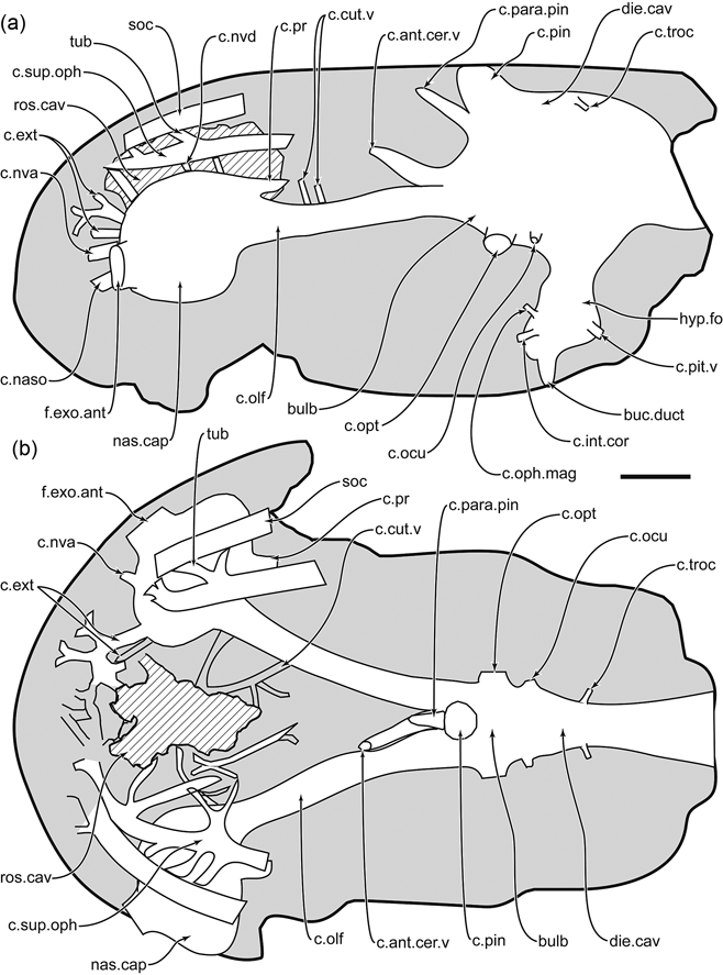
Reconstruction of the ethmosphenoid portion of the cranial cavity: (a) left lateral view; (b) dorsal view. Scale bar=2 mm. Abbreviations: buc.duct=buccohypophysial duct; bulb=bulbar portion of the braincase; c.ant.cer.v=canal for anterior cerebral vein; c.cut.v=canal for cutaneous blood vessel; c.ext=canal for r. externus narium; c.int.cor=canal for the internal carotid; c.naso=nasobasal canal; c.nva=anterior canal for nerve or vessel; c.nvd=canal for nerve of vessel perforating the roof of the nasal capsule; c.ocu=canal for oculomotor nerve; c.olf=canal for olfactory nerve; c.oph.mag=canal for the ophthalmica magna; c.opt=canal for optic nerve; c.para.pin=canal for parapineal organ; c.pin=canal for pineal organ; c.pit.v=canal for the pituitary vein; c.pr=canal for profundus nerve; c.sup.oph=canal for superficial ophthalmic nerve; c.troc=canal for troclear nerve; die.cav=cavity for the diencephalon; f.exo.ant=fenestra exonarina anterior; hyp.fo=hypophysial fossa; nas.cap=nasal capsule; ros.cav=rostral cavity; soc=supraorbital sensory line canal; tub=tubuli.

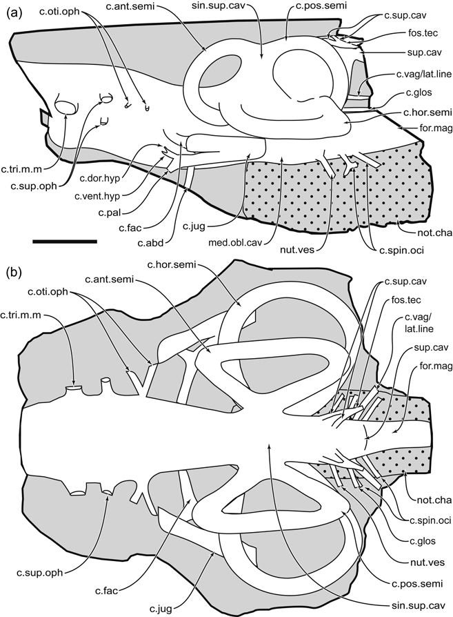
Reconstruction of the otico-occipital portion of the cranial cavity: (a) left lateral view; (b) dorsal view. Scale bar=4 mm. Abbreviations: c.abd=canal for abducens nerve; c.ant.semi=anterior semicircular canal; c.dor.hyp=dorsal opening for the hypotic ramus of the facial nerve; c.fac=canal for the main branch of the facial nerve; c.glos=canal for glossopharyngeal nerve; c.hor.semi=horizontal semicircular canal; c.jug=jugular canal; c.oti.oph=canal for otic ophthalmic ramus; c.pal=canal for the palatine nerve VII; c.pos.semi=posterior semicircular canal; c.spin.oci=canal for spino-occipital nerve; c.sup.cav=posterior canal exiting the lateral wall of the supraotic cavity; c.sup.oph=superficial ophthalmic ramus; c.tri.m.m=opening for the maxillary and mandibular components of the trigeminal nerve; c.vag/lat.line=canal for vagus, and presumably the lateral line nerve; c.vent.hyp=ventral opening for the hypotic ramus of the facial nerve; for.mag=foramen magnum; fos.tec=fossa tectosynotica; med.obl.cav=space for the medulla oblongata; not.cha=notochordal chamber; nut.ves=nutrient vessel; sin.sup.cav=cavity for the sinus superior; sup.cav=supraotic cavity.

Several difficulties make it untenable to include braincase characters from Gogonasus in a phylogenetic analysis of tetrapodomorph fishes at this time. Foremost is the large number of taxa for which detailed endocranial anatomy is unknown, as well as varying interpretations of morphology. For instance, reports of nasal capsule size, shape and the length of the olfactory canal differ between reconstructions of Ectosteorhachis in Romer (Reference Romer1937) and Thomson (Reference Thomson1964b), although both studies partially rely on the same grinding series (Thomson Reference Thomson1964b). It is tempting to suggest that the relatively short olfactory canal and elongate bulbar portion of the telencephalon region of the braincase in Eusthenopteron (Jarvik Reference Jarvik1980) could be derived character states exhibited by tristichopterids, and associated with the elongation of the snout. However, although Gogonasus lacks these features, they are present in Osteolepis (Thomson Reference Thomson1965).
Other potential hazards in defining character states are the numerous instances of asymmetry on either side of the midline in some tetrapodomorph fish taxa. The presence of both single and double internal openings (with the associated internal latitudinal ridge) for the trigeminal nerve in Gogonasus specimen NMV P221807, and the ophthalmicus superficialis in Medoevia (Lebedev Reference Lebedev1995a), suggest little phylogenetic importance. Asymmetry also occurs in the shape of the rostral cavity, the branching of the canals for the cutaneous blood vessels and the slightly laterally-slanted canal for the anterior cerebral vein in the midline of specimen NMV P221807. Differences in the number of openings for the vagus/lateral line nerves have also been reported in Gogonasus specimen ANU 49259 (Long et al. Reference Long, Barwick and Campbell1995, p. 51).
Several other features described from Gogonasus specimen NMV P221807 may represent ontogenetic states. These include the notch representing the glossopharyngeal nerve foramen in the posterodorsal border of the vestibular fontanelle, which superficially appears unique to specimen NMV P221807. This opening appears close to, but separate from, the vestibular fontanelle in other tetrapodomorph fish taxa (e.g. Lebedev Reference Lebedev1995a) and in Gogonasus specimen WAM 86.9.661 (TH pers. obs.). The larger size of the latter specimen suggests the margins of the vestibular fontanelle might have ossified later in development in Gogonasus, along with the area surrounding the glossopharyngeal nerve. Increasing braincase ossification with ontogeny may also affect the region around the abducens nerve opening, which is more complete in specimen WAM 86.9.661 than in specimen NMV P221807. The reported ‘suture’ possibly separating the ethmoid from the sphenoid portion of the braincase (sut; Fig. 21) in Gogonasus also needs to be further assessed. Long (Reference Long1985) and Long et al. (Reference Long, Barwick and Campbell1997) attributed this fissure to either an early ontogenetic state or a result of the fine preservation of Gogo material. It appears to acutely undulate beneath the intertemporal, then continue ventrally between the openings for the oculomotor and optic nerves, with a probable muscle scar (mus.scar; Fig. 21) positioned slightly anterior to the latter foramen. This feature is clearly visible in the holotype specimen ANU 21885, on the left side of specimen ANU 49259, and as a faint line on the right side of specimen NMV P221807, with all specimens being approximately equal in size. Long et al. (Reference Long, Barwick and Campbell1997, fig. 10) also figured this suture in WAM 86.9.661, the largest specimen of Gogonasus andrewsae, but it is less distinct in photographs of the same specimen (Long et al. Reference Long, Barwick and Campbell1997, fig. 11; TH pers. obs.). Thus, it is possible that this suture is strongly maintained only in the smaller specimens, indicative of an early growth state.
4.2. Resolving misinterpretations of tetrapodomorph fish morphology
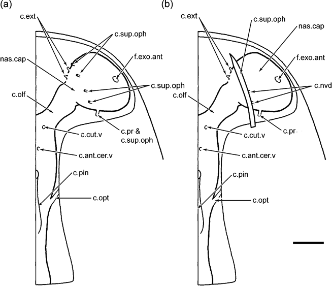
The description of the internal braincase anatomy of Gogonasus also potentially elucidates several differing interpretations of tetrapodomorph fish morphology, including that of the postnasal wall. This particularly relates to the opening for the superficial ophthalmic nerve and the fenestra endonarina posterior, the latter being clearly absent from the nasal capsule of Gogonasus and Cladarosymblema (Fox et al. Reference Fox, Campbell, Barwick and Long1995). In his work on Eusthenopteron foordi, Jarvik (Reference Jarvik1942, Reference Jarvik1980) described this canal as the conduit for nerves, blood vessels and an apparent posterior nasal tube, acting as a nasolacrimal duct serving the orbit. Although hesitant to assign such function with the lack of soft-tissue preservation, Thomson (Reference Thomson1964b) later identified an homologous posteroventral fenestra in the postnasal wall of Ectosteorhachis. Furthermore, the fenestra endonarina posterior was also identified as being present and continuous with the ‘fenestra endochoanalis’ in Porolepis by Jarvik (Reference Jarvik1942), although subsequent work has clearly shown the absence of choanae amongst the Porolepiformes (Clément Reference Clément2002). However, Ahlberg (Reference Ahlberg1991) highlighted the term ‘orbitonasal canal’ to describe a nasal capsule aperture in Youngolepis, Powichthys, porolepiforms, dipnoans and actinistians, apparently homologous to the “large opening in the lateral part of the postnasal wall of Eusthenopteron … interpreted as a nasolacrimal duct by Jarvik (Reference Jarvik1942, and elsewhere)”. Unfortunately, the term ‘orbitonasal canal’ also appears to have been used to describe a different, more medially positioned canal in other tetrapodomorph fishes. This ‘foramen orbitonasale’ was thought to carry the profundus and superficial ophthalmic nerves inside the nasal capsule in Osteolepis (Thomson Reference Thomson1965) and Gyroptychius (Thomson Reference Thomson1964a), therefore being radically different from the morphology of the postnasal wall of Eusthenopteron foordi, where these nerves apparently pass through independent canals, and not the fenestra endonarina posterior (Jarvik Reference Jarvik1980). A similar set of separate openings for the orbitonasal canal and profundus nerve/nerves are also present in the dipnomorphs Powichthys and Porolepis (Clément & Janvier Reference Clément and Janvier2004). Based on this data, a new interpretation of the postnasal wall perforations in Osteolepis (Thomson Reference Thomson1965) and Gyroptychius (Thomson Reference Thomson1964a) is presented here. In these two taxa, the ‘foramen orbitonasale’ is reinterpreted as a canal solely carrying the profundus nerve, and is not homologous to the fenestra endonarina posterior. Evidence for the passage of the superficial ophthalmic nerve inside the nasal capsule via the canal for the profundus nerve is presented by Thomson (Reference Thomson1964a, Reference Thomson1965, Reference Thomson1967). This erroneous hypothesis was based on the presence of multiple small canals exiting the roof of the nasal capsule, thought by Thomson (Reference Thomson1967) to represent branches of the superficial ophthalmic nerve travelling outwards to the dorsally-positioned lateral line canal (Fig. 24a). The description of the nasal capsule in Gogonasus specimen NMV P221807 clearly shows several similar dorsal openings which do not carry the main course of the superficial ophthalmic nerve, as it is positioned outside the nasal capsule. This morphology is likely present in Osteolepis (Fig. 24b) and Gyroptychius, with previous reconstructions (Thomson Reference Thomson1964a, Reference Thomson1965) based on incomplete natural moulds, in which the canal for the superficial ophthalmic nerve outside the nasal capsule is unpreserved. Such an interpretation would also be in line with the morphology present in other tetrapodomorph fishes (Thomson Reference Thomson1964b; Jarvik Reference Jarvik1980). It is worth noting that the apparent profundus nerve external to the nasal capsule of Spodichthys in Snitting (Reference Snitting2008) is interpreted here as a misidentified superficial ophthalmic nerve. Varying interpretations of morphology pertaining to the fenestra endonarina posterior amongst tetrapodomorph fishes has been addressed by Lebedev ((Reference Lebedev1995a)=orbitonasal canal) as having “no specific phylogenetic meaning”. In the absence of a large, laterally-positioned canal in the nasal capsule in Medoevia, a notch in the ventral margin of the postnasal wall has been interpreted as facilitating the passage of the orbitonasal canal (Lebedev Reference Lebedev1995a). This morphology has also been attributed to Shirolepis, Jarvikina, Eusthenopteron obruchevi and Gogonasus specimen ANU 21885 (Lebedev Reference Lebedev1995a). Although the ventral margin of specimen NMV P221807 is concave, it is not permissible to attribute to any specific canal to this area, as there is no acute shape for such lodging.
Reconstruction of the course of the superficial ophthalmic nerve in the ethmosphenoid of Osteolepis: (a) previous reconstruction of the path of the superficial ophthalmic nerve inside the nasal capsule from Thomson (Reference Thomson1967); (b) new reconstruction of the superficial ophthalmic nerve canal dorsal to the nasal capsule. Scale bar=1 mm. Abbreviations: c.ant.cer.v=canal for anterior cerebral vein; c.cut.v=canal for cutaneous blood vessel; c.ext=canal for r. externus narium; c.nvd=canal for nerve of vessel perforating the roof of the nasal capsule; c.olf=canal for olfactory nerve; c.opt=canal for optic nerve; c.pin=canal for pineal organ; c.pr=canal for profundus nerve; c.sup.oph=canal for superficial ophthalmic nerve; f.exo.ant=fenestra exonarina anterior; nas.cap=nasal capsule.

4.3. Electroreception
Finally, of the internal features of the ethmoid region, none is more curious than the potential electroreceptive properties of the intricate tubuli and large capsule underlying the median postrostral and rostral series. In her studies on Gogo Formation dipnoans, Cheng (Reference Cheng1989) suggested that the rostral tubuli of Chirodipterus and Griphognathus represented sunken neuromasts associated with the lateral line system, enclosed underneath external dermal bone. However, this work discounted possible electroreception, based on the apparent lack of homologous tubular structures relating to electroreception, and the chiefly seismosensory function of the lateral line system (Cheng Reference Cheng1989). This contrasts to the position taken by Campbell & Barwick (Reference Campbell and Barwick1986), who convincingly inferred rostral tubuli electroreception in Devonian lungfishes, based on the innervation from the profundus and superficial ophthalmic nerves, association with the lateral line system and the close proximity to the overlying cosmine pore canals. In regards to electroreception, this comparable arrangement in Gogonasus is increasingly compelling with the presence of the median ethmoid cavity. The similarly-placed cavity in the snout of actinistians houses the gelatinous-filled, electroreceptive rostral organ (Bemis & Hetherington Reference Bemis and Hetherington1982). Aside from the large associated canals perforating the skull roof, the relative position and innervation from the superficial ophthalmic nerve of this cavity seems homologous to that of Gogonasus. Interestingly, large internal cavities have been depicted in the snouts of the tetrapodomorph fish Platycephalichthys (Vorobyeva Reference Vorobyeva1962) and Panderichthys (Vorobyeva Reference Vorobyeva1962), although their veracity has been questioned by Thomson (Reference Thomson1967). These cavities differ dramatically in morphology from that of Gogonasus in seemingly being connected directly to the olfactory canal. A similarly connected hollow, known as the “forebrain division of the cranial cavity” has been reported from Porolepis (Jarvik Reference Jarvik1942). However, the presence of this structure in porolepiform fishes has been disputed by Thomson (Reference Thomson1967), who cites the absence of this cavity in other specimens of Porolepis (Kulczycki Reference Kulczycki1960) and in Glyptolepis (Stensiö Reference Stensiö1963). Perhaps these structures figured in Vorobyeva (Reference Vorobyeva1962) and Jarvik (Reference Jarvik1942) do not represent divisions of the forebrain, but misidentified cavities for an electroreceptive organ which have been damaged through preservation. This scenario seems plausible considering the fragile network of thin canals supporting the capsule in Gogonasus, and for this reason such characters may be difficult to include in a phylogenetic analysis. The description of this area as “solid” in Eusthenopteron (Jarvik Reference Jarvik1942, p. 428) and containing “no large cavities” in Ectosteorhachis (Thomson Reference Thomson1964b) may alternatively reflect a deficit in fine preservational detail. Along with the aforementioned sub-rostral structures in specimen NMV P221807, the rostral tubuli of dipnomorphs, including basal members such as Diabolepis, Powichthys and Youngolepis (Chang & Yu Reference Chang and Yu1997) and the rostral organ of actinistians, could suggest the presence of electroreceptive organs underneath the dermal bone of the snout as a plesiomorphy for crown-group sarcopterygians. It is important to note that possible electroreception in tetrapodomorph fishes have been previously proposed. The possibly electroreceptive-related tubuli described from Cladarosymblema by Fox et al. (Reference Fox, Campbell, Barwick and Long1995) appear similar to those of Gogonasus in lacking connections with the external surface via individual pores (an evidently derived condition present in lungfish), but are linked indirectly through association with the sensory line canal system. Work by Thomson (Reference Thomson and Andrews1977) had previously focused on the pore canal sensory system of other megalichthyids, and the possible peripheral integration of electric signal with ampullary receptors. Thomson (Reference Thomson1967) posited that “… electroreception is an ancient feature of all fishes”. This position is bolstered by the report of tubuli in the snout of an Emsian placoderm in the collections of the Australian National University (Campbell & Barwick Reference Campbell and Barwick2001) and the ampulla of Lorenzini in sharks and rays (Kalmijn Reference Kalmijn1971). Indeed, electroreception is a plesiomorphic feature amongst the Gnathostomata, with the condition lost in most teleost and tetrapod groups (Bullock et al. Reference Bullock, Bodznick and Northcutt1983). Within the Tetrapoda, rare instances of electroreceptive reinnervation are known from the hairless vibrissal crypts in the rostrum of the Guiana dolphin Sotalia guianensis (Czech-Damal et al. Reference Czech-Damal, Liebschner, Miersch, Klauer, Hanke, Marshall, Dehnhardt and Hanke2012) and the cutaneous glands in the bills of monotremes (Pettigrew Reference Pettigrew1999). In these instances, electroreception is permitted by innervation of the trigeminal nerve; in contrast to the condition in amphibians where electroreception is facilitated by innervation of the lateral line system (Fritzsch & Munz Reference Fritzsch, Munz, Bullock and Heiligenberg1986). It is worth noting that the presence of an electroreceptive organ has been hypothesised in the skull of the Carboniferous tetrapod Baphetes (Bjerring Reference Bjerring and Rocek1986), suggesting that electroreception was maintained during the evolution of early tetrapods from basal sarcopterygian stock.
4.4. Future research
It is important that the description of the internal braincase in Gogonasus is accompanied by future comparative studies of other tetrapodomorph fish endocrania. Further research is required on initial tomographic analysis of the braincase in Eusthenopteron (Ahlberg et al. Reference Ahlberg, Blom, Brazeau, Clément, Snitting, Ivanov and Young2005; Ahlberg Reference Ahlberg2006) to verify various aspects of the description by Jarvik (Reference Jarvik1980, including the apparent subdivision of the nasal capsule into three distinct regions), the presence of the processus dermintermedius overlying a separate processus intermedius, and the presence of a separate canal for the vena cerebralis anterior on each olfactory canal. Similar reinterpretations on comparative material in Ectosteorhachis could also help confirm the work by Romer (Reference Romer1937), with specimens such as AMNH 7271 making an excellent candidate for micro CT-scanning. Other potential sources for CT-scanning include the wealth of uncrushed and near complete tetrapodomorph braincase material from Russia, including material from Cryptolepis grossi (Lebedv Reference Lebedev1995b, figs 7, 8), Medoevia lata (Lebedev Reference Lebedev1995a, figs 2, 6), Thursius estoniscus (Vorobyeva Reference Vorobyeva1977, fig. 25), Megapomus heckeri (Vorobyeva Reference Vorobyeva1977, fig. 34) and Megistolepis klumentzi (Vorobyeva Reference Vorobyeva1977, fig. 35). Further coding of characters from these taxa may well provide a much better resolution of tetrapodomorph fish phylogeny.
5. Acknowledgements
I would like to give special thanks to John Long of the Los Angeles County Museum and Patricia Vickers-Rich of the School of Geosciences, Monash University, Clayton for their supervision and guidance during my PhD candidature; to Sarah McCaffrey of Museum Victoria for her photography; to Tim Senden and Ajay Limaye of the Department of Applied Mathematics, Research School of Physical Sciences and Engineering, Australian National University for their micro-CT scanning and Drishti tuition; and to David Pickering and Erich Fitzgerald of Museum Victoria for their encouragement and tutelage. Invaluable discussion surrounding relevant topics was also had with Daniel Snitting of the Sub-Department of Evolutionary Organismal Biology, Department of Physiology and Developmental Biology, Uppsala University, Sweden; Gavin Young of the Research School of Earth Sciences, Australian National University; Kate Trinajstic of Curtin University, Perth; John Maisey of the American Museum of Natural History, New York; Daniel Goujet of the Museum National d'Histoire Naturelle, Paris; Zhu Min of the Institute of Vertebrate Paleontology and Paleoanthropology, Beijing; and Mike Taylor of the National Museum of Scotland, Edinburgh. This research was supported by a Monash University Dean's Postgraduate Scholarship.