Introduction
Taenia solium is a taeniid cestode parasite that causes taeniasis in humans and cysticercosis in both humans and pigs. Neurocysticercosis occurs when metacestodes affect the central nervous system. Experimental models are excellent tools for understanding the host–parasite interactions of human cysticercosis. Experimental murine Taeniidae metacestode infections or inoculation with Taeniidae secretion products have been used to study structural gonadal tissues and reproductive functions. It has been observed that a substance secreted by Taenia taeniaeformis significantly reduced the in vitro testosterone production in rat Leydig cells (Rikihisa et al., Reference Rikihisa, Lin and Fukaya1985). Studies of larval T. taeniaeformis infection in female and male rats showed that mating, uterine implantation sites and the number of full-term pups were decreased in females 5 months after infection. Furthermore, serum and testicular testosterone levels were significantly decreased in male rats (Lin et al., Reference Lin, Rikihisa, Kono and Gu1990). Recently, it was reported that Taenia crassiceps infection disrupted the oestrous cycle and significantly decreased oestradiol levels in female mice (Arteaga-Silva et al., Reference Arteaga-Silva, Vargas-Villavicencio, Vigueras-Villaseñor, Rodríguez-Dorantes and Morales-Montor2009). Examination of the testis seminiferous epithelium from mice intraperitoneally infected with T. crassiceps metacestodes, or subcutaneously inoculated with a low molecular weight T. crassiceps metacestode factor (MF), revealed severe disruption and generalized apoptosis of the seminiferous tubule cells. Significant infiltration of macrophages in the tubular lumen was also observed (Zepeda et al., Reference Zepeda, Copitin, Solano, González, Fernandez, Tato and Molinari2011a, Reference Zepeda, Copitin, Solano, Fernandez, Tato and Molinarib). A low molecular weight ( < 3.5 kDa) T. solium MF has previously been isolated and shown to impair the proliferation of human and murine lectin-stimulated lymphocytes and reduce the inflammatory reaction surrounding subcutaneously implanted T. solium metacestodes in mice (Molinari et al., Reference Molinari, Tato, Reynoso and Cazares1990; Tato et al., Reference Tato, Castro, Rodriguez, Soto, Arechavaleta and Molinari1995). Subsequently, it was reported that this T. solium MF reduced in vitro interleukin (IL)-2, interferon (IFN)-γ, IL-4 and tumour necrosis factor (TNF)-α production from lipopolysaccharide (LPS)-stimulated macrophages (Arechavaleta et al., Reference Arechavaleta, Molinari and Tato1998) and also induced DNA damage in human lymphocytes in vitro (Herrera et al., Reference Herrera, Tato, Molinari, Perez, Dominguez and Ostrosky-Wegman2003). Additionally, excretion/secretion products of T. crassiceps larvae suppressed the T-cell proliferative responses in vitro as well as IFN-γ and IL-4 production early during the infection (Spolski et al., Reference Spolski, Corson, Thomas and Kuhn2000). The objective of this study was to determine whether infection of female mice with T. crassiceps metacestodes or inoculation with T. crassiceps MF leads to structural damage of the ovary.
Materials and methods
Parasitological procedures
Twenty-eight-day-old BALB/c female mice (20–22 g) were maintained under standard animal housing conditions with access to Purina chow and water ad libitum. Taenia crassiceps metacestodes (WFU strain) were maintained in our laboratory through peritoneal passage in female mice as described previously (Everhart et al., Reference Everhart, Kuhn and Zelmer2004).
Peritoneal fluid from both healthy (HPF) and T. crassiceps metacestode-infected female mice was obtained following the method of Zepeda et al. (Reference Zepeda, Copitin, Solano, Fernandez, Tato and Molinari2011b). Briefly, after 9 days of infection with T. crassiceps metacestodes, mice were anaesthetized and the peritoneal cavity of each mouse, including control mice, was washed with 8 ml sterile phosphate-buffered saline (PBS) through a small laparotomy. Peritoneal fluids were centrifuged at 4000 rpm at 4°C for 10 min, and supernatants from each group were pooled, lyophilized and stored at 4°C until use.
To obtain the T. crassiceps MF, lyophilized peritoneal fluids (15 mg dry weight) from infected mice were resuspended in 15 ml PBS, filtered through a 0.22 μm Millipore membrane and loaded on to a Sephadex G-50 M (90 × 1.5 cm) column (Pharmacia, Sweden) according to the method of Zepeda et al. (Reference Zepeda, Copitin, Solano, Fernandez, Tato and Molinari2011b). Fractions, each of 4 ml, were then collected and the eluted material was monitored at 280 nm. The eluted fractions containing the MF were pooled as well as the corresponding eluted fractions from the HPF. Pooled fractions were lyophilized and stored at 4°C until use. Protein and carbohydrate concentrations were determined in both pools using the Bradford (Reference Bradford1976) and Anthrona methods (Ashwell, Reference Ashwell1957), respectively.
Female mice were divided into each of three groups of six mice, namely control, MF-treated and mice infected with T. crassiceps. Control mice were inoculated subcutaneously (sc) every 4 days for up to 45 days with 0.1 ml HPF containing 50 μg carbohydrates previously filtered through a 0.22 μm Millipore membrane. MF-treated mice were inoculated subcutaneously every 4 days for up to 45 days with 0.1 ml MF containing 50 μg carbohydrates previously filtered through a 0.22 μm Millipore membrane. Infected mice were implanted intraperitoneally (ip) with 40 T. crassiceps metacestodes (2–3 mm diameter) resuspended in 0.2 ml PBS.
Microscopy and TUNEL assays
On day 45 post-treatment, all mice were euthanized and their ovaries were carefully removed and fixed with 2.5% paraformaldehyde in PBS for 12 h at room temperature. Ovaries were dehydrated using a graded series of ethanol concentrations ranging from 70 to 100% and then stored until further use. The left ovary of each mouse was embedded in paraffin, and 5-μm sections were prepared on a rotary microtome and subjected to haematoxylin/eosin (H/E) and TUNEL (terminal deoxynucleotidyl transferase (TdT)-mediated dUTP nick end labelling) staining (Roche, Salt Lake City, Utah, USA). The right ovary was prepared for transmission electron microscopy analysis.
Two H/E stained sections from each ovary were analysed using an IX71 Olympus light microscope, and all follicles and oocytes of each section were counted. To classify follicles, we used the following oocyte and follicle classification system proposed by Hsueh et al. (Reference Hsueh, Billig and Safriri1994): (1) primary follicles, with a single layer of cuboidal granulosa cells; (2) preantral or secondary follicles, with two or three layers of granulosa cells around the oocyte; (3) antral follicles, with several layers of granulosa cells, an antrum filled with fluid, and well-differentiated thecal cells outside the basement membrane; and (4) preovulatory or Graafian follicles, which are follicles that reach the maximal diameter and are responsive to the preovulatory surge of gonadotropins.
The other ovaries were postfixed in 1% buffered osmium tetroxide for 1.5 h at 4°C. Samples were transferred into 70% acetone solution, treated with 1% phosphotungstic acid and 1% uranyl acetate at 20°C for 1 h, dehydrated in acetone and subsequently embedded in Epon. Ultrathin sections were mounted on copper grids, contrasted with uranyl acetate and lead citrate, and viewed using a Jeol 100 S electron microscope. Micrographs were obtained with an Orius Sc1000A1 digital camera (Gata, Pittsburg, Pensylvania, USA).
The presence of DNA strand breaks in the paraffin-embedded mouse-ovary cross-sections was evaluated using TUNEL assays and an in situ Cell Death Detection Kit (Roche). Stained images were acquired under an FV 1000 Olympus inverted confocal microscope, and then analysed off-line with the Image Pro V 6.2 program (Media Cybernetics, Silver Spring, Maryland, USA) to determine the mean ± SD and P values of fluorescent objects/areas (DNA) ranging from 0.3 to 6 μm.
Data analysis
Two H/E-stained ovary cross-sections per mouse were selected randomly and completely scanned three times under a Nikon Alphaphot YS optic microscope at 400 × . During the first scan, all healthy and atretic follicles were carefully identified and counted, except the primordial and primary follicles, as well as the corpora lutea. Healthy antral stages and atretic antral follicles were counted during the second scan, and degenerated oocytes were counted during the third and final scan. TUNEL-stained ovary sections (two sections per ovary) were analysed with ImagePro V 6.2 using the TUNEL-positive objects/area parameter. Two ultrathin sections per mouse (12 sections per group) were evaluated with transmission electron microscopy. Micrographs of healthy and atretic primordial, primary and secondary follicles of each section were obtained for further analysis. All data were processed to determine means ± SD and P values according to the Mann–Whitney test using the GraphPadInStat software (San Diego, California, USA). P values less than 0.05 were considered statistically significant.
Results
Peritoneal fluids
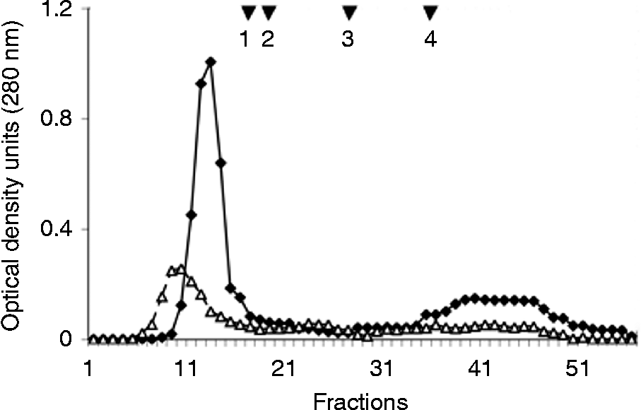
Peritoneal fluid samples from healthy or T. crassiceps metacestode-infected female mice were ultrafiltered using a Sephadex G-50 column. The elution profile of the peritoneal fluid from infected mice showed two peaks. Based on the molecular standards used to calibrate the column, the first peak was eluted with the void volume, but the second peak, which contained the T. crassiceps MF, eluted very close to vitamin B12 (molecular weight 1.3 kDa, standard 4). The elution profile of healthy peritoneal fluid also showed a first peak that was eluted with the void volume between fractions 7 and 18, and a lower absorbance peak that eluted between fractions 30 and 44 (fig. 1).
Elution profiles of the peritoneal fluids from mice infected with Taenia crassiceps (♦) and control mice (△) using a Sephadex G-50 column; molecular weight standards are 1 = 105.92 kDa, 2 = 36.84 kDa, 3 = 9.2 kDa, and 4 = 1.3 kDa.

Analyses of carbohydrate concentration in the second peaks of Sephadex G-50 showed that there was twice as much carbohydrate in the peritoneal fluid of infected mice (55.5 μg/mg dry weight) compared with healthy mice (27.6 μg/mg dry weight).
Histology and ultrastructure
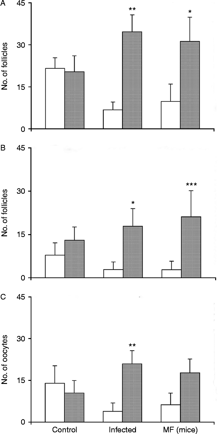
Analyses of the H/E-stained ovary sections revealed a significant increase of atretic follicles, mainly at the antral stage of development, from mice infected with T. crassiceps metacestodes and those inoculated with MF, compared with control mice (fig. 2A, B). The number of degenerated oocytes in both the infected group and the MF group was higher than in the control group (fig. 2C), although this difference was significant only in infected mice. Finally, a decrease of corpora lutea was found in the ovaries of the mice of infected and MF-treated groups compared with those of the mice of the control group, even though this was only significant in the MF group (0.5 ± 0.83 for MF-treated mice versus 3.6 ± 0.89 for control mice, P= 0.01). Additionally, there were evident degenerative alterations of numerous granulosa cells and oocytes of primary and secondary follicles from infected and MF-treated mice. In a large number of mice, the degenerative alterations were not very evident because all degenerative follicles could not be defined sharply in the examined sections. On the contrary, primary and secondary follicles of control mice were intact (data not shown).
The mean number ( ± SD) of atretic (grey bars) and non-atretic follicles (white bars): (A) total atretic and non-atretic follicles; (B) antral atretic and non-atretic follicles. (C) The degenerated (grey bars) and non-degenerated oocytes (white bars) in the ovaries of mice infected with Taenia crassiceps, metacestode factor-treated mice (MF) and controls. P< 0.05(*), P< 0.01(**) and P< 0.001(***).
There was an intense inflammatory reaction caused primarily by mononuclear cells in the adipose tissue surrounding the ovaries of infected mice, though inflammation was not observed around of the ovaries of MF-treated and control mice (data not shown).
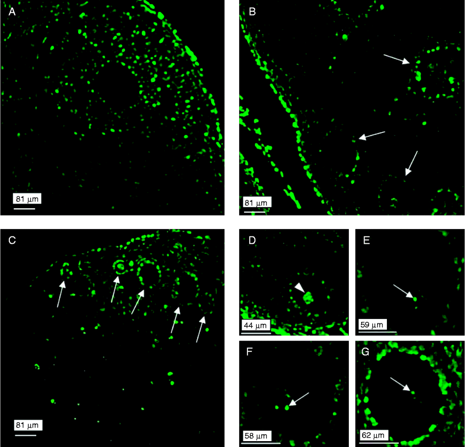
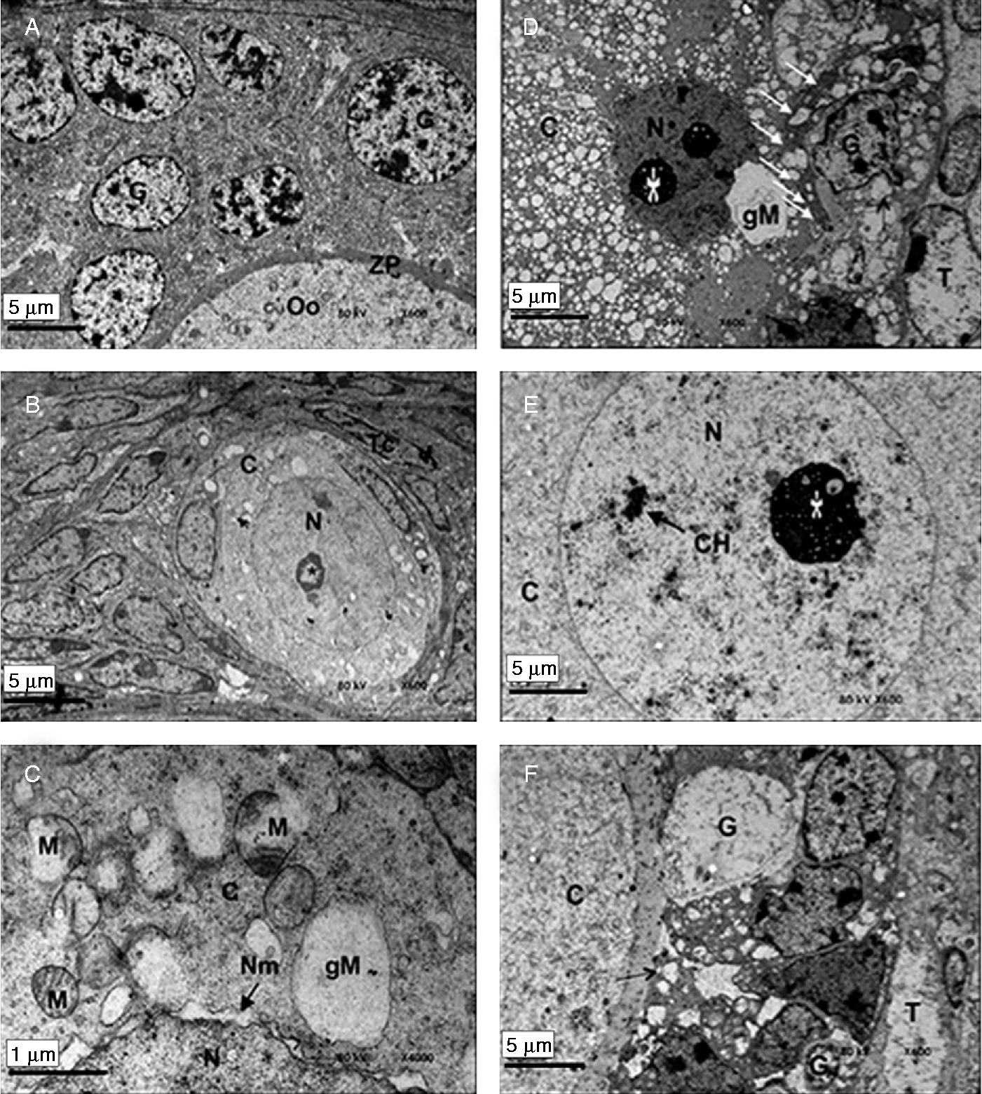
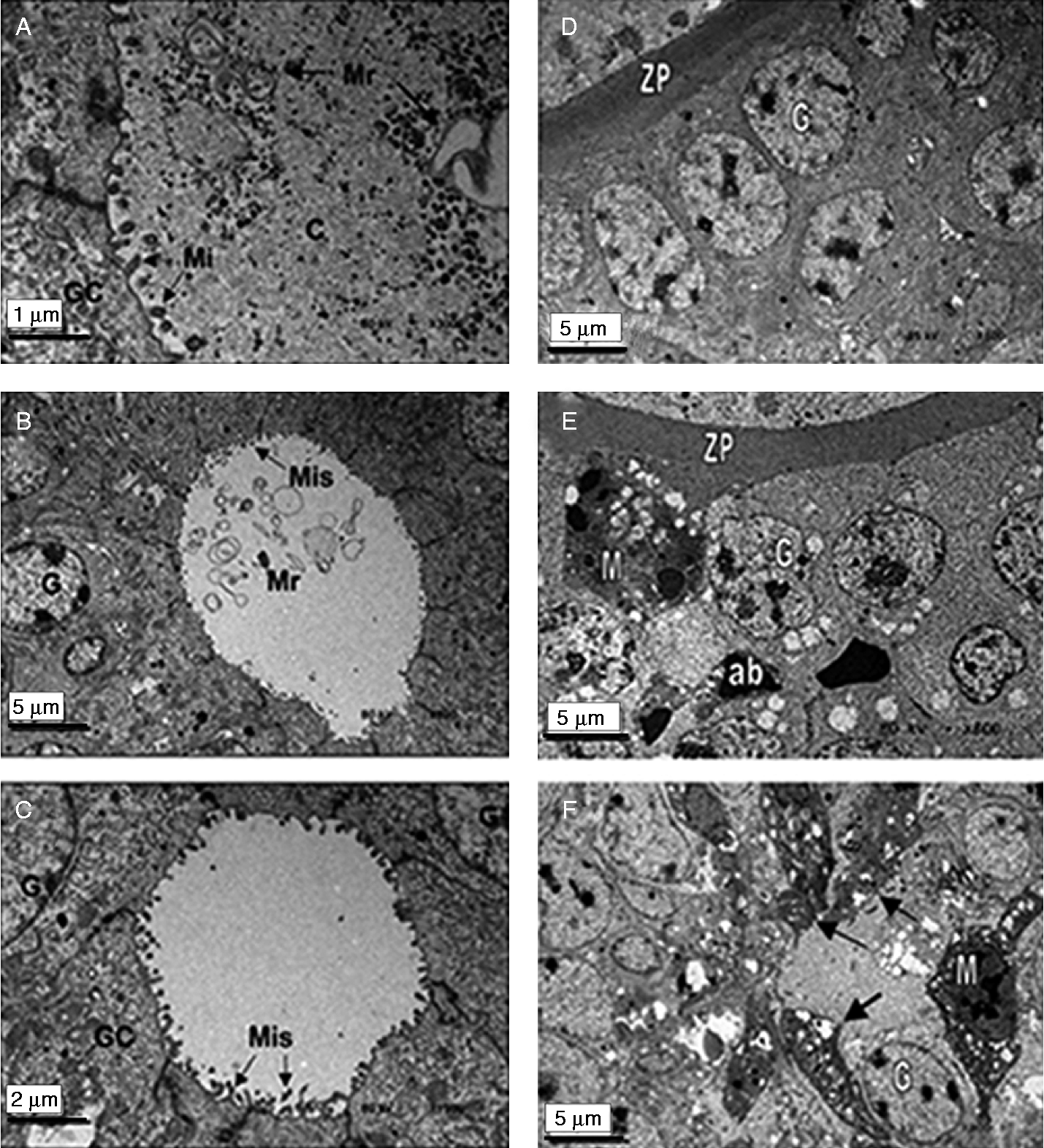
To determine whether apoptosis occurs in the follicles during the early stages of development, TUNEL staining was used on ovary sections. Images of TUNEL-stained ovary sections were obtained using confocal microscopy. Analysis of ovary sections from control mice did not show fluorescence of granulosa cells or oocytes in the early stages of development (fig. 3A). In contrast, sections of ovaries from infected mice or those inoculated with MF showed that granulosa cells (the first line) in primary and secondary stages of development, and some oocytes exhibited positive TUNEL staining (fig. 3B and C). In some cases, fragmented oocyte nuclei (fig. 3D), and particularly oocyte nucleoli from follicles in several stages of development, were intensely stained using TUNEL assays (figs 3E–G). In all ovary sections, some fluorescent cells were observed on the periphery of the ovary, which may be a natural phenomenon because the ovarian surface epithelium must be renewed to repair minor damage caused by ovulation. The mean ± SD of fluorescent objects was significantly higher in the TUNEL-stained ovary sections of MF-treated mice compared with control mice (546 ± 73 for MF-treated mice versus 198 ± 165 for control mice; P= 0.0143 for MF-treated mice versus control mice). Ultra-structural changes in the primordial, primary and secondary follicles of the ovaries in female mice intraperitoneally infected with T. crassiceps metacestodes and in MF-treated mice after 45 days of treatment were confirmed and enhanced using transmission electron microscopy analysis. Grouped together, the means ± SD of degenerated primary and secondary follicles in ovaries from infected (0.833 ± 0.408) and MF-treated mice (0.833 ± 0.408) were significantly higher compared to controls that showed no degenerated follicles at this stage of development (P< 0.05 control versus infected or MF mice). On the other hand, in a large number of mice, healthy follicles were not observed in the examined sections. Granulosa cells and oocytes in primary and secondary follicles of ovaries of control mice appeared to be intact (fig. 4A). In contrast, in infected mice, granulosa cells and oocytes of primordial, primary and secondary follicles exhibited several abnormalities. A generalized degeneration of the mitochondria in the oocyte cytoplasm was observed, as well as deformity and separation of the nuclear membrane bilayers of oocytes in primordial and primary follicles (fig. 4B and C). Although some primary follicles showed large vacuolar structures, which were most likely formed through the fusion of several mitochondrial membranes, they likely contained mitochondrial membrane remnants. In ovary sections of MF-treated mice, almost all theca and granulosa cells were degenerated. The latter cell type showed severe signs of degeneration, such as numerous enlarged and empty mitochondria (fig. 4D). Some oocytes of primary and secondary follicles exhibited amorphous condensation of heterochromatin (fig. 4D and E). A secondary follicle showed pyknotic granulosa and theca cells with peripheral chromatin and empty cytoplasmic membranes (fig. 4F). In the same MF-treated mice sections, one peculiar finding was an apparent retraction of the oocyte cytoplasm observed in one cell, which left a space between the granulosa cells and the oocyte cytoplasm, allowing observation of microtubules joining the granulosa cells with their oocyte (fig. 5A). No oocyte was observed in the primary follicles and only microtubule stumps at the periphery of the granulosa cells remained (fig. 5B and C). In one of these follicles, only membrane remnants were observed instead of a complete oocyte (fig. 5B). Finally, in some sections with T. crassiceps infection and with MF-treated mice, macrophages were observed among the granulosa cells in nearly all the atretic follicles (fig. 5E and F). In control mice macrophages were not observed (fig. 5D).
Apoptosis in ovaries from control (A), Taenia crassiceps-infected (B), and MF-treated mice (C, D–G) to show (A) an atretic anthral follicle and some ovary peripheral fluorescent cells; (B) and (C) fluorescent (TUNEL-positive) granulosa cells in primary and secondary follicles (arrows); (D–G) fluorescent granulosa cells, a fragmented oocyte nucleolus (arrowhead) and oocyte nucleoli (arrows).
Alterations to the ovary of Taenia crassiceps-infected mice (A, B, C) and MF-treated mice (D, E, F): in (A) intact secondary follicle, oocyte (Oo), zona pellucida (ZP), and granulosa cells (G), 600 × ; in (B) degenerated primordial follicle, oocyte nucleus (N), oocyte cytoplasm (C), nucleolus (*), theca cell (Tc) 600 × ; in (C) degenerated mitochondria (M) in the oocyte cytoplasm, an empty giant mitochondrion (gM), separation of the bilayer of the nuclear membrane (Nm), and a portion of the nucleus (N) (magnification of a section of (B)) 4000 × ; in (D) a degenerated primary follicle, a macrophage surrounding a granulosa cell (white arrows), enlarged and empty mitochondria (black arrow), hypervacuolated oocyte cytoplasm, giant mitochondrion in a granulosa cell (gM), degenerated engulfed granulosa cell (G), degenerated theca cell (T), 800 × ; in (E) an oocyte nucleus, precipitated chromatin (CH), 800 × ; in (F) a degenerated secondary follicle, degenerated granulosa cells (G, one is pyknotic and the other is an empty cytoplasmic membrane) and a degenerated theca cell (T), 600 × .
Degeneration of ovaries from MF-treated (A, B, C, F), control (D) and Taenia crassiceps-infected mice (E) to show: (A–C) degenerated primary follicles, degenerated oocyte cytoplasm (C), oocyte membrane remnants (Mr), microtubules (Mi), granulosa cell cytoplasm (GC), granulosa cell (G), and microtubule stumps (Mis), 3000 × , 600 × and 1500 × , respectively; (D) a control intact follicle zona pellucida (ZP), 800 × ; (E) an atretic follicle with a macrophage (M) among several morphologically altered granulosa cells (G) and some apoptotic bodies (ab) inside macrophage cytoplasm, 800 × ; (F) an atretic follicle with several macrophages (M and arrows) interspersed among altered granulosa cells (G), 800 × .
Discussion
The present results identify the histopathological effects induced by intraperitoneal Taenia crassiceps metacestode infection or subcutaneous inoculation with a low molecular weight T. crassiceps MF in the ovaries of the mice. The increase in the carbohydrate concentration in the Sephadex G-50 MF second peak compared with the HPF suggests that at least half of this concentration could be secreted by intraperitoneal metacestodes.
Zepeda et al. (Reference Zepeda, Copitin, Solano, Fernandez, Tato and Molinari2011b) showed that subcutaneous inoculation of peritoneal fluid or T. crassiceps MF was successfully used and hence this procedure was chosen in this study despite the fact that parasites were implanted intraperitoneally.
Using light microscopy, inflammation was evident around the ovaries of T. crassiceps infected mice but not in control or MF-treated mice. Such inflammation is likely to occur in response to the presence of parasites in the peritoneum (Zepeda et al., Reference Zepeda, Solano, Copitin, Fernández, Hernández, Tato and Molinari2010). No inflammation was observed in the ovaries of any of the mice. Taenia crassiceps infection and secretion products of the parasite induce immunosuppression in mice (Spolski et al., Reference Spolski, Corson, Thomas and Kuhn2000; Zepeda et al., Reference Zepeda, Solano, Copitin, Fernández, Hernández, Tato and Molinari2010). Therefore, the alterations observed in ovaries from T. crassiceps infected and MF-treated mice may have been caused by the biological activity of the MF. So, further studies should be performed to elucidate the immune regulatory mechanisms of this process. H/E-stained ovary sections from these mice showed a significant increase of follicular atresia and oocyte degeneration, and a decrease of corpora lutea compared with control mice inoculated with peritoneal fluid fractions from healthy mice. After 45 days of treatment, a significant increase of follicular atresia was observed, predominantly in the antral stage of development. Another finding was the significant increase of atresia of primary and secondary follicles, together with oocyte degeneration (P< 0.05) in mice infected with T. crassiceps metacestodes or treated with MF. Ovarian follicular atresia of primordial, primary and secondary follicles observed in experimental mice in this study contrasts with previous reports because follicular atresia in the early stages of development has rarely been detected (Hirchfield, Reference Hirchfield1988; Teilmann, Reference Teilmann2005). The increase of follicular atresia observed at the different stages of development, along with the decrease of corpora lutea, suggested a high probability of alteration of reproductive function in infected or MF-treated mice. It has been reported that follicles that reach the early antral stage of development when levels of follicle stimulating hormone (FSH) are high can complete the final round of cell division and initiate the phenotypic differentiation that transforms them into preovulatory follicles (Hirchfield, Reference Hirchfield1988). Significant levels of naturally occurring apoptosis take place throughout fetal development and postnatal life in the female gonad (Tilly, Reference Tilly1996). Atresia occurs continuously after birth until the follicle pool is completely depleted (Hirchfield, Reference Hirchfield1991; Hsueh et al., Reference Hsueh, Billig and Safriri1994; Kaipia & Hsueh, Reference Kaipia and Hsueh1997). It has been suggested that follicles that degenerate may have failed to develop adequate receptors for specific hormones or they may have been deprived of critical hormonal support at a crucial stage of development. Apoptosis is the mechanism underlying follicular atresia in the mammalian ovary. Fas and Fas ligand expression, which are the proximal members of the death receptor pathway, have been observed in murine ovarian follicles (Dharma et al., Reference Dharma, Kelkar and Nandedkar2003). The term ‘apoptosis’ was coined by Keer et al. (Reference Keer, Wyllie and Currie1972) to describe a specific morphological aspect of cell death. Apoptosis is accompanied by cell rounding-up, cytoplasmic prolongation retraction, reduced cellular volume, chromatin condensation (pyknosis), nuclear fragmentation (karyorrhexis), plasma membrane blebbing, with maintenance of integrity until the final stages, and engulfment by resident phagocytes in vivo. Hence, the term ‘apoptosis’ can be applied exclusively to cell death-events that manifest several of these morphological features according to the Nomenclature Committee on Cell Death (Kroemer et al., Reference Kroemer, Galluzzi, Vandenabeele, Abrams, Alnemri, Baehrecke, Blagosklonny, El-Deiry, Goldstein and Green2009). Another remarkable finding was the significant increase of degenerated oocytes in the ovaries of infected mice. Oocyte degeneration can lead to a decrease or loss of the expression of factors that stimulate cumulus expansion, such as growth differentiation factor 9 (GDF-9) and bone morphogenetic protein (BMP15). Also, epidermal growth factor receptor (EGFR) expression in cumulus cells can be lost following oocyte degeneration (Elvin et al., Reference Elvin, Changning and Matzuk2000; Su et al., Reference Su, Sugiura, Li, Wigglesworth, Matzuc and Eppig2010). Cumulus expansion is one of several key processes that must occur in preovulatory follicles to enable ovulation. If cumulus expansion is suppressed, the ovulation rate is greatly reduced (Chen et al., Reference Chen, Russell and Larsen1993).
The physical and chemical characteristics of the low molecular weight ( < 1.350 kDa) T. crassiceps MF, its absorbance at 260 nm wavelength (Zepeda et al., Reference Zepeda, Copitin, Solano, Fernandez, Tato and Molinari2011b) and the presence of an oligosaccharide (Cassani et al., unpublished data) resemble the T. solium MF (Molinari et al., Reference Molinari, Tato, Reynoso and Cazares1990). The T. crassiceps MF resembles substances such as glycosphingolipids (ceramide mono-, di-, tri- and tetrasaccharides) that have been found in T. crassiceps metacestodes (Dennis et al., Reference Dennis, Baumeister, Geyer, Peter-Katalinic, Hartmann, Egge, Geyer and Weigandt1992) and, also, a ceramide that induces apoptosis in Jurkat cells (Mizushima et al., Reference Mizushima, Koike, Kohsaka, Kushi, Handa, Yagita and Miyasaka1996).
Both T. solium (Molinari et al., Reference Molinari, Tato, Reynoso and Cazares1990) and T. crassiceps factors depress lymphocyte proliferation, suggesting that they could systemically depress host immune responses. The ability of T. solium MF to reduce IL-2, IFN-γ, IL4 and TNF-α production from LPS-stimulated macrophages in vitro (Arechavaleta et al., Reference Arechavaleta, Molinari and Tato1998), and the ability of the T. crassiceps MF to significantly decrease splenic CD4+ cells in vivo (Zepeda et al., unpublished data) may be related to the observation that T. crassiceps metacestode infection in mice significantly depresses humoral responses to Salmonella typhimurium antigens (Rubio et al., Reference Rubio, Tato, Govezensky and Molinari1998). Currently, the mechanism by which T. crassiceps MF induces gonadal damage in both sexes is under investigation.
Transmission electron microscopy analysis detected high phagocytic activity, most likely by macrophages interspersed among the granulosa cells, particularly in antral/preovulatory follicles of infected and MF-treated mice. Macrophages are important regulators in the ovary because they modulate cellular proliferation, differentiation and apoptosis (van der Hoek et al., Reference van der Hoek, Maddocks, Woodhouse, van Rooijen, Robertson and Norman2000; Wu et al., Reference Wu, van der Hoek, Ryan, Norman and Robker2004).
The present findings may have important implications for understanding the problems of ovarian dysfunction and infertility in female hosts suffering from T. solium cysticercosis, particularly in neurocysticercotic patients parasitized by multiple cysts. A recent study showed that at 30 days post-infection with T. crassiceps metacestodes, female mice (n= 5) were unable to become pregnant (Molinari et al., unpublished data).
Human T. solium neurocysticercosis is a major public health problem in developing countries, and pathological findings presented here have not been described previously. Cardenas et al. (Reference Cardenas, Valdez, Saenz, Bottasso, Fragoso, Sciutto, Romano and Fleury2012) recently reported a decrease of sex hormones in neurocysticercotic patients (oestrogen and progesterone in women and testosterone in men). This is likely, because structural changes in the ovary and in the testis of mice were reported by Zepeda et al. (Reference Zepeda, Copitin, Solano, González, Fernandez, Tato and Molinari2011a, b), similar to the disease reported in this study. Structural damage to the gonads of mice induced by T. crassiceps infection or inoculation with the T. crassiceps MF could lead to a decrease in the production of hormones and infertility. Cardenas et al. (Reference Cardenas, Valdez, Saenz, Bottasso, Fragoso, Sciutto, Romano and Fleury2012) suggested that chronic stress during human cysticercosis could result in such endocrine changes. Bothelo et al. (Reference Bothelo, Soares, Vale, Ribeiro, Camilo, Almeida, Medeiros, Gomes, Machado and Correia da Costa2010) showed that an oestradiol (E-2)-related molecule produced by Schistosoma haematobium had an antagonistic effect on oestradiol in vitro by inactivating the oestrogen receptor (ER). The present study reveals that the pathological changes in the ovary may lead to temporary castration, allowing host resources to be used for the growth and fecundity of T. crassiceps.
Acknowledgements
We are grateful to R. Paredes and Juan M. Barbosa for performing the electron microscopy analyses and generating the figures, respectively. We also thank Veronica Rodriguez-Mata for technical support, and Silvia Tato and William L. Crothers for English language review.
Financial support
This work was financed by the Universidad Nacional Autónoma de México.
Conflict of interest
The authors declare that they have no conflicts of interest.
Ethical standards
The authors assert that all procedures contributing to this work comply with the ethical standards of the laws of Mexico on the care and use of laboratory animals. Animal studies were performed under an institutional protocol similar to that of the US Public Health Service: Guide for the Care and Use of Laboratory Animal from the Secretariat of Agriculture (SAGARPA NOM-062-ZOO-1999).