INTRODUCTION
Siboglinidae (Annelida: Vestimentifera) represent one of the most intriguing clades within polychaetes, from both a phylogenetic and an ecological point of view (e.g. Schulze, Reference Schulze2003; Schulze & Halanych, Reference Schulze and Halanych2003; Southward et al., Reference Southward, Schulze and Gardiner2005). Vestimentifera, within Siboglinidae, are some of the most typical organisms associated with reducing habitats such as hydrothermal vents and cold seeps, due to their obligate symbiosis with chemosynthetic bacteria. Vestimentifera are represented by ten formerly described genera and ~18 species (Pleijel et al., Reference Pleijel, Dahlgren and Rouse2009). The genus Lamellibrachia shows the highest species radiation with six described species and several species that remain un-described but can be identified at the molecular level (Kojima et al., Reference Kojima, Ohta, Yamamoto, Yamaguchi, Miura, Fujiwara, Fujikura and Hashimoto2003, Reference Kojima, Watanabe, Tsuchida, Fujikura, Rowden, Takai and Miura2006; McMullin et al., Reference McMullin, Hourdez, Schaeffer and Fisher2003; Miura & Kojima, Reference Miura and Kojima2006). In the Mediterranean Sea, Lamellibrachia sp. have recently been reported in the eastern basin from mud volcanoes off the coast of Crete (Olu-LeRoy et al., Reference Olu-LeRoy, Sibuet, Fiala-Medioni, Gofas, Salas, Mariotti, Foucher and Woodside2004), from a deep shipwreck off the coast of Crete (Hughes & Crawford, Reference Hughes and Crawford2008) and in the Nile Deep-sea Fan (Bayon et al., Reference Bayon, Loncke, Dupré, Caprais, Ducassou, Duperron, Etoubleau, Foucher, Fouquet, Gontharet, Henderson, Huguen, Klaucke, Masde, Migeon, Olu-Le Roy, Ondreas, Pierre, Sibuet, Stadnitskaia and Woodside2009).
Recently another record of Vestimentifera was reported from a pockmark in the Alboran Sea (west Mediterranean) (Hilário et al., in press) and other Siboglinidae species from mud volcanoes in the Gulf of Cadiz (Hilário et al., Reference Hilário and Cunha2008, Reference Hilário, Johnson, Cunha and Vrijenhoek2010). The Mediterranean specimens of Lamellibrachia from the eastern basin belong to a new species, currently under description (Southward et al., in press).
Previous records of Lamellibrachia in the Mediterranean Sea mainly refer to reducing habitats (pockmark, mud volcanoes; Hilário et al., in press, Southward et al., in press), the most typical biotope where the obligate endosymbiotic bacteria of these giant worms can perform chemosynthesis. However, Hughes & Crawford (Reference Hughes and Crawford2008) discovered Lamellibrachia sp. on a shipwreck (SS ‘Persia’) and suggested that these worms may rely on the paper ‘cellulose’ transported by the ship. This finding, together with another record from a shipwreck in the Atlantic (Dando et al., Reference Dando, Southward, Southward, Crawford and Crawford1992), highlight the important role of shipwrecks as possible stepping stones to allow large scale dispersal of Vestimentifera into more suitable and characteristic reducing habitats, which generally are widespread over large distances. Due to their peculiar biology, extreme habitat requirements and widespread, but scattered distribution in the deep sea, a further record of Vestimentifera is an interesting finding, especially when associated with another shipwreck. Here we report the presence of the genus Lamellibrachia (Vestimentifera) on the shipwreck of the Italian passenger liner and steamship ‘Catania’, sunk at 490 m depth by a German U-boat in 1917 (during World War I) off the coast of Cetraro (southern Tyrrhenian Sea, Italy), where these giant worms were observed and sampled by a remotely operating vehicle (ROV). We also attempt identification of our specimens through both morphological and genetic (DNA) analysis.
MATERIALS AND METHODS
The shipwreck was localized at 39°28.515′N 015°41.553′E off the coast of Cetraro (Calabria, southern Tyrrhenian Sea, Italy) at 490 m depth.
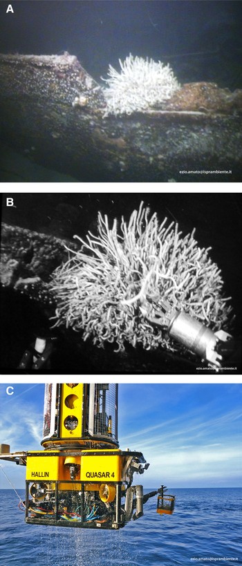
Videos showing tubes were digitally recorded in situ with a working class ROV ‘QUASAR 4’. Such videos, where giant tubes are visible (Figure 1), were provided and accessible on-line by the Italian Ministry of the Environment (MiATTM) for a limited period and are now available at: www.youtube.com/watch?=Ve89AT3z0Yw ‘View of the internal holds of the shipwreck’ (at 8 minutes 29 seconds), and at www.youtube.com/watch?v=W51XMiuJgcw ‘Sampling of external sediments’ (from 0.42 minutes to 7 minutes 15 seconds). Tubes were collected utilizing the grabber of the ROV equipped with an improvised collecting basket (Figure 1). A video of the collecting phase is available at: www.youtube.com/watch?v=KOYfMAESqBA.
(A) A bush-like group of Lamellibrachia sp. tubes on the side of the shipwreck ‘Catania’ at 490 m depth; (B) a phase of tube sampling with the arm of the ROV; (C) the working class ROV ‘QUASAR 4’ with the basket in the phase of recovering the tubes on the deck of the mother ship.

Once recovered on the ship, the tubes were fixed in 50% ethanol (all that was available on-board when sampled), then shortly passed into 4% formalin and then back to 70% ethanol.
In the laboratory, tubes were measured in diameter and length and were inspected for the presence of living animals inside. Only two tubes were found to host specimens of Lamellibrachia. The worms were then gently removed from the tubes and analysed under a stereo-microscope to check the main morphological characters and compare with the available literature.
A small piece of the vestimentum from both specimens was used for DNA extraction and sequencing of a fragment of the mitochondrial cytochrome c oxidase subunit I (COI) gene.
For DNA extractions, we followed the protocol by Schander & Halanych (Reference Schander and Halanych2003) for formalin-fixed tissues, except that we omitted the last step in the protocol (treatment with GeneReleaserTM). The measurement of 3 µl of the extractions was used in polymerase chain reaction (PCR) reactions. The PCR reactions to amplify COI were carried out in 25 µl volume using standard protocols with an annealing temperature of 45°C. We used the forward primer LCO1490 5′ GGTCAACAAATCATAAAGATATTGG 3′ (Folmer et al., Reference Folmer, Black, Hoeh, Lutz and Vrijenhoek1994) and the reverse primer CO-D 5′ TCNGGRTGNCCRAANARYCARAA 3′ (Saito et al., Reference Saito, Kojima and Endo2000). The PCR reactions were cleaned with ExoSapIT (USB Corporation). Cycle sequencing was performed in 10 µl volume in forward and reverse direction, using 3 µl H2O, 2 µl BigDye Terminator v. 3.1 (Applied Biosystems), 2 µl 5X sequencing buffer (Applied Biosystems), 1 µl primer (10 µM) and 2 µl of the cleaned PCR products. Sequence reactions were cleaned with PrepEase® Sequencing Dye Clean-Up Kit (USB Corporation). Sequences were analysed on an ABI3130 Genetic Analyser.
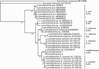
Sequencher 4.8 (GeneCodes) was used to edit the sequences and to assemble forward and reverse fragments. The two new Lamellibrachia sequences are available in GenBank (www.ncbi.nlm.nih.gov/) under Accession Nos. HQ154525 (Lamellibrachia no. 1) and HQ154526 (Lamellibrachia no. 2). They were aligned with other sequences of Lamellibrachia spp. from GenBank as well as Escarpia laminata and Sclerolinum contortum (see Figure 4 for GenBank accession numbers) as outgroups. The COI sequences are available for five of the six described Lamellibrachia species. The only species for which there are no sequence data is L. victori, known only from its type location off the coast of Uruguay (Mane-Garzón & Montero, Reference Mane-Garzón and Montero1986). Sequence alignment was conducted using Clustal W (Thompson et al., Reference Thompson, Higgins and Gibson1994) as implemented in MEGA 4.1 (Tamura et al., Reference Tamura, Dudley, Nei and Kumar2007). The alignment contained 630 nucleotide positions and had no ambiguities.
A phylogenetic analysis was conducted using Bayesian Inference in MrBayes 3.1.2 (Ronquist & Huelsenbeck, Reference Ronquist and Huelsenbeck2003) under a GTR + I + G model. Sclerolinum contortum was used as the outgroup and the temperature parameter was set to 0.1. Two runs with four Monte Carlo Markov Chains each were run simultaneously for 5,000,000 generations and every 100th tree retained. The trees resulting from the first 2,500,000 of each run were discarded as burn-in and the remaining trees from each run were combined in a 50% majority rule consensus tree (Figure 4).
RESULTS AND DISCUSSION
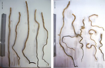
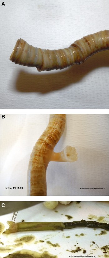
In the videos of the shipwreck examined, vestimentiferan tubes appeared grouped in dense bush-like aggregates, ‘thickets’ of approximately 100–150 tubes (Figure 1A, B). The tubes were mainly present in the interior of the hold, on the main deck of the shipwreck and around the ship's mast, apparently settled on hard substrates, possibly the wood deck pavement and/or material transported by the ship stored in the hold. The whole shipwreck seemed covered by a thin layer of mud. Twenty-seven entire tubes were collected by the grabber of the ROV, from the side border on the upper part of the bow of the shipwreck (Figure 1A, C). Tubes ranged from 23 to 60 cm in length and from 0.6 to 13 mm in diameter; they were shaped from straight vertical to very curled (Figure 2). Clear annulations were present near the tube openings, while on various tubes the scleractinian Desmophyllum cristagalli was settled (Figure 3A, B). Only two specimens of Lamellibrachia sp. were found, both of the same size and lacking most of the posterior portion (including part of the trophosoma and the segmented portion, the opisthosoma); the obturaculum was 13 mm long in both specimens, and had the typical reverse cone shape with winged sides; it was surrounded by three sheath lamellae and eight branchial lamellae in specimen no. 1, and six sheath lamellae and 14 branchial lamellae in specimen no. 2. Branchial filaments are smooth in both individuals. The vestimentum was 8.1 cm long and 6.5 mm in diameter for specimen no. 1 (Figure 3C) and 8.3 cm and 6.8 mm in diameter for specimen no. 2, so the ratio between obturaculum and vestimentum length was 0.16 and 0.15, and that between vestimentum length and diameter was 12.4 and 12.2, respectively. The portion of the trophosoma of both specimens was of a dark green colour and full of granular material (likely bacteriocytes with symbiont bacteria); the epithelium was thin and finely covered by very small papillae.
Some of the collected tubes of the vestimentiferan Lamellibrachia sp. on the shipwreck ‘Catania’. Note the occurrence of both very vertical and straight tubes (left side) and very curled ones (right side). Scale bar: 30 cm length.

(A) Detail of the final portion (~3 cm) and opening of the tube of the vestimentiferan worm Lamellibrachia sp. which hosted a specimen (note the clear annulations); (B) detail of the madreporarian Desmophyllum cristagalli (1 cm length) settled on the tube of a vestimentiferan Lamellibrachia sp.; (C) specimen no. 1 of Lamellibrachia sp. out of its tube, note the cephalic part (indicated by the pins), the vestimentum (8.1 cm length) and the anterior part of the trunk (trophosoma).

Due to the low numbers of specimens collected, their relatively large size, and the incomplete conditions (lack of part of the opisthosoma and trophosoma), morphological comparisons with other species of Lamellibrachia are difficult and may have a limited diagnostic value. However, the few characters measured, showed that the length of the obturaculum (13 mm) and its shape, as well as the dimensions of the vestimentum (length and diameters) are within the range of variability observed for other Lamellibrachia species, and especially for L. luymesi, which should be the closest geographic taxon (van der Land & Nørrevang, Reference Land, van and Nørrevang1975; Gardiner & Hurdez, Reference Gardiner and Hourdez2003; Southward et al., in press). With respect to specimens of a newly described species, collected in the eastern Mediterranean, our specimen showed higher length and diameter (Southward et al., in press). The number of both sheath and branchial lamellae are in the lower range of the whole variability of Lamellibrachia species, and comparable to both L. luymesi, and the newly described species (Southward et al., in press). The ratio between obturaculum and vestimentum length in our specimens is lower than the mean value reported in other Lamellibrachia species, while that between vestimentum length and diameter resulted to be higher (Schulze, Reference Schulze2003); this may be due to size or partially to fixation.
The COI sequences of our two specimens differ from each other by only two transitions and no transversions. They form a highly supported clade with a specimen collected from the eastern Mediterranean (GenBank Accession No. EU046616), and now corresponding with the newly described species by Southward et al. (in press). The average genetic distances among the three Mediterranean specimens is 0.4% (Figure 4). The species most closely related to this clade is Lamellibrachia cf. luymesi, a species from the Gulf of Mexico. The average genetic distance between members of the L. cf. luymesi clade and the Mediterranean clade is 2.9% (Kimura-2-parameter distance) (Table 1). The L. barhami and L. satsuma clades also have high branch support, as does L. columna; the latter includes two previously unnamed Lamellibrachia species from Japan (Kojima et al., Reference Kojima, Segawa and Ohta1997). Lamellibrachia juni is weakly supported as a clade. Another weakly supported clade contains three unnamed Lamellibrachia species from the western Pacific (see Kojima et al., Reference Kojima, Ohta, Yamamoto, Miura, Fujiwara and Hashimoto2001, Reference Kojima, Ohta, Yamamoto, Yamaguchi, Miura, Fujiwara, Fujikura and Hashimoto2003).
The 50% majority rule consensus tree based on the Bayesian inference analysis of the COI sequence data. The tree shows high branch support (Bayesian posterior probability) for the Mediterranean clade, as well as for the sister group relationship with Lamellibrachia cf. luymesi. Percentages to the right of the tree indicate average pairwise genetic distances (Kimura 2-parameter) among the members of each clade.

Average genetic distances (Kimura-2-parameter) among the Lamellibrachia clades as defined in Figure 4.

This pattern suggests that our specimens, genetically very close to the EU046616, belong to the same new species described from the eastern Mediterranean (Southward et al., in press).
The close relationship between the Mediterranean and the Atlantic clade seems geographically consistent, suggesting a differentiation of the Mediterranean clade, likely after the Messinian crisis (approximately 5–6 My BP), when the Mediterranean dried out and was then refilled and flooded by the Atlantic waters (Bianchi & Morri, Reference Bianchi and Morri2000; Taviani, Reference Taviani2002, Reference Taviani2003).
It needs to be discussed and clarified whether there is a sulphide source in the shipwreck or in its vicinity to allow chemosynthesis of the obligate symbiotic bacteria of these worms. Another possibility is that the symbionts degrade complex organic molecules such as cellulose. The presence of various benthic fish and fouling invertebrates, documented in ROV videos and settled on the porthole brought to the surface, testify an oxidized environment around the ‘Catania’ shipwreck, or at least not influenced by sulphur emissions. The occurrence of living scleractinians (D. cristagalli) settled on the tubes also demonstrates that at least the mid- and upper portions of the tubes are well oxygenated.
A search about the characteristics of the wrecked ship ‘Catania’ was performed on the Archive by the AIDMEN Association (http://www.aidmen.org/index.php). This confirmed that the ‘Catania’, in accordance with the construction features at the time of ship building (1906), had the deck and other structural parts made of wood. The ship was sunk by a German U-boat on 18 March 1917 (during World War I) while it was sailing from Mumbai (India) to Genova (Ligurian Sea, Italy), and 81 people died in this act of war. The ‘Catania’ cargo was holding 1000 tons of manganese, and 2500 tons of cotton and oil seeds. The source of energy to sustain the obligate bacterial endosymbionts of these giant worms may be related to the degradation of the ship's wooden structures and/or of the above mentioned cargo material.
Other records of Lamellibrachia and vestimentiferans on shipwrecks were in fact associated with material transported by the ship, as for the SS ‘Persia’ wreck, where the tubes were embedded in sacks of cellulose for newspapers (Hughes & Crawford, Reference Hughes and Crawford2008), or in the Atlantic wreck ‘Francois Vieljeux’, where bales of seeds were hosting some tubes (Dando et al., Reference Dando, Southward, Southward, Crawford and Crawford1992). It is known that some Siboglinidae (e.g. frenulates) are able to tolerate relatively low sulphide levels (Schulze & Halanych, Reference Schulze and Halanych2003; Sommer et al., Reference Sommer, Linke, Pfannkuche, Schleicher, Scheider, Deimling, Reitz, Haekel, Flogel and Hensen2009), while others rely on different kinds of symbiotic bacteria (e.g. Osedax spp.), not restricted to sulphide oxidation (Pleijel et al., Reference Pleijel, Dahlgren and Rouse2009); this may be the case for some vestimentiferans as well, in particular Lamellibrachia. Investigations to identify the bacterial populations associated with our Lamellibrachia sp. are in progress.
The presence of dense aggregates of giant worms in the ‘Catania’ shipwreck demonstrates the importance of these unusual and scattered habitats for the propagation of vestimentiferan species. Shipwrecks, which seem to be relatively frequent and randomly scattered on the sea bottom, especially when they hold suitable material or have wooden structures, may function as stepping stone habitats that allow spread of colonizers, typical of reducing biotopes, over large scales and into potentially more suitable environments such as deep cold seeps and other sulphide-rich habitats. The vestimentiferans on the ‘Catania’, therefore, may represent a connection of the Alboran Sea populations with those off the eastern Mediterranean. Shipwrecks therefore, hold much potential for future investigations of marine species' dispersal in the deep sea.
ACKNOWLEDGEMENTS
We wish to thank Sara Marzialetti for help in worm dissection. Professors Helmut Zibrowius and Stefano Piraino confirmed the identification of the scleractinian Desmophyllum cristagalli. Eve Southward kindly allowed us to check her paper in press and the drawings and description of a new species of Lamellibrachia in the eastern Mediterranean Sea. Ana Hilário provided information on the record of vestimentiferans in the Alboran Sea. We also wish to thank Stefano Di Muccio, and associates of the AIDMEN (Italy) who provided information about the steamship ‘Catania’: Stefano Ruia, Luigi Griva, Giorgio Spazzapan and Nereo Castelli. Two anonymous referees provided useful comments to improve the manuscript.