Introduction
Cestodes, or tapeworms, are segmented worms which primarily infest dogs and other canine species. Grazing animals such as sheep, cattle, pigs, horses and camels are intermediate hosts in the life cycle of these worms. Occasionally, humans ingest food or water contaminated with tapeworm eggs and become intermediate hosts, leading to echinococcosis or hydatid disease.Reference White, Weller, Kasper, Braunwald, Fauci, Hauser, Longo and Jameson1, Reference Strohl, Rouse, Fisher, Strohl, Rouse and Fisher2
The literature contains anecdotal reports of hydatid cysts in the head and neck region. We report a case of hydatid cyst located in the sublingual region, and present a review of relevant literature.
Case report
A 23-year-old man presented to the ENT services at St John's Medical College Hospital, Bangalore, a tertiary referral hospital in South India. He had a four-month history of a swelling in the midline of the sublingual region, which had been progressively increasing in size.
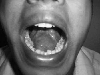
On examination, the swelling was oval in shape, approximately 4 × 3 cms in size, nontender, soft and fluctuant. The oral mucosa was free from the swelling and there were no signs of inflammation (Figure 1). On neck examination, there was no swelling in the submental region. On abdominal examination, neither the liver nor the spleen was enlarged. The diagnoses considered were ranula, sublingual dermoid and lymphangioma.
Clinical photograph of the swelling in the sublingual region.

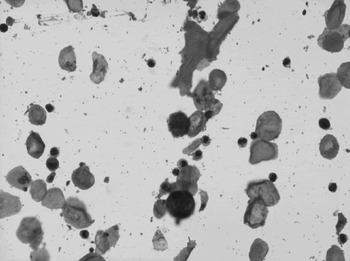
Photomicrograph of fine needle aspiration cytology smear, showing hydatid scolices (H&E; ×400).

Ultrasonography of the neck revealed a well defined, hypoechoic lesion in the sublingual region, measuring 5.3 × 3 × 3 cms, with an approximate volume of 29 ml and containing a calcific focus.
Fine needle aspiration of cyst contents was performed via the sublingual route, yielding scant, whitish fluid. Smears were prepared from the aspirate and stained with haematoxylin and eosin, Papanicolaou and May-Grunwald-Giemsa stains. The smears showed numerous round to oval structures resembling brood capsules, with scolices and occasional hooklets. A few acellular membranous fragments were also seen. The surrounding area showed a minimal inflammatory cell response. A diagnosis of a parasitic cyst, most probably hydatid, was made.
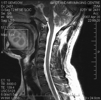
T1- and T2-weighted, multiplanar magnetic resonance imaging (MRI) showed a well defined, multiloculated, 5.5 × 3 × 3 cm lesion in the sublingual region. The lesion showed a hypointense rim on all imaging sequences (Figure 3). The contents appeared iso- to mildly hyper-intense on T1-weighted images and hypo-intense on T2-weighted images (Figure 3). These imaging features were diagnostic of hydatid cyst.
Sagittal, T2-weighted magnetic resonance imaging scan, showing multiloculated lesion in the sublingual region with a hypointense rim. Note that the contents appear iso- to mildly hyper-intense.

Surgical excision of the cyst was planned. However, as the patient was unwilling to undergo surgery, he was commenced on oral albendazole 400 mg twice daily for three months. The swelling regressed partially in response to this treatment. After three months, the patient was lost to follow up.
Discussion
Echinococcus granulosus (canine tapeworm) is the causative parasite of hydatid disease. Its life cycle involves two hosts. The primary host (dogs and other canine species) is defined as one in which the adult stage lives or in which the sexual mode of reproduction takes place. The intermediate host (other grazers) is the organism in which the larval stage of the parasite lives or in which asexual multiplication takes place.Reference White, Weller, Kasper, Braunwald, Fauci, Hauser, Longo and Jameson1, Reference Strohl, Rouse, Fisher, Strohl, Rouse and Fisher2 The oncosphere is the fully developed egg, and contains an embryo with six hooklets (hexacanth embryo). When humans ingest food or water contaminated with oncospheres, they become an intermediate host of the tapeworm.
After ingestion, the parasite emerges from the cyst, penetrates the small intestine wall, and is carried to the liver and other organs via the bloodstream. The larvae lodge within capillaries of various organs and invoke an inflammatory response of mononuclear cells and eosinophils. While many of the larvae are destroyed, a few survive by forming a slow-growing, thick-walled cyst inside which they divide.Reference McAdam, Sharpe, Kumar, Abbas and Fausto3 However, the life cycle, i.e. formation of the adult tapeworm, cannot be completed unless one of the canine species, its primary host, ingests these cysts.
The echinococcus cyst is usually unilocular; however, up to 30 per cent may be multilocular. Cysts may be present in the same organ or in multiple organs,Reference Kammerer and Schantz4 particularly the liver and lungs, occasionally the central nervous system (CNS) and heart, and rarely the musculoskeletal system. In the CNS and musculoskeletal systems, cysts may grow primarily from direct implantation of oncospheres or secondarily from metastatic dissemination of visceral cysts from the liver or lungs.Reference Nath, Prabhakar and Nagar5 The cyst has a wall comprising three layers. The outermost layer, called the pericyst, is composed of compressed host tissue. The intermediate layer, termed the laminar layer, is derived from the parasite. The innermost layer, termed the germinal layer, comprises the live cellular parasite tissue and is responsible for cyst growth and production of protoscolices, brood capsules (i.e. budded-off pieces of germinal layer with attached protoscolices) and daughter cysts.Reference Morris and Richards6
Parasitic diseases are rarely seen in the head and neck region. In a 1998 review by Prousalidis et al. of 49 Greek patients with hydatid cysts located in various organs other than the liver and lungs, none was located in the cervical region.Reference Prousalidis, Tzardinoglou, Sgouradis, Katsohis and Aletras7 Primary hydatid cysts located in the sublingual region are extremely rare even in endemic areas, and very few cases have been reported to date.
The diagnosis of hydatid disease may be established on the basis of clinical presentation, radiography and imaging techniques such as ultrasonography (US), computed tomography (CT) and MRI. Laboratory tests, the intradermal Casoni's test and serological examinations have a limited role in diagnosis as these have poorer diagnostic sensitivity and specificity.Reference Prousalidis, Tzardinoglou, Sgouradis, Katsohis and Aletras7 If spillage of the cyst contents occurs in the pre- or peri-operative periods, the lesion is very likely to transform into untreatable, multiple hydatidosis.Reference Akal and Kara8 Thus, pre-operative diagnosis of hydatid disease is of great clinical significance.
Ultrasonography and CT examination may demonstrate internal septae and daughter cysts.Reference Akal and Kara8–Reference Soylu, Aydogan, Kiroglu, Javadzadeh and Tuncer10 Hydatid cysts have certain characteristic MRI features. The cyst wall appears hypointense on all sequences due to its collagen-rich pericyst.Reference Gupta, Rathi and Bhargava11 The daughter cyst contents appear hypointense on T1-weighted images, compared with the parent cyst, and hyperintense on T2-weighted images.Reference Singh and John12–Reference Singh, Korah, Gibikote, Shyam, Nair and Korula14 However, the imaging features depend on the stage of the disease. Signal intensity may change with coexistent infection, calcification or haemorrhage.Reference Singh and John12, Reference Singh, Korah, Gibikote, Shyam, Nair and Korula14
In the present case, the disease was of a long duration and may have been secondarily infected, giving rise to turbidity of the fluid contents and causing the iso- to hyper-intensity seen on T1-weighted images and hypo-intensity seen on T2-weighted images.
There are numerous reports of hydatid cysts occurring in unusual sites in the body, often first diagnosed on fine needle aspiration biopsy.Reference Das and Choudhury15–Reference Giuffre, Mondello, Inferrera, Furchi, Gentile and Speciale18 Fine needle aspiration biopsy in hydatid disease is potentially risky, as spillage of embryos causes contamination and possible fatal anaphylactic reaction; however, the pathologist may encounter hydatid cysts, with typical cytomorphological features, in unexpected sites. Hence, a high index of suspicion is warranted.
The surgical management of a hydatid cyst consists of removal of the cystic material, including the germinative layer, and subsequent obliteration of the potential space occupied by the cyst; this is referred to as a cystotomy and capitonnage.Reference Morris and Richards6 If the germinative layer is left behind, it could lead not only to recurrence but also to suppuration of the cyst cavity. Cystectomy or so-called enucleation is an alternative surgical procedure.Reference Morris and Richards6, Reference Sennaroglu, Onerci, Turan and Sungur19
Alternative therapies advocated in the management of patients with recurrence or high risk of contamination include puncture, aspiration, injection and reaspiration with non-toxic scolicidal agents (such as 20 per cent hypertonic saline, 0.5 per cent silver nitrate, 95 per cent sterile ethanol, absolute alcohol and mebendazole 2.4 µg/ml),Reference Taylor and Morris20, Reference Russell, Williams, Bulstrode and Chiodini21 or combination chemotherapy using imidazole derivatives, particularly albendazole given orally in a dose of 15 mg/kg (as two divided doses) administered in cycles of 28 days' treatment interrupted by 14 days' rest.Reference White, Weller, Kasper, Braunwald, Fauci, Hauser, Longo and Jameson1
Conclusion
The case of a slow-growing cyst in the sublingual region is presented. Although the diagnosis was suggested by fine needle aspiration, without any complications, aspiration cytology should preferably be avoided if hydatid cyst is suspected until radiological imaging studies are complete, as cyst puncture and escape of hydatid fluid may lead to anaphylactic shock and also to secondary cyst formation.Reference Amice, Sparfel, Petillon, Amice, Jezequel and Riviere22 A high index of clinical suspicion is necessary in order to diagnose hydatid disease in unusual locations.