Incessant or persistent atrial tachycardia may lead to tachycardia-induced cardiomyopathy.Reference Ge, Li, Liu and Jiang1, Reference Medi, Kalman and Haqqani2 Especially in older children, ectopic atrial tachycardia is less likely to resolve spontaneously, and antiarrhythmic medications are often ineffective.Reference Salerno, Kertesz, Friedman and Fenrich3 Thus, radiofrequency catheter ablation should be considered early in the course of treatment for these patients, and surgical intervention may be warranted. We present a 4-year-old boy who underwent minimally invasive video-assisted thoracoscopic left atrial appendectomy for refractory atrial tachycardia originating from the left atrial appendage.
Case report
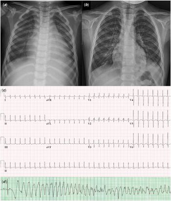
A 4-year-old boy visited the emergency department due to prolonged abdominal pain and lethargy. On admission, his blood pressure, pulse, and respiratory rate were 96/56 mmHg, 208 beats/min, and 40 breaths/min, respectively. His weight was 22 kg, which had recently increased by 2 kg. On chest X-ray, the cardiothoracic ratio was 62% and bilateral pleural effusions were present (Fig 1a). Electrocardiography showed narrow QRS tachycardia with invisible normal P-waves (Fig 1c). During atrioventricular conduction block by adenosine infusion, ectopic P-waves were apparent; therefore, we suspected ectopic atrial tachycardia. Echocardiography showed dilation of the four cardiac chambers and a left ventricular ejection fraction of 33%. The plasma level of B-type natriuretic peptide was 3296 pg/ml.
(a) Initial chest X-ray. (b) Chest X-ray taken 2 months after the surgery. (c) Initial electrocardiography. (d) Torsade de pointes caused by amiodarone infusion.

Intravenous digoxin was initially started for rate control, but it was ineffective. A very low dose of esmolol was administered, but his blood pressure dropped to 70/49 mmHg prompting discontinuation of the medication. Intravenous amiodarone infusion was started for sustained atrial tachycardia; however, it remained uncontrolled. Finally, amiodarone was discontinued because of recurrent Torsade de pointes and non-sustained ventricular tachycardia (Fig 1d).
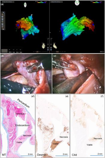
Since the atrial tachycardia could not be controlled with antiarrhythmic drugs, electrophysiologic study and radiofrequency catheter ablation were planned. Activation mapping with the CARTO® navigation system (Biosense Webster, Diamond Bar, California, United States of America) demonstrated an eccentric atrial activation from the distal portion of the left atrial appendage (Fig 2a). Temporary terminations of atrial tachycardia were achieved several times during the ablation, but the tachycardia frequently recurred at multiple sites in the left atrial appendage. Even after the radiofrequency catheter ablation, his pulse increased to 215 beats/min and his systolic blood pressure decreased to 50 mmHg.
(a) Electroanatomical activation mapping demonstrated an eccentric atrial activation from the distal portion of the left atrial appendage and multiple ablation sites (red dots). (b) Thoracoscopic video showed several scars on the surface of the left atrial appendage caused by the radiofrequency ablation (asterisks). (c) An endoscopic vascular stapler was placed at the left atrial appendage base and the appendage was excised. (d) Pathologic specimen of the left atrial appendage tip stained with Masson’s trichrome showing multiple areas of radiofrequency ablation, extending from the endocardium to the subepicardium. (e) Desmin immunohistochemical staining showing remnants of viable cardiomyocytes (brown staining). (f) Immunohistochemical staining for C4d showing necrotic cardiomyocytes (light brown staining).

Finally, we decided to perform surgical atrial appendectomy due to intractable atrial tachycardia and worsening heart failure. Instead of using the traditional left thoracotomy or median sternotomy approach, we chose a video-assisted thoracoscopic approach despite the small size of the patient. Minimal manipulation of the patient’s unstable heart and less pain from this minimal invasive video-assisted thoracoscopic approach were among some of the considerations in our decision. Under general anesthesia, in the right decubitus position, three ports were placed: the anterior port in the fourth intercostal space at the anterior axillary line, the camera port in the seventh intercostal space at the median axillary line, and the posterior port in the fifth intercostal space at the posterior axillary line. After CO2 inflation, pericardiotomy was performed 4–5 mm posterior to the phrenic nerve to expose the left atrial appendage. Several reddish scars caused by the radiofrequency catheter ablation were visible on the surface of the left atrial appendage (Fig 2b). An endoscopic vascular stapler, ECHELON FLEXTM PVE35A 35 mm/320 mm (Ethicon, Cincinnati, Ohio, United States of America) was applied at the base of the left atrial appendage and the left atrial appendage was excised under guidance of a videoscope (Fig 2c). Immediately after the appendectomy, the persistent atrial tachycardia was terminated, and his rhythm restored to sinus rhythm while in the operating room. The surgery was uneventful, and the patient was extubated the day after the surgery. His left ventricular ejection fraction increased to 60% within 1 week and he was discharged. His cardiothoracic ratio on chest X-ray decreased to 47% (Fig 1b), and plasma B-type natriuretic peptide level decreased to 31 pg/ml within 2 months. He had no arrhythmia and maintained normal left ventricular function during the subsequent 9-month follow-up period.
Pathologic evaluation of the left atrial appendage showed multiple round lesions, caused by the radiofrequency catheter ablations, and intervening non-ablative myocardium. The ablative lesions showed acute myocardial coagulative necrosis, haemorrhage, and neutrophilic infiltration (Fig 2d–f).
Discussion
In children, tachycardia can be asymptomatic or present with atypical symptoms such as abdominal pain and lethargy. In a previous report of ectopic atrial tachycardia in children, only 5.7% of the patients complained of paroxysmal palpitations and the most patients with incessant tachycardia denied palpitation symptoms.Reference Toyohara, Fukuhara, Yoshimoto, Ozaki and Nakamura4 Incessant atrial tachycardia is frequently complicated by tachycardia-induced cardiomyopathy in both adults and children; however, decreased ventricular function at the time of ectopic atrial tachycardia diagnosis is more common in children than in adults.Reference Ge, Li, Liu and Jiang1–Reference Cummings, Mahle and Strieper5 Therefore, early suspicion of tachycardia as a potential cause of heart failure is emphasised in children.
Because atrial appendage sites are known to be foci associated with a high incidence of incessant tachycardia and left ventricular dysfunction,Reference Medi, Kalman and Haqqani2 radiofrequency catheter ablation should be considered early in the course of the illness, even in children.Reference Toyohara, Fukuhara, Yoshimoto, Ozaki and Nakamura4 However, atrial tachycardia originating from the atrial appendages is difficult to treat with catheter ablation because of the anatomical complexity of the atrial appendages. They have thick pectinate muscles with intervening thin-walled myocardium, an elongated morphology with a multi-lobulated apex, and a comparatively narrow orifice.Reference Guo, Zhang and Ma6 Therefore, for the treatment of refractory atrial tachycardia originating from atrial appendages, a surgical approach should be considered. A previous study has reported thoracoscopic ligation of the left atrial appendage after epicardial ablation in a 10-year-old childReference Perez-Caballero-Marinez, Pita-Fernandez and Gonzalez-Lopez7; however, the epicardial approach is technically difficult and limited in a small, 4-year-old child. We decided to eliminate the left atrial appendage because it is functionally insignificant and presents a risk for thromboembolism after extensive ablation.
When compared with traditional open thoracotomy, video-assisted thoracoscopic surgery is less invasive and has the advantages of fewer complications, lesser post-operative pain, and a shorter hospitalisation period.Reference Yamada, Ajiro and Shoda8 To our knowledge, the patient in our case is the youngest reported patient to undergo successful minimally invasive thoracoscopic atrial appendectomy through the joint efforts of a paediatric intensivist, electrophysiologist, and cardiac surgeon.
Conclusions
Tachycardia-induced cardiomyopathy can be improved by aggressive treatment of arrhythmias. Minimally invasive thoracoscopic surgery can be applied, instead of the traditional thoracotomy approach, even in a small child who has focal atrial tachycardia originating from an atrial appendage that is not responding to drugs or radiofrequency ablation.
Acknowledgements
None.
Financial Support
This research received no specific grant from any funding agency, commercial or not-for profit sectors.
Conflicts of Interest
None.
Ethical Standards
This article does not involve experimentation on human or animals.