Implication
Our results indicate that the inclusion of chitooligosaccharides (COSs) in piglet diets may moderate several gut health parameters that contribute to some of the common problems that occur after weaning in the absence of in-feed antibiotics. It was observed that COSs with a molecular weight (MW) of 5 to 10 kDa were more effective in reducing Escherichia coli populations while a MW of 10 to 50 kDa enhanced the intestinal structure.
Introduction
The weaning period imposes profound social and environmental stresses on the piglet such as removal from the sow, change in diet and mixing of piglets from different litters. Numerous studies have reported that there is a reduction in villous height (villous atrophy) and an increase in crypt depth (crypt hyperplasia) after weaning, which leads to increased susceptibility to intestinal gut dysfunction (Spreeuwenberg et al., Reference Spreeuwenberg, Verdonk, Gaskins and Verstegen2001; Pierce et al., Reference Pierce, Sweeney, Brophy, Callan, Fitzpatrick, McCarthy and O'Doherty2006). The post-weaning period is characterised by a reduction in feed intake, poor growth rates, diarrhoea and an increased risk of disease (Lalles et al., Reference Lalles, Bosi, Smidt and Stokes2007). These negative effects on piglet growth during the weaning period were managed by growth-promoting antibiotics. However, the European Union placed a total ban on the use of in-feed antibiotic growth promoters on the 1st January 2006 due to public concerns regarding bacterial resistant and human health issues.
Chitooligosaccharides (COS) may be a potential viable alternative to traditional antimicrobials in animal production. Chitosan is a natural biopolymer derived by alkaline deacetylation of chitin, which is the principal component of protective cuticles of crustaceans such as crabs, shrimps, prawns, lobsters and cell walls of some fungi such as aspergillus (Qin et al., Reference Qin, Li, Xiao, Liu, Zhu and Du2006). Both chitin and chitosan are biopolymers composed of glucosamine and N-acetylated glucosamine (2-acetylamino-2-deoxy-d-glucopyranose) units linked by β (1 to 4) glycosidic bonds (Koide, Reference Koide1998). Low molecular weight (MW) COS is a water-soluble derivative of chitosan due to shorter chain lengths (Kim and Rajapakse, Reference Kim and Rajapakse2005). Recently, both chitosan and its derivatives have generated considerable interest due to their biological activities, including antimicrobial, antitumour, immunoenhancing effects and the acceleration of wound healing (No et al., Reference No, Young Park, Ho Lee and Meyers2002; Liu et al., Reference Liu, Chen, Park, Liu, Liu, Meng and Yu2006) There is considerable variation in the literature on the biological properties of COS (Jeon et al., Reference Jeon, Park and Kim2001; Liu et al., Reference Liu, Chen, Park, Liu, Liu, Meng and Yu2006). Most of this variation is partly due to the widely different MW used across studies. It is hypothesised that the biological properties of COS may be influenced by its MW and COS will enhance selected indices of health in weaned piglets.
Material and methods
All procedures described in this experiment were conducted under an experimental licence from the Irish Department of Health in accordance with the cruelty to Animals Act 1876 and the European Communities (Amendments of the Cruelty to Animals Act 1976) Regulations.
Experimental diets
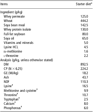
The experiment was designed as a complete randomised block design and comprised four dietary treatments. The dietary treatments were as follows: (1) control diet (0 ppm COS), (2) control diet plus 5 to 10 kDa COS, (3) control diet plus 10 to 50 kDa COS and (4) control diet plus 50 to 100 kDa COS. The COS was sourced from Kitto Life Co. Ltd (Kyungki-do, Seoul, Korea) and was supplemented in the experimental diets at a concentration of 250 ppm. The diets were fed for 8 days ad libitium, after which time the pigs were humanely sacrificed. The diets were formulated to have similar digestible energy (16 MJ/kg) and standardised ileal digestible (SID) lysine (14 g/kg) contents. All amino acids requirements were met relative to SID lysine (National Research Council, 1998). The ingredient composition and chemical analysis of the dietary treatments are presented in Table 1.
Composition and chemical analysis of experimental diets (as-fed basis)

DM = dry matter; GE = gross energy.
Starter diet provided (mg/kg completed diet): Cu, 175; Fe, 140; Mn, 47; Zn, 120; I, 0.6; Se, 0.3; retinol, 1.8; cholecalciferol, 0.025; alpha-tocopherol, 67; phytylmenaquinone, 4; cyanocobalamin, 0.01; riboflavin, 2; nicotinic acid, 12; pantothenic acid, 10; choline chloride, 250; thiamine, 2; pyridoxine, 0.015.
*COS was included in dietary treatments T2–T4 at a rate of 250 mg/kg.
†Calculated for tabulated nutritional composition Sauvant et al., (Reference Sauvant, Perez and Tran2004).
Animals and management
A total of 28 piglets (progeny of large white × (large white × landrace sows)) were selected from a commercial pig unit at 24 days of age. The piglets had a weaning weight of 9.1 kg (s.d. = 0.80) and were blocked on the basis of litter of origin and live weight (n = 7). The piglets were individually housed in fully slated pens (1.7 m × 1.2 m). They were individually fed and had ad libitum access to feed and water. The house temperature was thermostatically controlled at 30°C throughout the experiment. This study was not a growth performance study but some performance data were recorded. The piglets were weighed at the beginning of the experiment (day 0) and at the end of the experiment (day 8). Food was available up to the final weighing and all remaining food was weighed back for the purpose of calculating feed efficiency. Pigs were observed for clinical signs of diarrhoea and a scoring system was applied to indicate the presence and severity of this as described by Pierce et al. (Reference Pierce, Sweeney, Brophy, Callan, Fitzpatrick, McCarthy and O'Doherty2006). Faeces scores were assigned daily for individual pigs from day 0 and continued until day 8. The following faeces scoring system was used: 1 = hard faeces, 2 = slightly soft faeces in the pen, 3 = soft, partially formed faeces, 4 = loose, semi-liquid faeces and 5 = watery, mucous-like faeces.
Gut morphological analysis
The piglets were humanely sacrificed on day 8 by a lethal injection of Euthatal (pentobarbitone sodium BP – Merial Animal Ltd, Sandringham House, Essex, UK) at a rate of 1 ml/1.4 kg BW. On removal of the digestive tract, sections of the duodenum (10 cm from the stomach), the jejunum (60 cm from stomach) and the ileum (15 cm from caecum) were excised and fixed in 10% phosphate-buffered formalin. The preserved segments were prepared using standard paraffin-embedding techniques. The samples were sectioned at 5 μm thickness and stained with haemotoxylin and eosin (Pierce et al., Reference Pierce, Sweeney, Brophy, Callan, Fitzpatrick, McCarthy and O'Doherty2006). Villous height and crypt depth were measured on the stained sections (4× objective) using a light microscope fitted with an image analyser (Image Pro Plus, Media Cybernetics, Buckinghamshire, UK). Measurements of 15 well oriented and intact villi and crypts were taken for each segment. Villous height was measured from the crypt–villous junction to the tip. Crypt depth was measured from the crypt–villous junction to the base. Results were expressed as the mean villous height or crypt depth in micrometres.
Intestinal microflora
For microbial analysis, digesta samples (∼10 ± 1 g) were aseptically recovered from the proximal colon of each pig immediately post slaughter. Digesta samples were stored in sterile containers (Sarstedt, Wexford, Ireland), placed on ice and transported to the laboratory within 2 h. A 1.0 g sample was removed from the digesta sample, serially diluted (1 : 10) in 9.0 ml aliquots of maximum recovery diluents (Oxoid, Basingstoke, UK) and spread plated (0.1 ml aliquots) onto selective agars, as follows: Lactobacillus spp. were isolated on de Man, Rogosa and Sharp (MRS) agar (Oxoid) with an overnight (18 to 24 h) incubation at 37°C in an atmosphere enriched with 5% CO2, as recommended by the manufacturers (Oxoid). The Escherichia coli species were isolated on MacConkey agar (Oxoid) following aerobic incubation at 37°C for 18 to 24 h (O'Doherty et al., Reference O'Doherty, Dillon, Figat, Callan and Sweeney2010). Target colonies of Lactobacilli and E. coli were identified by Gram stains and colony morphology (Salanitro et al., Reference Salanitro, Blake and Muirhead1977). The API 50 CHL (BioMerieux, Biomerieux, Craponne, France) kit was used to confirm suspect Lactobacilli spp. Suspect E. coli colonies were confirmed with API 20E (BioMerieux, France). This API system identifies the suspect colonies by measuring their ability to produce cytochrome oxidase. Typical colonies of each bacteria on each agar were counted, log transformed and the numbers of bacteria were expressed per gram of digesta after being serially diluted.
Volatile fatty acid (VFA) analysis
Samples of digesta from individual pigs were taken from the caecum and the proximal colon to measure the VFA concentration and molar proportions of VFAs. The VFA concentrations in the digesta were determined using gas liquid chromatography according to the method described by Pierce et al. (Reference Pierce, Callan, McCarthy and O'Doherty2007). A 1-g sample was diluted with distilled water (2.5 × weight of sample) and centrifuged at 1400 × g for 4 min (Sorvall GLC – 2B laboratory centrifuge, Dupont, Wilmington, DE, USA). Then, 1 ml of the subsequent supernatant and 1 μl of internal standard (0.5 g 3-methyl-n-valeric acid in 1 l of 0.15 mol/l oxalic acid) were mixed with 3 ml of distilled water. Following centrifugation to remove the precipitate, the sample was filtered through Whatman 0.45 μm polyethersulphone membrane filters into a chromatographic sample vial. A 1-μl sample was injected into a model 3800 Varian gas chromatograph with a 25 m × 0.53 mm i.d. megabore column (coating CP-Wax 58 (FFAP) – CB (no. CP7614)) (Varian, Middelburg, the Netherlands).
RNA extraction and complementary DNA (cDNA) synthesis
Tissue samples were collected from the mesenteric side of the colon, rinsed with ice-cold sterile phosphate-buffered saline (Oxoid) and stripped of overlying smooth muscle cells. Approximately 1 to 2 g of the porcine colon tissue was cut into small pieces and placed in tubes containing 15 ml of RNAlater (Applied Biosystems, Foster City, CA, USA) and immediately stored at −20°C pending RNA extraction. Total RNA was extracted from colon tissue samples (25 mg) using a GenElute Mammalian Total RNA Miniprep Kit (RTN70, Sigma-Aldrich, St Louis, MO, USA) according to the manufacturer's instructions. To eliminate possible genomic DNA contamination, total RNA samples were subjected to DNAse I (AMPD1, Sigma-Aldrich) treatment according to the manufacturer's protocol. Then RNA purification was performed using a phenol–chloroform extraction method (Chomczynski and Sacchi, Reference Chomczynski and Sacchi2006). The total RNA was quantified using a NanoDrop-ND1000 Spectrophotometer (Thermo Fisher Scientific, Wilmington, DE, USA) and the purity was assessed by determining the ratio of the absorbance at 260 and 280 nm. All total RNA samples had 260/280 nm ratios above 1.8. In addition, RNA integrity was verified by visualisation of the 18 and 28 S ribosomal RNA bands stained with ethidium bromide after gel electrophoresis on 1.2% agarose gels (Egel, Invitrogen Inc., Carlsbad, CA, USA). Total RNA (1 μg) was reverse transcribed (RT) using the RevertAid H minus first strand cDNA synthesis kit (Fermentas GmbH, St Leon-Rot, Germany) with oligo dT primers. The final RT product was adjusted to a volume of 120 μl using nuclease-free water.
Real-time quantitative PCR
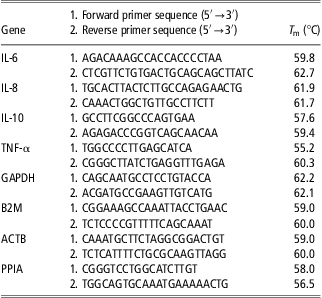
All primers for the selected cytokines, genes such as Interleukin-1α (IL-1α), IL-6, IL-10, tumour necrosis factor (TNF-α) and the reference genes β-actin (ACTB), β2-microglobin (B2M), glyceraldehyde-3-phosphate dehydrogenase (GAPDH) and peptidylprolyl isomerise A (PPIA) are presented in Table 2. Amplification was carried out in a reaction volume of 20 μl containing 10 μl SYBR Green Fast PCR Mastermix (Applied Biosystem), forward and reverse primer mix (1 μl), 8 μl DEPC treated water and 1 μl of template cDNA. Quantitative real-time PCR was carried out using an ABI PRISM 7500 Fast sequence detection system for 96-well plates (Applied Biosystem). The thermal cycling conditions were as follows: an initial denaturation step at 95°C for 10 min, 40 cycles of 95°C for 15 s, followed by 60°C for 1 min. Dissociation analyses of the PCR product were performed to confirm the specificity of the resulting PCR products. All samples were run in triplicate. The cycle threshold value (C t) is defined as the fractional cycle number at which fluorescence passes the fixed threshold. The mean C t values of triplicates of each sample were used for calculations.
Porcine-specific primers used for real-time PCR

IL = interleukin; TNF = tumour necrosis factor; GAPDH = glyceraldehyde-3-phosphate dehydrogenase; B2M = β2-microglobin; ACTB = genes β-actin; PPIA = peptidylprolyl isomerise A.
Primers were designed using Primer Express™ software and were synthesised by MWG Biotech (Milton Keynes, UK).
Normalisation of quantitative PCR data
Normalisation of the C t values obtained from real-time PCR was performed by (i) transforming the raw C t values into relative quantities using the formula, relative quantities = (PCR efficiency)ΔCt, where ΔC t is the change in the C t values of the sample relative to the highest expression (minimum C t value), (ii) using geNorm, a normalisation factor was obtained from the relative quantities of four most stable housekeeping genes (GAPDH, B2M, ACTB and PPIA) and (iii) the normalised fold change or the relative abundance of each of the target genes was calculated by dividing their relative quantities by the normalisation factor.
Statistical analysis
The experimental data were analysed as a randomised block design using the GLM procedure of SAS (2004). The individual pig served as the experimental unit. Food intake was included as a covariate in the model for villous height, crypt depth and villous height to crypt depth ratio in the digestive tract. The microbial counts were log transformed. The data were checked for normality using the Proc Univariate function of SAS. The means were separated using the Tukey–Kramer Test. Probability values of <0.05 were used as the criterion of statistical significance. All results are presented in the tables as least square means ± standard error of the means (s.e.).
Results
Performance and faecal scoring
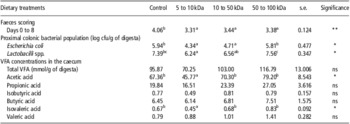
The average faecal scores of the pigs are presented in Table 3. The supplementation of different MW of COS had no significant effect on the growth performance of the pig during the 8-day experimental period (P > 0.05). However, the inclusion of COS at all MW in the diet significantly reduced faecal scores compared with the control treatment (P < 0.01).
Effect of COS supplementation at different MW on faecal scoring, selected microbial populations in the proximal colon and the total VFA concentration and the proportions of VFAs in the caecum of the weaned pig (least square means and s.e.; n = 7)

COS = chitooligosaccharide; MW = molecular weight; VFA = volatile fatty acid.
Probability of significance: *P < 0.05; **P < 0.01; ns, P < 0.05.
Means with the same superscript alphabets within rows are not significantly different (P > 0.05).
Microbiology
The effect of COS supplementation at different MW on selected microbial populations in the colon of the pig is shown in Table 3. Pigs offered diets containing 5 to 10 kDa COS had a lower E. coli number compared with the control (P < 0.05) and the 50 to 100 kDa COS (P < 0.05) treatments. The 10 to 50 kDa treatment had a neumerically lower E. coli number compared with the control group (P = 0.09). Pigs offered diets containing 5 to 10 kDa COS had a significantly lower population of lactobacilli in the colon compared with the control group (P < 0.05) and the 50 to 100 kDa COS diet (P < 0.01). Pigs offered 50 to 100 kDa COS had a higher lactobacilli number than pigs offered 10 to 50 kDa COS (P < 0.05). The supplementation of different MW of COS had no significant dietary effect on the lactobacilli : E. coli ratio in the colon of the pig.
Volatile fatty acids
The effects of COS supplementation at different MW on the VFA concentrations in the caecum are shown in Table 3. The supplementation of different MW of COS had a significant effect on the concentrations of acetic acid (P < 0.05) and isovaleric acid (P < 0.05) in the caecum. Pigs fed 5 to 10 kDa COS had lower levels of acetic acid and isovaleric acid compared with the control (P < 0.05), 10 to 50 kDa COS (P < 0.05) and 50 to 100 kDa COS (P < 005). There was no significant effect of MW on VFA concentrations (P > 0.05) in the proximal colon (data not shown).
Gut morphology
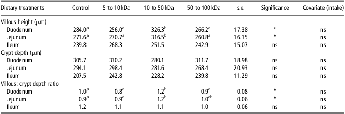
The effects of varying COS MW on villous height, crypt depth and the villous height : crypt depth ratios in the gastro-intestinal tract are shown in Table 4. Pigs fed the 10 to 50 kDa COS had a higher villous height in the duodenum and the jejunum compared with the control group (P < 0.05), 5 to 10 kDa COS (P < 0.01) and 50 to 100 kDa COS diets (P < 0.05). There was no effect of dietary treatment on crypt depth in the duodenum (P > 0.05). Pigs offered the 10 to 50 kDa COS had a higher villous height : crypt ratio in the duodenum and the jejunum compared with the control group (P < 0.05) and the 5 to 10 kDa COS diet (P < 0.01).
Effect of COS supplementation at different MW on villous height, crypt depth and the villous height : crypt depth ratio in the gastro-intestinal tract of the weaned pig (least square means and s.e.)

COS = chitooligosaccharide; MW = molecular weight.
Probability of significance: *P < 0.05; **P < 0.01; ns, P < 0.05.
Means with the same superscript alphabets within rows are not significantly different (P > 0.05).
Cytokine gene expression analysis
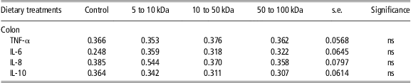
The effects of COS supplementation on the immune response in colon tissues of the pig are shown in Table 5. The supplementation of different MW of COS had no significant effect on the expression of the cytokines TNF-α, IL-6, IL-8 and IL-10 (P > 0.05) in the gastro-intestinal tract of the pig.
Effect of COS supplementation at different MW on the immune response in unchallenged proximal colon tissues (least square means of fold change in normalised relative gene expression with their s.e.; n = 7 animals)

COS = chitooligosaccharide; MW = molecular weight; TNF = tumour necrosis factor; IL = interleukin.
Probability of significance: *P < 0.05; **P < 0.01; ns, P < 0.05.
Discussion
The hypothesis of the current experiment is that the biological properties of COS may be influenced by its MW and COS will enhance selected indices of health in weaned piglets. It was demonstrated in the current study that the lower MW of 5 to 10 kDa possessed antibacterial activity while the higher MW of 10 to 50 kDa was optimum for enhancing intestinal structure.
Dietary supplementation of COS at the low MW of 5 to 10 kDa decreased both lactobacilli and E. coli counts, while the 10 to 50 kDa COS numerically decreased E. coli populations in the colon of the pig. In a study by Liu et al. (Reference Liu, Piao, Kim, Wang, Shen, Lee and Li2008), COS supplementation at different concentrations reduced E. coli concentrations in the caecum of the weanling pig. E. coli is considered to be one of the most important causes of post-weaning diarrhoea in weaned pigs; therefore, a reduction in E. coli populations may reduce the incidence of diarrhoea in post-weaned pigs (Fairbrother et al., Reference Fairbrother, Nadeau and Gyles2005). Although many species of E. coli are commensal, high levels of specific E. coli (like ETEC) will increase the risk of disease. Unfortunately, ETEC numbers were not measured in the current study. In the current study, the faecal score was decreased in pigs fed the COS diets compared with the control. These results suggest that the supplementation of the 5 to 10 kDa and 10 to 50 kDa COS reduces E. coli populations in the colon, resulting in a lower faecal score in the post-weaning period. The 50 to 100 kDa COS led to a reduced diarrhoea score but no reduction in E. coli populations; therefore, this MW of COS may be working as a bulking agent to affect the faecal score. The 50 to 100 kDa COS may retard the rate of passage through the intestine and may have the ability to absorb water.
In the current study, it was demonstrated that supplementation of 5 to 10 kDa COS had the strongest antimicrobial effect against both lactobacilli and E. coli. This is in agreement with other studies in which low MW COS (5 to 10 kDa) were shown to possess strong antibacterial properties compared with higher MW COS and the antibacterial properties of COS increased at a low MW of <5 kDa against Gram-negative such as E. coli (Zheng and Zhu, Reference Zheng and Zhu2003; Kittur et al., Reference Kittur, Vishu Kumar, Varadaraj and Tharanathan2005). In a study by Liu et al. (Reference Liu, Piao, Thacker, Zeng, Li, Wang and Kim2010), COS supplementation decreased E. coli populations compared with the control in the caecum of weaned pigs, while Jeon et al. (Reference Jeon, Park and Kim2001) observed a antimicrobial effect of COS against Gram-positive bacteria such as Lactobacilli under in-vitro conditions.
To explain COS antibacterial activity, two mechanisms have been proposed. The first mechanism is that the positively charged COS reacts with negatively charged molecules at the microbial cell surface, thereby altering cell permeability (Chung and Chen, Reference Chung and Chen2008). Therefore, COS may interact with the membrane of the cell to alter cell permeability. However, as evident from the current study, this activity may differ with varying MW as the 50 to 100 kDa group had no inhibitive effect on the selected microbial populations, while the MWs of 5 to 10 kDa and 10 to 50 kDa COS had the strongest inhibitive effect. The other antibacterial mechanism is the binding of COS with DNA to inhibit RNA synthesis (Liu et al., Reference Liu, Du, Wang and Sun2004). It has been proposed that COS penetrates the nuclei of the bacteria and interferes with RNA and protein synthesis. It is noteworthy that all the COS samples used in the current study were soluble in aqueous solutions. Kim and Rajapakse (Reference Kim and Rajapakse2005) found that COS with a MW of >30 kDa were not effective as antibacterial agents due to their poor solubility in aqueous solutions at a neutral pH.
Volatile fatty acids are the major end products of bacterial metabolism in the large intestine (Macfarlane and Macfarlane, Reference Macfarlane and Macfarlane2003). Both protein and carbohydrate fermentation contribute to the production of acetic acid; however, branched-chain fatty acids such as isovaleric acid are produced from protein fermentation (Mackie et al., Reference Mackie, Stroot and Varel1998). In the current study, the 5 to 10 kDa group had the lowest selected microbial populations while also reducing isovaleric acid and acetic acid concentrations in the caecum. The shift in the production of the fermentation end products is reflected in the reduction of the selected microbial populations. The quantity of VFA produced depends on the amount and composition of the substrate and on the type of microbes present in the large intestine (van Beers-Schreurs et al., Reference van Beers-Schreurs, Nabuurs, Vellenga, van der Valk, Wensing and Breukink1998). Reduced VFA concentrations indicate that lower amounts of substrate were fermented as a result of a lower microbial activity in the caecum (Htoo et al., Reference Htoo, Araiza, Sauer, Rademacher, Zhang, Cervantes and Zijlstra2007).
Villous height is generally reduced and crypt depth is increased, which may explain the increased occurrence of diarrhoea and reduced growth after weaning (Pluske et al., Reference Pluske, Thompson, Atwood, Bird, Williams and Hartmann1996). The inclusion of 10 to 50 kDa COS in the present study was found to increase the villous height and villous : crypt depth ratio in the duodenum and also in the jejunum compared with the control group. Very little data have been published on the effects of COS MW on gut morphology in weaned piglets; thus, the exact mechanism for the increase in villous height and villous : crypt depth ratio is unclear. It may be hypothesised that low MW COS has the potential to promote intestinal morphology through cell proliferation. The COS has been shown to influence colonic cell proliferation, crypt depth and crypt circumference in mice (Torzsas et al., Reference Torzsas, Kendall, Sugano, Iwamoto and Rao1996).
A study carried out by Liu et al. (Reference Liu, Piao, Kim, Wang, Shen, Lee and Li2008), on different concentrations of COS, demonstrated that 200 mg/kg of COS increased villous height and villous : crypt ratio in the jejunum and ileum (Liu et al., Reference Liu, Piao, Kim, Wang, Shen, Lee and Li2008). The possible explanation for this improved intestinal structure was that COS is composed of N-acetyl glucosamine (Kim and Rajapakse, Reference Kim and Rajapakse2005), which may bind to certain types of bacteria and possibly interfere with their adhesion to the gut tissue of host animals (Ofek et al., Reference Ofek, Hasty and Sharon2003; Liu et al., Reference Liu, Piao, Kim, Wang, Shen, Lee and Li2008). This result is in agreement with Mourão et al. (Reference Mourão, Pinheiro, Alves, Guedes, Pinto, Saavedra, Spring and Kocher2006), who reported that an increase in villi length in the ileum of weaned rabbits was correlated to a lower intestinal microflora. A decrease in bacteria load has been shown to increase the proliferation of epithelial cells, which leads to an improved intestinal morphology and increased villous height (Mourão et al., Reference Mourão, Pinheiro, Alves, Guedes, Pinto, Saavedra, Spring and Kocher2006). In the present study, in pigs fed the lower MW of 5 to 10 kDa COS, a strong antimicrobial effect on both Lactobacilli and E. coli populations was observed, with no effect on villous structure, while the higher MW of 10 to 50 kDa resulted in a reduction in E. coli numbers in comparison with the control and was optimum for improving villous integrity.
There were no effects of COS supplementation in colon tissue on any of the cytokines analysed. This overall lack of an effect on these inflammatory cytokines implies that COS inclusion in the diet had no effects on immune gene expression of the pigs. Mori et al. (Reference Mori, Okumura, Matsuura, Ueno, Tokura, Okamoto, Minami and Fujinaga1997) also demonstrated that chitin and its derivatives do not stimulate the production of IL-6, IL-1 and TNF-α by fibroblasts. In our study, no differences were observed on growth performance between days 0 and 8 post-weaning.
In conclusion, MW is an important factor to consider when investigating the biological properties of COS. On the basis of the current study, the lower MW of 5 to 10 kDa possessed antibacterial activity while the higher MW of 10 to 50 kDa was optimum for enhancing intestinal structure. The COS supplementation exerted no deleterious effects on immune gene expression or growth performance (days 0 to 8) of the pigs while reducing the incidence of diarrhoea. However, further studies are required to investigate the exact mechanism by which COS supplementation of varying MW affects gut microflora and growth performance of the weaned pig. Further studies should also explore the potential of these diets to prevent the negative impact of a pathogen challenge.
Acknowledgements
Funding for this research was provided under the Marine Functional Food Research Consortium (NutraMara) by the Marine Institute and the Department of Agriculture, Fisheries and Food, Agriculture House, Co. Dublin, Ireland.