Introduction
The family Felidae consists of 37 species (Bristol-Gould and Woodruff, Reference Bristol-Gould and Woodruff2006). Most of them are included in the International Union for the Conservation of Nature (IUCN) Red List of endangered species (IUCN, 2016) except for the domestic cat (Bristol-Gould and Woodruff, Reference Bristol-Gould and Woodruff2006). Therefore, there is worldwide concern about their preservation. One conservation effort involves the development of assisted reproductive techniques that can be potentially integrated into the genetic management of small populations in breeding centres, zoos and animal parks (Silva et al., Reference Silva, Silva, da Silvada and Comizzoli2019). However, in addition to the lack of physiological information for some species, access to wild felid reproductive tissues for experimental purposes is extremely restricted (Leonel et al., Reference Leonel, Vilela, Carrilho and Lucci2018). Therefore, the domestic cat is a valuable model for development of techniques of assisted reproduction that can be applied to parallel studies of wild and endangered felids (Wildt et al., Reference Wildt, Swanson, Brown, Sliwa, Vargas, MacDonald and Loveridge2010). In this context, preantral follicle in vitro culture is an emerging fertility preservation technique.
Primordial follicles (PFs) constitute the ovarian reserve (Monniaux et al., Reference Monniaux, Clément, Dalbiès-Tran, Estienne, Fabre, Mansanet and Monget2014). The process of recruitment of PFs into the growing pool, termed PF activation, is the first major event of folliculogenesis (Grosbois et al., Reference Grosbois, Devos and Demeestere2020). Studies on in vitro activation and subsequent in vitro growth and maturation of PFs from fresh and cryopreserved ovarian cortical tissue has gained much attention as a potential source of mature oocytes that are capable of fertilization (Li et al., Reference Li, Kawamura, Cheng, Liu, Klein, Liu, Duan and Hsueh2010; Kawamura et al., Reference Kawamura, Cheng, Suzuki, Deguchi, Sato, Takae, Ho, Kawamura, Tamura, Hashimoto, Sugishita, Morimoto, Hosoi, Yoshioka, Ishizuka and Hsueh2013; McLaughlin et al., Reference McLaughlin, Albertini, Wallace, Anderson and Telfer2018). Several autocrine and paracrine factors contribute to control PFs recruitment (Grosbois et al., Reference Grosbois, Devos and Demeestere2020), including members of the FGF family.
Fibroblast growth factor-2 (FGF-2), also called basic-FGF, has been implicated in ovarian function and follicle development (reviewed by Chaves et al., Reference Chaves, de Matos, Buratini and de Figueiredo2012 and Price, Reference Price2016), promoting granulosa cell proliferation and decreasing apoptosis and steroidogenesis (Gospodarowicz and Bialecki, Reference Gospodarowicz and Bialecki1979; Baird and Hsueh Reference Baird and Hsueh1986; Tilly et al., Reference Tilly, Billig, Kowalski and Hsueh1992; Lavranos et al., Reference Lavranos, Rodgers, Bertoncello and Rodgers1994). Substantial evidence indicates that FGF-2 plays a key role in in vitro follicle activation in some species (rat: Nilsson et al., Reference Nilsson, Parrott and Skinner2001; goat: Matos et al., Reference Matos, Van Den Hurk, Lima-Verde, Luque, Santos, Martins, Báo, Lucci and Figueiredo2007; human: Garor et al., Reference Garor, Abir, Erman, Felz, Nitke and Fisch2009; macaque monkey: Lu et al., Reference Lu, Yan, Zhi, Xia, Wang, Yan, Yu, Ding, Gao, Li and Qiao2015).
Although ovarian cortex culture has been studied in a wide variety of animals, there are still few reports using domestic cats as a research model (Fujihara et al., 2012; Reference Fujihara, Comizzoli, Keefer, Wildt and Songsasen2014) and little information is known about what regulates PF activation in any felid species. To our knowledge, only two studies found evidence of in vitro follicle activation in the cat ovary after 7 (Fujihara et al., Reference Fujihara, Yamamizu, Comizzoli, Wildt and Songsasen2018) and 14 days of culture (Thuwanut et al., Reference Thuwanut, Comizzoli, Wildt, Keefer and Songsasen2017). However, the effect of FGF-2 on the survival and in vitro activation of cat PFs has not been investigated. Therefore, the aims of this study were to analyze the effects of FGF-2 on the survival and PF activation after in vitro culture of domestic cat ovarian tissue.
Materials and methods
All chemicals were purchased from Sigma-Aldrich (St Louis, MO, USA) unless otherwise indicated.
Ovaries (n = 12) were collected from six prepubertal domestic cats (age 3–6 months) that underwent routine ovariosalpingohysterectomy. Immediately after surgery, ovaries were washed in 70% alcohol (Dinâmica, São Paulo, Brazil) and in Minimum Essential Medium (MEM-HEPES) supplemented with 100 μg/ml penicillin and 100 μg/ml streptomycin. The ovaries were transported within 1–6 h to the laboratory in MEM-HEPES and antibiotics at 4°C (Chaves et al., Reference Chaves, Martins, Saraiva, Celestino, Lopes, Correia, Verde, Matos, Báo, Name, Campello, Silva and Figueiredo2008; Fujihara et al., Reference Fujihara, Comizzoli, Keefer, Wildt and Songsasen2014).
Ovarian cortical slices (1 mm thickness) were sectioned in fragments (∼3 mm × 3 mm). For each animal, one fragment of ovarian cortex was fixed in 10% buffered formalin (Dinâmica) for histological analysis (fresh control). The remaining fragments were incubated individually in 1 ml of culture medium in 24-well culture dishes at 38.5°C in 5% CO2 for 7 days. The culture medium was MEM supplemented with 10 ng/ml insulin, 5.5 μg/ml transferrin, 5.0 ng/ml selenium, 2 mM glutamine, 2 mM hypoxanthine, 50 μg/ml ascorbic acid and 0.1% polyvinyl alcohol, which is referred to as MEM+ (adapted from Fujihara et al., Reference Fujihara, Comizzoli, Keefer, Wildt and Songsasen2014). This culture medium was then supplemented with no factor (control) or with 10, 50 or 100 ng/ml FGF-2. The FGF-2 concentrations were selected based on a study of ovarian tissue culture in caprine species (Matos et al., Reference Matos, Van Den Hurk, Lima-Verde, Luque, Santos, Martins, Báo, Lucci and Figueiredo2007). The culture medium was exchanged every 48 h. Each treatment was repeated six times.
Tissues from the fresh control and cultured treatments were fixed in 10% buffered paraformaldehyde (Dinâmica) for 18 h, embedded in paraffin and 5-μm sections were cut. Tissues were stained with haematoxylin and eosin (H&E) and evaluated using light microscopy (Nikon, Tokyo, Japan; ×400 magnification) with the assessor blinded to experimental conditions. Follicles were classified as morphologically normal if no clear signs of degeneration were noted, which included shrunken oocytes, disorganization of the granulosa cell layer, condensed nuclear chromatin and/or cell swelling. Follicles were classified as primordial (one layer of flattened granulosa cells around the oocyte) or developing follicles (transitional: a single mixed layer of flattened and cuboidal granulosa cells around the oocyte; primary: a single layer of exclusively cuboidal granulosa cells around the oocyte; or secondary: two or more layers of cuboidal granulosa cells) (Fujihara et al., Reference Fujihara, Yamamizu, Comizzoli, Wildt and Songsasen2018). To analyze follicular activation, only morphologically normal follicles with a visible oocyte nucleus were counted and the proportion of primordial and developing follicles was calculated in the different treatments. Furthermore, oocyte and follicle diameters were measured using Image Pro-Plus® software (Media Cybernetics Inc., Silver Spring, MD, USA). Overall, 180 follicles were evaluated for each treatment.
Data from normal follicles and activation were compared using chi-squared test. Data from follicle and oocyte diameters were evaluated using the Shapiro–Wilk test. Therefore, analysis of variance (ANOVA) and the Tukey’s tests were applied for comparison among treatments. The results of growth were expressed as the mean ± standard deviation (SD). The differences were considered to be statistically significant when P < 0.05.
Results and Discussion
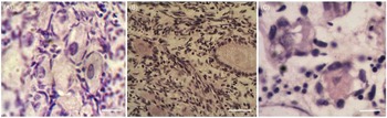
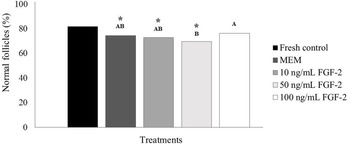
Most of the follicles (81.6%) were morphologically normal (Fig. 1A) at the onset of collection, which was consistent with an earlier study (>80% of viable follicles in the fresh control; Fujihara et al., Reference Fujihara, Yamamizu, Comizzoli, Wildt and Songsasen2018). Only treatment with 100 ng/ml FGF-2 (Fig. 1B) resulted in maintenance (P > 0.05) of the percentage of normal follicles (76.1%) within a physiological range, i.e. similar to fresh control (Fig. 2). This significant portion of normal follicles was greater than 71.8% of viable follicles observed previously after culture of cat ovarian tissue for 7 days (Thuwanut et al., Reference Thuwanut, Comizzoli, Wildt, Keefer and Songsasen2017). Furthermore, follicle survival was greater (P < 0.05) after culture in 100 ng/ml FGF-2 than in 50 ng/ml FGF-2 (69.4%; Fig. 1C). Previously, FGF-2 has been reported to maintain follicle integrity and prevent apoptosis in follicular cells after in vitro culture of ovarian cortex (Matos et al., Reference Matos, Van Den Hurk, Lima-Verde, Luque, Santos, Martins, Báo, Lucci and Figueiredo2007; Santos et al., Reference Santos, Menezes, Barberino, Macedo, Lins, Gouveia, Barros, Santos, Gonçalves and Matos2014). Culture of buffalo granulosa cells with FGF-2 at different concentrations decreased the mRNA expression of BAX, indicating that FGF may promote granulosa cells survival through autocrine and paracrine manner (Mishra et al., Reference Mishra, Thakur, Somal, Parmar, Reshma, Rajesh, Yadav, Bharti, Bharati, Paul, Chouhan, Sharma, Singh and Sarkar2016). In addition, FGF-2 activates PKC, which stimulates calcium efflux, maintaining normal basal calcium levels and, ultimately, granulosa cell viability (Peluso et al., Reference Peluso, Pappalardo and Fernandez2001). In bovine granulosa cells, FGF-2 increases the mRNA levels of cell cycle regulator GADD45B (Jiang et al., Reference Jiang, Ripamonte, Buratini, Portela and Price2011). Results from another study indicated that reduced GADD45B mRNA levels were associated with an increase in apoptosis (Portela et al., Reference Portela, Machado, Buratini, Zamberlam, Amorim, Gonçalves and Price2010). GADD45B may therefore be a target gene of FGF-2 in cat granulosa cells.
Histological sections of cat ovarian fragments. Normal follicles in the fresh control (A) and after in vitro culture in 100 ng/ml FGF-2 (B) and abnormal follicles cultured in 50 ng/ml FGF-2 (C). Retracted oocytes and swollen and disorganized granulosa cells could be often observed after culture in 50 ng/ml FGF-2. GC, granulosa cells; O, oocyte. Scale bars: 25 μm (×400).

Percentages of morphologically normal follicles in the fresh control, after in vitro culture in α-MEM+or in 10, 50 and 100 ng/ml FGF-2. *Differs significantly from the fresh control (P < 0.05). A,BDifferent letters denote significant differences among treatments (P < 0.05).

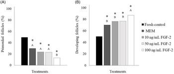
The percentage of PFs decreased (P < 0.05; Fig. 3A) and the percentage of developing follicles increased (P < 0.05; Fig. 3B) in all treatments compared with fresh tissue. However, the presence and concentration of FGF-2 influenced follicle activation after 7 days of culture. The proportion of developing follicles increased (P < 0.05) in tissues incubated with 100 ng/ml FGF-2 (86.87%) compared with control medium (70.15%) and other FGF-2 concentrations (76.34% and 76.8% for 10 and 50 ng/ml FGF-2, respectively). Furthermore, culture in 10 or 100 ng/ml FGF-2 resulted in increased (P < 0.05) follicle and oocyte diameters compared with fresh tissues and MEM+ (Table 1). Only one study has shown that in vitro culture of cat ovaries for 7 days in retinoic acid resulted in PF activation (Fujihara et al., Reference Fujihara, Yamamizu, Comizzoli, Wildt and Songsasen2018). Follicle activation in cats was also reported, but only after 14 days of ovarian tissue culture in medium containing stem cell factor (Thuwanut et al., Reference Thuwanut, Comizzoli, Wildt, Keefer and Songsasen2017). Therefore, the results from the present study revealed the significant contribution of using FGF-2 for promoting cat PF activation and growth during a short-term culture. Previous studies have indicated that FGF-2 stimulated in vitro follicle activation (rat: Nilsson et al., Reference Nilsson, Parrott and Skinner2001; goat: Matos et al., Reference Matos, Van Den Hurk, Lima-Verde, Luque, Santos, Martins, Báo, Lucci and Figueiredo2007; human: Garor et al., Reference Garor, Abir, Erman, Felz, Nitke and Fisch2009; monkey: Lu et al., Reference Lu, Yan, Zhi, Xia, Wang, Yan, Yu, Ding, Gao, Li and Qiao2015) and increased follicle and oocyte diameters (goat: Matos et al., Reference Matos, Van Den Hurk, Lima-Verde, Luque, Santos, Martins, Báo, Lucci and Figueiredo2007; sheep: Santos et al., Reference Santos, Menezes, Barberino, Macedo, Lins, Gouveia, Barros, Santos, Gonçalves and Matos2014). Furthermore, FGF-2 acts on the ovary to promote granulosa cell proliferation (Gospodarowicz and Bialecki, Reference Gospodarowicz and Bialecki1979; Lavranos et al., Reference Lavranos, Rodgers, Bertoncello and Rodgers1994). In bovine granulosa cells, FGF-2 stimulated MAPK3/1 and protein kinase-B (Akt) phosphorylation (Jiang et al., Reference Jiang, Ripamonte, Buratini, Portela and Price2011). Noteworthy, the phosphatidylinositol-3-kinase/Akt (PI3K/Akt) signalling pathway has been implicated in the regulation of survival and activation of PFs after in vitro culture of ovarian cortex in a variety of species (mice: Zhao et al., Reference Zhao, Ma, Sun, Ye, Zhang, Sun, Xu, Wang and Li2014; human: Grosbois and Demeestere, Reference Grosbois and Demeestere2018; ovine: Barberino et al., Reference Barberino, Santos, Lins, Menezes, Monte, Gouveia, Palheta and Matos2020), including domestic cat, in which stem cell factor promotes follicle development by upregulating AKT phosphorylation (Thuwanut et al., Reference Thuwanut, Comizzoli, Wildt, Keefer and Songsasen2017). Although more investigations are needed, the results from the present study suggest that, by reducing apoptosis and increasing cell proliferation, FGF-2 at 100 ng/ml maintains follicle survival and promotes PF activation. Nevertheless, the least concentrations of FGF-2 (10 and 50 ng/ml) may not be as effective as 100 ng/ml for promoting activation. Further studies to evaluate if higher concentrations of FGF-2 could have greater effects on the in vitro follicle development are warranted.
Percentages of normal primordial (A) and developing (B) follicles in the fresh control, after 7 days of in vitro culture in α-MEM+ or in 10, 50 and 100 ng/ml FGF-2. *Differs significantly from fresh control (P < 0.05). A,BDifferent letters denote significant differences among treatments (P < 0.05).

Mean follicular and oocyte diameters (mean ± standard deviation) in the fresh control and after in vitro culture of cat preantral follicle in different concentrations of fibroblast growth factor-2 (FGF-2)

* Differs significantly from fresh control (P < 0.05).
A,B,CDifferent letters denote significant differences among treatments (within the column; P < 0.05).
In conclusion, FGF-2 at 100 ng/ml maintains follicle survival and promotes the in vitro activation and growth of cat PFs. These findings provide important information about the factors that control early follicle biology in cats and indicate that FGF-2 could be a potential option for further oocyte maturation and in vitro production of embryos. This result also may be applied to prepubertal individuals that die before having the opportunity to reproduce and also to other wild and endangered feline species.
Acknowledgements
A.R. Silva and M.H.T. Matos are supported by a grant from the National Council for Scientific and Technological Development (CNPq), Brazil.
Financial support
This study did not receive any funding.
Conflict of interest
None of the authors have any conflict of interest to declare.
Ethical standards
The approval of the ethics committee was not required because the research involved tissues of slaughtered animals.