The intestinal epithelial barrier, composed of intestinal epithelial cells and intercellular tight junctions, is an important part of maintaining intestinal health and immune function(Reference Chelakkot, Ghim and Ryu1). However, due to the influence of stunting and environmental factors, the intestine of piglets is highly susceptible to oxidative stress, which triggers intestinal inflammation to release the pro-inflammatory cytokines, ultimately leading to the intestinal epithelial barrier injury(Reference Liu2,Reference Mavrommatis, Giamouri and Tavrizelou3) . It has been found that Nucleotide-binding Oligomerization Domain (NOD)-like receptor thermal protein domain associated protein 3 (NLRP3) inflammasome is a therapeutic target for inflammatory bowel disease, and its abnormal activation is one of the important causes of intestinal inflammation(Reference Song, Zhao and Ma4). In a study of weaned piglets, a large activation of the NLRP3 inflammasome in the colon led to massive release of the pro-inflammatory cytokines (IL-1β and IL-18), which impaired the intestinal epithelial barrier integrity(Reference Xia, Zhong and Wu5). Another in vitro study found that dihydromyricetin effectively alleviated lipopolysaccharide-induced inflammation of chicken intestinal epithelial cells by inhibiting NLRP3 inflammasome activation(Reference Chang, Jiang and Ji6). In addition, plant active ingredients such as L-theanine(Reference Chen, Luo and Jia7), ginsenoside Rg1(Reference Kang, Zhou and Zhu8) and eugenol(Reference Wang, Chen and Yu9) can all inhibit the activation of the NLRP3 inflammasome. This suggested that we could reduce intestinal inflammation to protect the intestinal epithelial barrier by inhibiting the activation of NLRP3 inflammasome with some plant active ingredients.
Caffeic acid (CA) is a phenolic acid compound that is widely present in plants and possesses various biological activities. It exists not only in some common fruits and vegetables (such as black chokeberries, apples, potatoes, lingonberries, carrots, etc.) but also in coffee, red wine and some herbs of the mint family (such as oregano, sage, thyme, etc.)(Reference Espíndola, Ferreira and Narvaez10,Reference Pavlíková11) . As a common plant active ingredient, CA exhibits excellent anti-inflammatory effects(Reference Pavlíková11,Reference Kim, Jung and Kim12) . Studies have found that dietary CA supplementation can reduce colonic inflammation in weaned piglets(Reference Chen, Qiao and Mao13) and improve dextran sulfate sodium-induced colitis in mice(Reference Wan, Zhong and Wang14). In vitro study also found that CA alleviated the inflammation and injury of human colorectal adenocarcinoma cells caused by hydrogen peroxide (H2O2)(Reference Zhao, Shin and Satsu15). As a type of reactive oxygen species (ROS), H2O2 is highly oxidative and can directly oxidise various biomolecules (such as lipids, proteins and deoxyribonucleic acid) in cells, thereby inducing oxidative stress(Reference Gautam, Misro and Chaki16) and intestinal epithelial cell injury(Reference Li, Huang and Jia17). However, whether CA can attenuate H2O2-induced intestinal epithelial injury and the underlying mechanism still requires further research.
At present, IPEC-J2 cells derived from jejunum are one of the best models for studying the immune response of porcine intestinal epithelial cells in vitro (Reference Tang, Zeng and Xiong18). The previous study has shown that CA can inhibit NLRP3 inflammasome activation(Reference Castro, Assmann and Stefanello19). For the above considerations, this study took IPEC-J2 cells as the research object and NLRP3 inflammasome as the research focus to explore the effect of CA on the inflammatory response of H2O2-induced IPEC-J2 cells and its molecular mechanism.
Materials and methods
Cell culture and treatment
IPEC-J2 cells were grown in DMEM/F12 (Cat. No. C11330500BT, Gibco, Shanghai, China) medium supplemented with penicillin-streptomycin (1 %) (Cat. No. 15140122, Gibco, USA) and fetal bovine serum (10 %) (Cat. No. 10099-141, Gibco, Australia) at 37℃ under 5 % CO2. During the culture period, the cell status was monitored using an inverted microscope (Cat. No. CKX53, Olympus, China). After the density of IPEC-J2 cells reached about 80 %, the cells were treated accordingly to explore the role of CA and H2O2 on the cells and their molecular mechanisms. The treatments were divided into the following conditions: (1) different levels of H2O2 (0 mM, 0·2 mM, 0·4 mM, 0·6 mM, 0·8 mM, 1·0 mM) (Cat. No. 7722-84-1, Xilong Scientific, Sichuan, China) treated the cells for 2 h; (2) different levels of CA (purity ≥ 98 %) (0 μM, 5 μM, 25 μM) (Cat. No. C0625-2G, Sigma, St. Louis, MO, USA) treated the cells for 24 h; (3) CA (5 μM) treated the cells for 22 h and H2O2 (1·0 mM) was added for 2 h; (4) MCC950 (10 μM) (Cat. No. HY-12815, MedChemExpress, Shanghai, China) pretreated the cells for 1 h and CA (5 μM) was added for 22 h, then H2O2 (1·0 mM) was added for 2 h and (5) CA (5 μM) and pyrrolidine dithiocarbamate (PDTC) (5 μM) (Cat. No. S1808, Beyotime, Shanghai, China) treated the cells for 22 h, then H2O2 (1·0 mM) was added for 2 h.
Lactate Dehydrogenase (LDH) activity analysis
After the cells’ treatment, the culture supernatant was collected for analysis of LDH activity. The kit from the Nanjing Institute of Biological Engineering (Cat. No. A020-2-2, Nanjing, China) was employed to detect LDH release according to the manufacturer’s instructions. The LDH activity represented the amount of LDH released by the cells.
Transepithelial electrical resistance (TEER) determination
The upper and lower chambers of 12-well Transwell culture plate (Cat. No. 3401, Corning Incorporated, NY, USA) were supplemented with 0·5 ml complete medium and 1·5 ml complete medium. After the IPEC-J2 cells adhesion in the upper chamber, the TEER was measured by Millicell ERS-2 resistance metre (Millipore, Molsheim, USA) every 24 h until the resistance value was stable. Then the cells were treated accordingly and the resistance value of each hole was determined separately.
Fluorescein Isothiocyanate-Dextran (FD4) permeability determination
After TEER determination, FD4 (Cat. No. 60842-46-8, Sigma, St. Louis, MO, USA) was added to 12-well Transwell culture plate to determine FD4 permeability. That is, the concentration of FD4 per hole in the upper chamber was 200 μg/ml and the cells were incubated for 2 h at 37℃. FD4 fluorescence intensity was determined with a fluorescent microplate reader (SpectraMax M2, Molecular Devices, SV, CA, USA) (excitation/emission wavelength: 485/528 nm).
Inflammatory cytokine determination
After treatment, we collected the cell culture supernatant. IL-1β content was measured using a porcine ELISA kit from Shanghai Enzyme-linked (Cat. No. ml581129, Shanghai, China) following the provided instructions. Finally, the absorbance was analysed using the Spectramax 190 microplate reader (Molecular Devices, SV, CA, USA) at 450 nm.
ROS determination
ROS level was measured using ROS Assay Kit (Cat. No. S0033M, Beyotime, Shanghai, China). In simple terms, the culture supernatant was aspirated at the end of cell treatment, and 1 ml 2’,7’-Dichlorodihydrofluorescein diacetate (DCFH-DA) probe diluted in DMEM/F12 (DMEM/F12: DCFH-DA = 1000:1) was added to each well. The cell culture plate was then placed in an incubator at 37℃ for 20 min before the cells were washed three times with serum-free DMEM/F12. Finally, at the emission wavelength of 525 nm and the excitation wavelength of 488 nm, the fluorescence intensity was determined with a fluorescent microplate reader (SpectraMax M2, Meigu Molecular Instrument Co., LTD, Shanghai, China). The obtained DCFH-DA fluorescence intensity was the intracellular ROS level.
Real-Time quantitative PCR
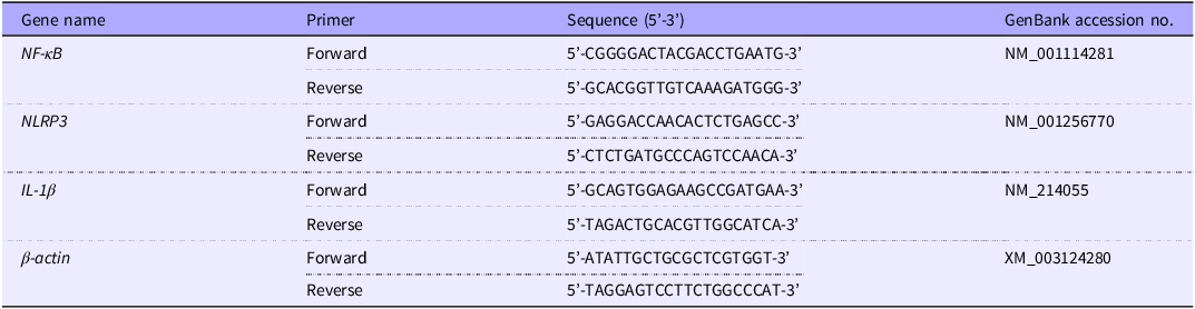
After the cell treatment, we used RNAiso Plus reagents to extract total RNA, and then transcribed the RNA into cDNA using Primescript RT reagent kit. Finally, relative gene expression was measured according to TB Green®Premix Ex Taq™ instructions. PCR amplification conditions were predenaturation at 95℃ for 30 s, followed by 40 cycles (5 s at 95℃ and 30 s at 60℃). All relevant reagents were from TaKaRa (Dalian, China). We used β-actin mRNA as a control and calculated target gene expression by the 2–ΔΔCt method(Reference Li, Huang and Jia17). The primer sequences were listed in Table 1.
Primer sequences used for real-time quantitative PCR

Western blot
The cells’ protein expression levels were measured as previously mentioned(Reference Huang, Che and Chen20). In short, the blot signal was observed by BeyoECL Plus (Cat. No. P0018M, Beyotime, Shanghai, China). The total protein internal reference was β-actin, and the nuclear protein internal reference was histone. Primary antibodies included nuclear factor kappa-B (NF-κB) (1:1000, Cat. No. AF0246, Beyotime, Shanghai, China), NLRP3 (1:1000, Cat. No. AF2155, Beyotime, Shanghai, China), Cleaved-Caspase1 (1:1000, Cat. No. HY-P80622, Medchemexpress), IL-1β (1:0000, Cat. No. AG2258, Beyotime, Shanghai, China), β-actin (1:3000, Cat. No. HC201-01, TransGen Biotechnology, Beijing, China), histone H3 (1:1000, Cat. No. AF0009, Beyotime, Shanghai, China).
Molecular docking analysis
Molecular docking is a widely used bioinformatics method for drug discovery and development that predicts interactions between ligands and receptors(Reference Ferreira, Dos Santos and Oliva21). When the binding energy is < –5 kJ/mol (–1·2 kcal/mol), it indicates that the small molecule ligand has a good affinity with the protein receptor(Reference Li, Liu and Yang22). The 3D structure of NF-κB was sourced from the Protein Data Bank (https://www.uniprot.org), while the 3D structure of CA was sourced from PubChem. Molecular docking analysis was performed using the AutoDock 4.2.6 programme to assess the interaction between the ligand (CA) and the target protein (NF-κB). Visualisation of the docking results was conducted using PyMOL 2.6.0 software.
Statistical analysis
All data were visualised using histograms, and the Shapiro–Wilk test was applied to assess their normality. Then we used SPSS 28·0 software (SPSS Inc.) to perform one-way ANOVA on the data, and applied the Dunnett method for multiple comparisons. P value less than 0·05 was considered a significant difference. All bar charts were made with GraphPad Prism 8·0·2 software (Graphpad Software Inc., La Jolla, CA, USA).
Results
H2O2 triggers inflammation and injury in IPEC-J2 cells
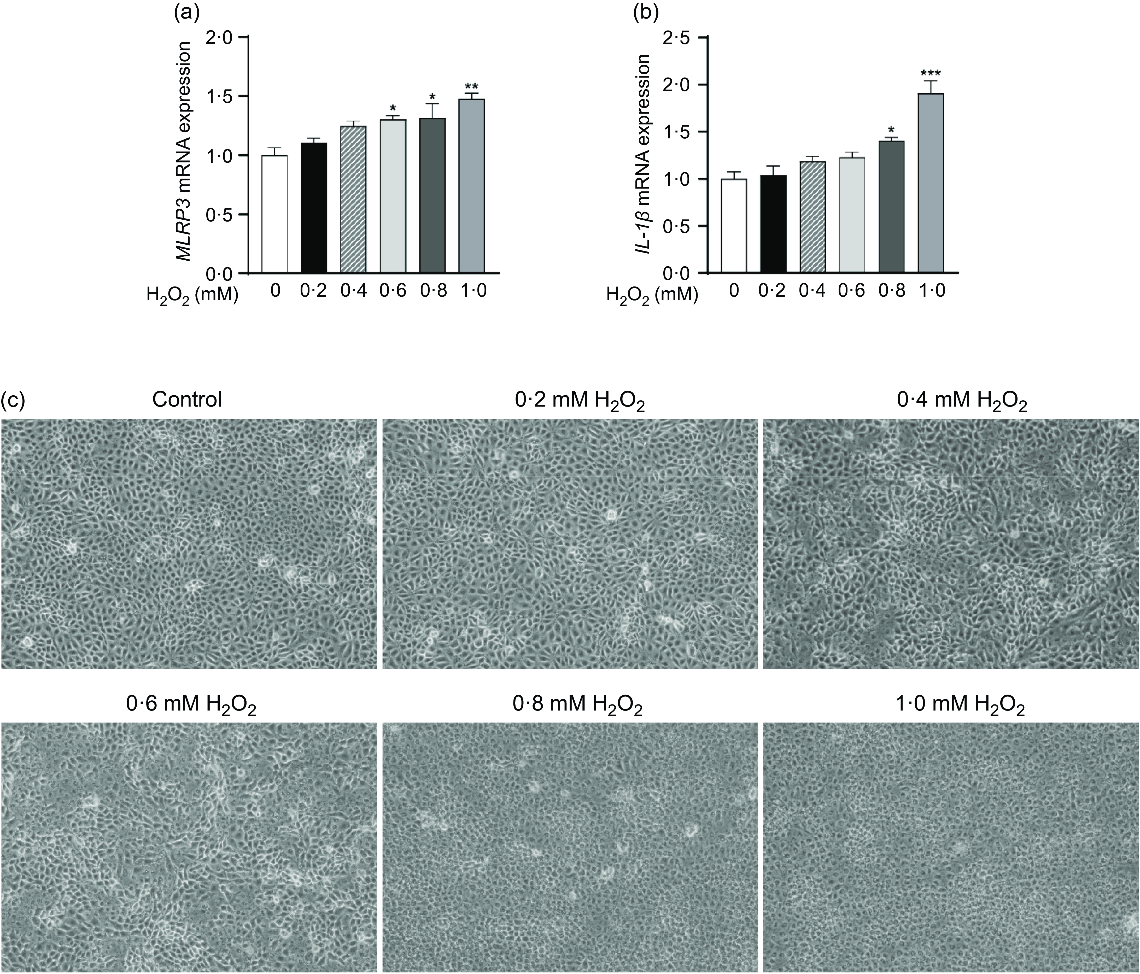
To construct the inflammation model of IPEC-J2 cells, we treated the cells with different levels of H2O2. The results showed that H2O2 increased NLRP3 and IL-1β mRNA levels in a concentration-dependent manner: compared with the control group, 0·6 mM H2O2 significantly increased NLRP3 mRNA level (P < 0·05), and 0·8 mM H2O2 and 1·0 mM H2O2 significantly increased NLRP3 (Figure 1(a)) and IL-1β mRNA levels (P < 0·05) (Figure 1(b)). In Figure 1(c), the cell morphology in the control group showed regular round spindle shape and tight intercellular connection. However, as the concentration of H2O2 increased, the outline of the cells gradually became unclear. When the concentration of H2O2 reached 0·8 mM and 1·0 mM, the morphology completely changed to a shrunked round shape, which indicated that the normal morphology of the cells was destroyed. Synthesising these results, 1·0 mM H2O2 exhibited the best effect. Therefore, 1·0 mM H2O2 was used for follow-up tests.
H2O2 triggers inflammation and injury in IPEC-J2 cells. (a) NLRP3 mRNA expression; (b) IL-1β mRNA expression; (c) IPEC-J2 cells morphology. Data were presented as means ± sem (n 3). *P < 0·05, **P < 0·01 and ***P < 0·001 for comparison with the control group.

CA inhibits H2O2-induced NLRP3 inflammasome activation in IPEC-J2 cells
We next screened CA treatment concentration by treating cells with different levels of CA. The results showed that treatment of cells with 5 μM CA significantly reduced the protein and mRNA expression of NLRP3 and IL-1β (P < 0·05) (Figure 2(a)–(c)). However, 25 μM CA had no significant effect on the expression of NLRP3 and IL-1β, possibly because CA level exceeded the optimal acting concentration. We then treated the cells with 5 μM CA and 1·0 mM H2O2 and found that CA reduced the H2O2-induced increase in the protein expression, including IL-1β, Cleaved-Caspase1 and NLRP3 (P < 0·05) (Figure 2(d)–(e)). Meanwhile, CA significantly reduced the levels of IL-1β mRNA, NLRP3 mRNA (P < 0·01) (Figure 2(f)) and ROS (P < 0·01) (Figure 2(g)). These results suggested that CA attenuated NLRP3 inflammasome activation and inflammation induced by H2O2.
CA inhibits H2O2-induced NLRP3 inflammasome activation in IPEC-J2 cells. IPEC-J2 cells were treated with different levels of CA: (a) Representative Western Blot images of NLRP3 and IL-1β; (b), (c) The protein and mRNA expression of NLRP3 and IL-1β. IPEC-J2 cells were treated with CA and H2O2: (d) Representative Western Blot images of NLRP3, Cleaved-Caspase1 and IL-1β; (e) The protein expression of NLRP3, Cleaved-Caspase1 and IL-1β; (f) The mRNA expression of NLRP3 and IL-1β; (g) Fluorescence intensity of DCFH-DA. Data were presented as means ± sem (n 3). *P < 0·05, **P < 0·01 and ***P < 0·001 for comparison with the control group. # P < 0·05, ## P < 0·01 and ### P < 0·001 for comparison with the H2O2 group.

MCC950 attenuates H2O2-induced IPEC-J2 cells inflammation and injury
Next, we investigated the effect of inhibiting NLRP3 expression on IPEC-J2 cells. It was found that MCC950 (NLRP3 specific inhibitor) and CA had almost identical effects on cells: compared with the H2O2 group, they all significantly inhibited IL-1β, Cleaved-Caspase1 and NLRP3 protein expression (P < 0·01) (Figure 3(a)–(b)), and reduced IL-1β and NLRP3 mRNA levels (P < 0·01) (Figure 3(c)). They also reduced IL-1β content (P < 0·001) (Figure 3(d)), LDH activity (P < 0·05) (Figure 3(e)), FD4 permeability (P < 0·01) (Figure 3(f)) and increased TEER (P < 0·001) (Figure 3(g)). All these suggested that CA attenuated the inflammation and injury of IPEC-J2 cells by inhibiting NLRP3 inflammasome activation.
MCC950 attenuates H2O2-induced IPEC-J2 cells inflammation and injury. (a) Representative Western Blot images of NLRP3, Cleaved-Caspase1 and IL-1β; (b) The protein expression of NLRP3, Cleaved-Caspase1 and IL-1β (n 4); (c) The mRNA expression of NLRP3 and IL-1β (n 3); (d) IL-1β content (n 3); (e) LDH activity (n 6); (f) FD4 permeability (n 3); (g) TEER (n 3). Data were presented as means ± sem. *P < 0·05, **P < 0·01 and ***P < 0·001 for comparison with the control group. # P < 0·05, ## P < 0·01 and ### P < 0·001 for comparison with the H2O2 group.

CA reduces NF-κB expression in IPEC-J2 cells induced by H2O2
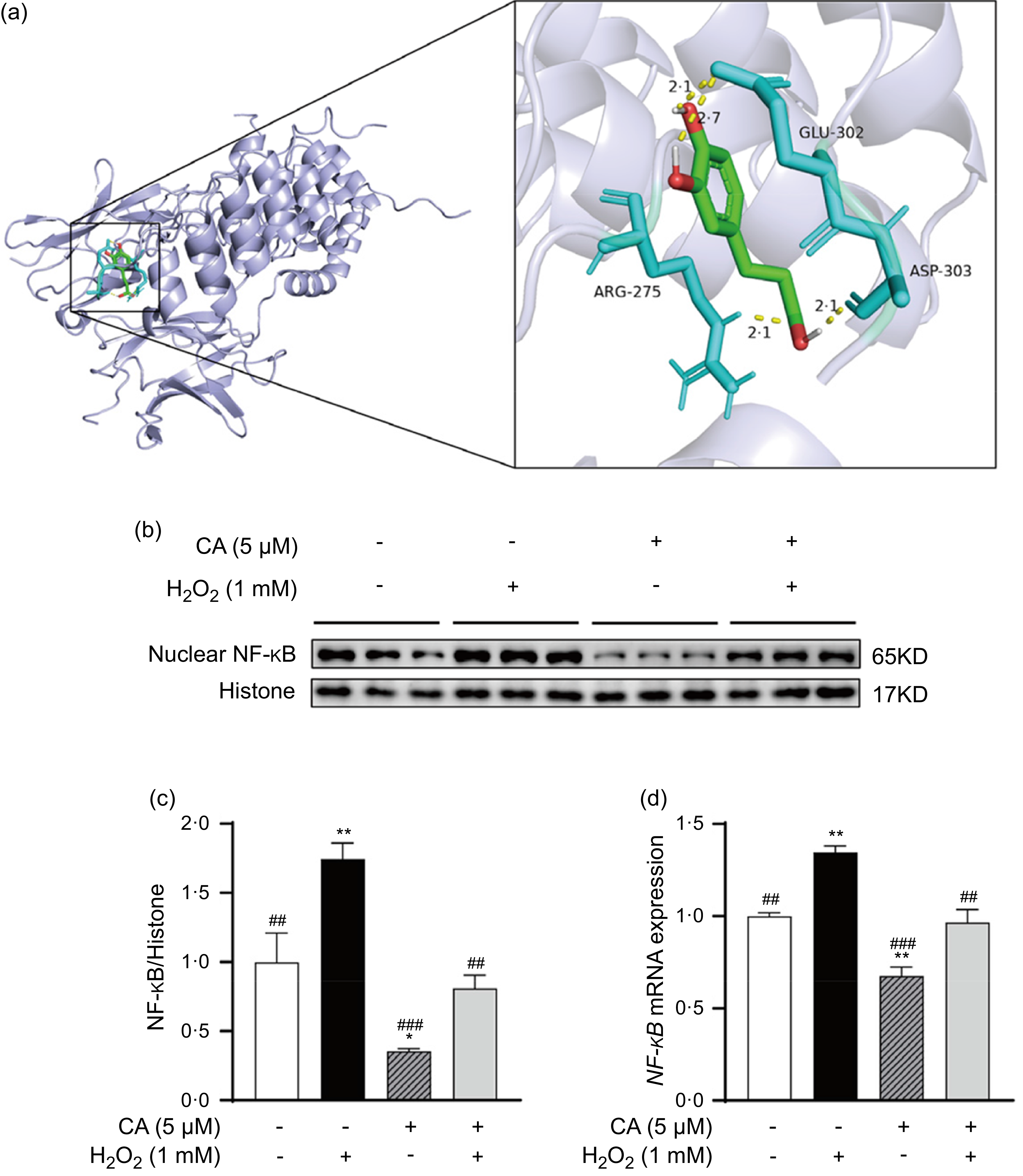
To explore the pathway by which CA inhibited NLRP3 inflammasome activation, we further investigated the effect of CA on NF-κB. The molecular docking between CA and NF-κB protein showed that CA formed four hydrogen bonds with the residues of NF-κB protein (Figure 4(a)). The binding energy between them was −5·1 kcal/mol, which indicated that they could interact well. Moreover, it was found that CA significantly decreased NF-κB protein and mRNA expression induced by H2O2 when compared with the H2O2 group (P < 0·01) (Figure 4(b)–(d)). These suggested that CA inhibited NF-κB expression.
CA reduces NF-κB expression in IPEC-J2 cells induced by H2O2. (a) Molecular docking images of CA and NF-κB protein. The amino acid residues of NF-κB protein were shown in light blue, CA was shown in green and hydrogen bond was shown in yellow; (b) Representative Western Blot images of NF-κB; (c), (d) The protein and mRNA expression of NF-κB. Data were presented as means ± sem (n 3). *P < 0·05, **P < 0·01 and ***P < 0·001 for comparison with the control group. # P < 0·05, ## P < 0·01 and ### P < 0·001 for comparison with the H2O2 group.

The inhibitory effect of CA on NLRP3 inflammasome activation in IPEC-J2 cells induced by H2O2 is mediated by NF-κB
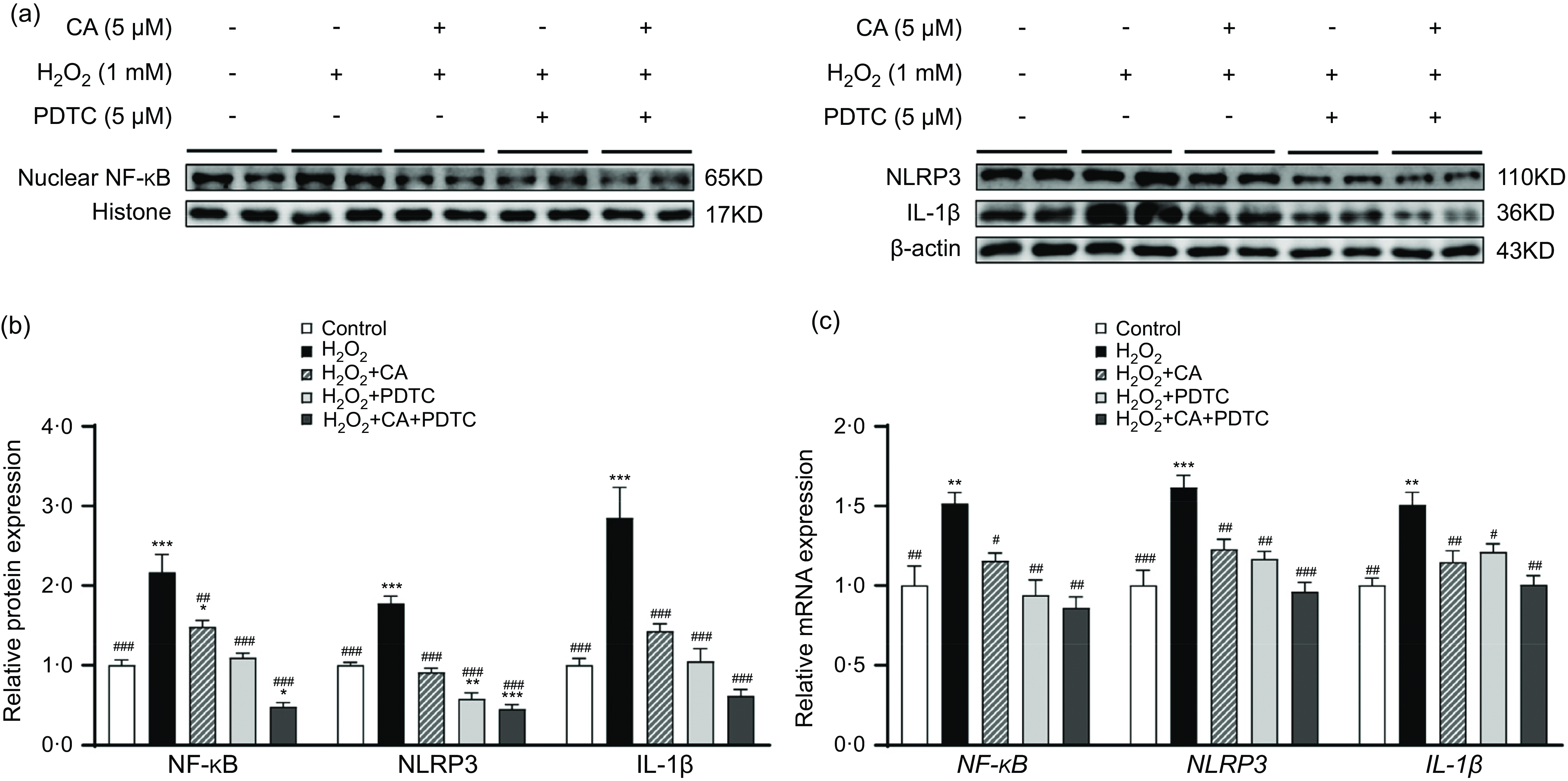
To verify whether CA inhibited NLRP3 inflammasome activation by regulating NF-κB expression in our study, we further treated cells with PDTC (an inhibitor of NF-κB). Results showed that compared with the H2O2 group, the PDTC + H2O2 group significantly decreased the protein and mRNA expression of IL-1β, NLRP3 and NF-κB (P < 0·05) (Figure 5), and these effects of PDTC were consistent with CA. It suggested that CA might attenuate the inflammation by regulating NF-κB-mediated NLRP3 inflammasome activation, thereby alleviating the injury of IPEC-J2 cells induced by H2O2.
The inhibitory effect of CA on NLRP3 inflammasome activation in IPEC-J2 cells induced by H2O2 is mediated by NF-κB. (a) Representative Western Blot images of NF-κB, NLRP3 and IL-1β; (b), (c) The protein (n 4) and mRNA (n 3) expression of NF-κB, NLRP3 and IL-1β. Data were presented as means ± sem. *P < 0·05, **P < 0·01 and ***P < 0·001 for comparison with the control group. # P < 0·05, ## P < 0·01 and ### P < 0·001 for comparison with the H2O2 group.

Discussion
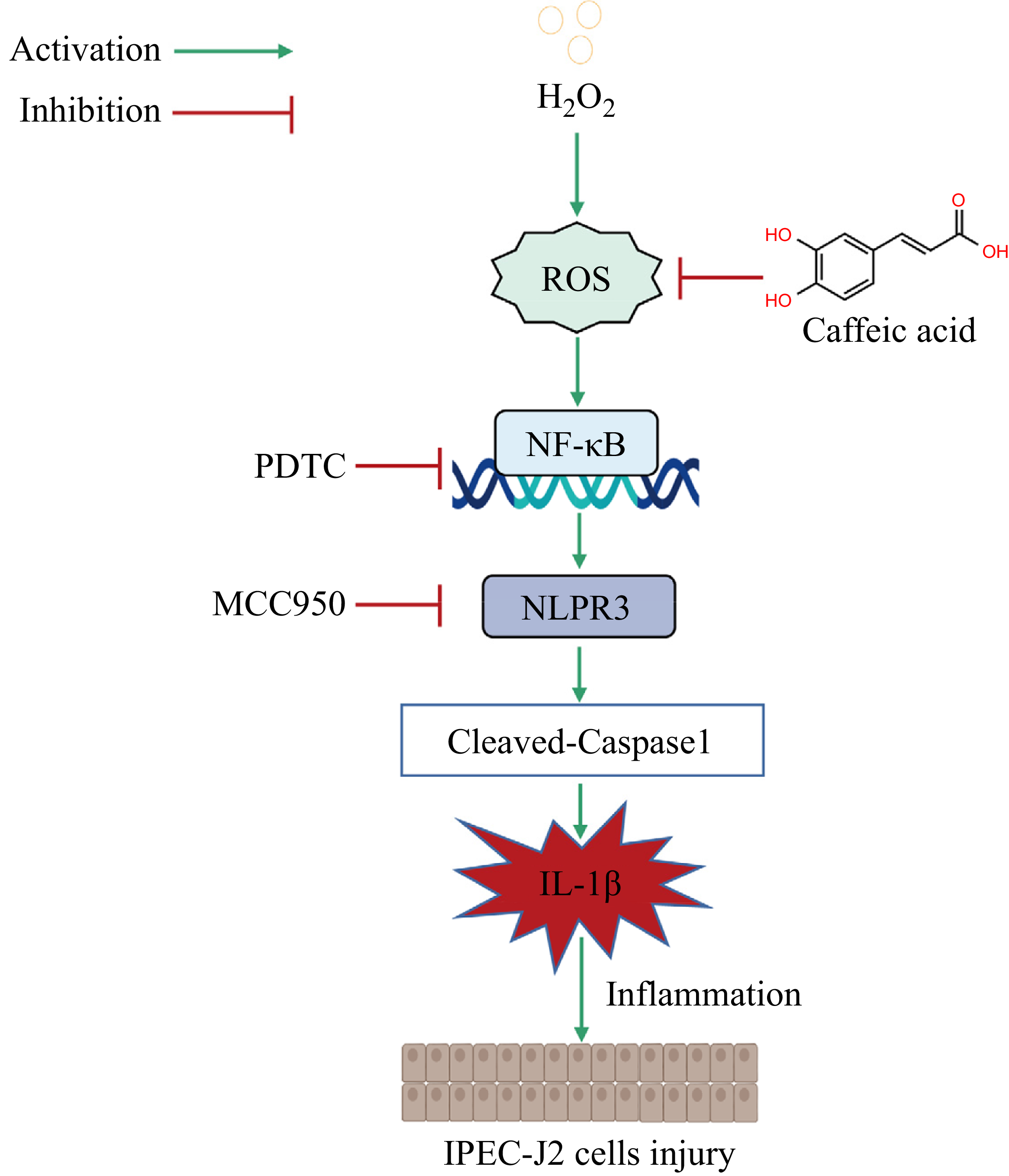
CA is considered a functional food with great application value(Reference De Araújo, De Paulo Farias and Neri-Numa23,Reference Khan, Bamunuarachchi and Tabassum24) . Some recent studies on piglets also found that CA had the potential to relieve intestinal inflammation(Reference Chen, Qiao and Mao13,Reference Wen, Wan and Wu25,Reference Wen, Wan and Wu26) , but the anti-inflammatory mechanism of CA on the intestine of piglets has not been reported. In this study, we found that CA might inhibit NLRP3 inflammasome activation by regulating the expression of NF-κB to attenuate the inflammation and injury of IPEC-J2 cells caused by H2O2 (Figure 6).
The mechanism of CA attenuating H2O2-induced IPEC-J2 cells inflammation. CA attenuated H2O2-induced inflammation by inhibiting NF-κB-mediated NLRP3 inflammasome activation, thereby alleviating IPEC-J2 cells injury.

In general, excessive ROS produced by oxidative stress is able to induce intestinal inflammation(Reference Wang, Chen and Zhang27). H2O2 is an important ROS that can easily penetrate the cell membrane. When a certain concentration of H2O2 enters the cells, it leads to the accumulation of ROS, which interferes with intracellular signalling pathways to release inflammatory factors (IL-1β, etc.), thereby increasing cell permeability and causing irreversible injury to the intestinal mucosa(Reference Ofoedu, You and Osuji28). FD4 permeability and TEER can reflect cell permeability. When FD4 permeability increased and TEER decreased, cell permeability increased(Reference De Gregorio, Serafino and Krasnowska29). Increased LDH release is one of the key features of cell injury, and LDH is released to the outside of the cell in a large amount when the cell is injured(Reference Abutaha30). In the previous study, H2O2 caused severe inflammation in rat intestine, and a large number of pro-inflammatory cytokines caused serious injury to intestinal morphology(Reference Lan, Chang and Wei31). Our study showed that H2O2 significantly increased ROS level, IL-1β level, LDH release as well as cell permeability, and caused significant changes in IPEC-J2 cell morphology. All these indicated that H2O2 caused the inflammation and injury of IPEC-J2 cells in our study. At present, a variety of plant active ingredients have been proved to reduce the excessive inflammatory response of intestinal cells, thereby alleviating intestinal injury(Reference Xiao, Wen and Shen32,Reference Hossen, Hua and Ting33) . A recent study showed that CA alleviated the expression of inflammation-related factors and reduced the damaged intestinal mucosa of lipopolysaccharide-challenged piglets(Reference Wen, Wan and Wu25). In our study, CA reduced the levels of ROS and IL-1β and alleviated IPEC-J2 cells injury. These results suggested that CA might attenuate the inflammatory response to reduce the levels of pro-inflammatory cytokines in some way, thus alleviating the injury of IPEC-J2 cells induced by H2O2.
NLRP3 inflammasome plays a vital role in regulating the inflammatory response and is crucial for maintaining intestinal homeostasis(Reference Xue, Yuan and Hou34). Once NLRP3 inflammasome is overactivated, it triggers self-cleavage and activation of Caspase-1. Then the formation of Cleaved-Caspase1 can lead to the maturation and release of large amounts of IL-1β, which eventually triggers the inflammatory cascade to damage the intestinal barrier(Reference Kelley, Jeltema and Duan35,Reference Man and Kanneganti36) . Therefore, Cleaved-Caspase1 and NLRP3 levels are usually used to evaluate NLRP3 inflammasome activation(Reference Tsai, Chiang and Hung37,Reference Tamura, Ishikawa and Yoshie38) . The recent research has identified that a great many of plant active ingredients (such as ginsenoside Rg1, curcumin, baicalin) can inhibit NLRP3 inflammasome activation(Reference Kang, Zhou and Zhu8,Reference Spano, Di Matteo and Ingallina39–Reference Lu, Wang and Wen41) and alleviate intestinal inflammatory damage(Reference Kang, Zhou and Zhu8,Reference Wang, Zheng and Men40,Reference Lu, Wang and Wen41) . We found that CA could also effectively inhibit NLRP3 inflammasome activation, but whether CA alleviated IPEC-J2 cells’ inflammation by inhibiting NLRP3 inflammasome activation was unclear. Previous study found that phenethyl caffeate (the derivative of CA) could inhibit NLRP3 inflammasome activation to prevent colitis(Reference Dai, Jiang and Sun42). Therefore, we speculated that CA might attenuate IPEC-J2 cells’ inflammation by inhibiting NLRP3 inflammasome activation. MCC950 is a specific inhibitor of NLRP3 and can specifically inhibit NLRP3 expression(Reference Li, Guan and Liang43). To further investigate the mechanism of CA action, we treated the cells with MCC950. The results showed that MCC950 had the same effect as CA. They not only inhibited the expression of NLRP3 and Cleaved-Caspase1 but also reduced cell permeability, LDH release and the expression and release of IL-1β. These suggested that CA attenuated the inflammation and injury of H2O2-induced IPEC-J2 cells by inhibiting NLRP3 inflammasome activation.
Studies have shown that NF-κB can initiate NLRP3 inflammasome activation, thereby regulating the occurrence and development of inflammatory diseases(Reference Boaru, Borkham-Kamphorst and Van De Leur44,Reference Wang, Lin and Yuan45) . When excessive ROS leads to the activation of NF-κB, the NLRP3 inflammasome is initiated and assembled(Reference Huang, Xu and Zhou46). In the mice study, ROS drove NF-κB to initiate NLRP3 inflammasome activation, which led to increased expression of pro-inflammatory cytokines, thereby worsening the duodenal inflammation(Reference He, Li and Xu47). A study showed that CA inhibited NF-κB expression to alleviate skeletal muscle injury in rats(Reference He, He and Wang48). Our findings aligned with previous study, which showed that CA inhibited NF-κB expression. However, whether the inhibition of CA on NLRP3 inflammasome induced by H2O2 was related to NF-κB was still unclear. PDTC is a potent inhibitor of NF-κB(Reference Qin, Cao and Li49), so we further investigated the effect of CA on the NLRP3 inflammasome by treating cells with PDTC. The results showed that the effect of PDTC on the cells was consistent with that of CA, which significantly reduced the expression of IL-1β and NLRP3. It suggested that CA reduced H2O2-induced pro-inflammatory cytokine levels by inhibiting NF-κB-mediated activation of NLRP3 inflammasome, thereby attenuating IPEC-J2 cells inflammation.
Conclusion
In conclusion, this study demonstrated that CA could attenuate H2O2-induced inflammation and injury in IPEC-J2 cells by inhibiting NF-κB-mediated NLRP3 inflammasome activation. It is worth noting that molecular docking results have shown that CA has a good affinity with NF-κB protein, but whether CA directly targets NF-κB to protect against intestinal injury needs to be further investigated. In addition, this study conducted only in vitro experiments, and further animal experiments will be needed in the future to confirm the results of this study.
Acknowledgements
This work was supported by the Natural Science Foundation of Sichuan Province (No. 2025ZNSFSC0281).
X. Y. Z.: Investigation, Conceptualisation, Validation, Formal analysis, Data curation, Writing-original draft. X. X. R.: Investigation. Z. Q. H.: Methodology, Conceptualisation, Resources. H. Z.: Software, Resources. G. J.: Software, Resources. X. L. C.: Methodology, Supervision, Funding Acquisition, Project administration, Writing-editing and review.
The authors declare no conflicts.
The data will be made available upon request.