Cardiac strangulation is a rare complication of permanent epicardial pacemakers in children. It can occur as a result of progressive compression of cardiac structures by the pacing leads with growth of the child,Reference Riede, Kostelka and Dahnert1–Reference Brenner, Gaines, Cordier, Reiner, Haney and Gundry3 and was reported to cause cardiac failureReference Riede, Kostelka and Dahnert1 and sudden death.Reference Eyskens, Mertens, Moerman, Ector, Daenen and Gewilling2 We describe a 3-year-old girl who presented with suspected cardiac strangulation on echocardiography and underwent successful pacemaker revision.
Clinical summary
Our patient had an antenatal diagnosis of complete congenital cardiac block secondary to maternal lupus antibody. Foetal echocardiography showed atrioventricular dyssynchrony with a ventricular rate of 50 beats per minute, but otherwise normal cardiac anatomy. The baby was born at term by caesarean section, weighing 3.2 kilograms, and was in low cardiac output for which she required an isoproterenol infusion. On the second day of life, she underwent implantation of epicardial bipolar steroid-eluting leads (25 centimetres, Model 4965, Medtronic Inc., Minneapolis, Minnesota, United States of America) and a dual-chamber pacemaker via a lower median sternotomy. The excess leads were positioned in the anterior pericardium with the generator in the left rectus sheath. Her post-operative course was uncomplicated and she was discharged home where she remained asymptomatic for 38 months. At 15-month follow-up, her echocardiogram showed normal right ventricular size and mild tricuspid regurgitation, with normal ventricular function. Throughout follow-up, her pacemaker system maintained excellent sensing, capture, and impedance.
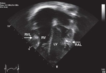
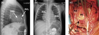
At 3 years of age, she was incidentally found to have a new pansystolic cardiac murmur and she had no clinical symptoms. Her echocardiography showed moderate tricuspid regurgitation from anterior leaflet prolapse with an enlargement of the right ventricle and right atrium. Dense extra-cardiac structures (Fig 1) were also observed to cause constriction of the posterolateral wall of the left ventricle and the right ventricular free wall in diastole, findings that were not previously observed. A chest X-ray was performed, which showed the epicardial leads to loop anteriorly and, diving posteroinferiorly on each side of the heart, to attach to the right atrium and ventricle, respectively (Fig 2a and b). These combined findings were suspicious of cardiac strangulation, and she was scheduled for urgent pacemaker leads revision.
Echocardiogram frame showing dense structures impinging on both ventricles in diastole (RVL = right ventricular lead; RAL = right atrial lead; RV = right ventricle; LV = left ventricle).

Lateral and anteroposterior chest X-rays (a and b, respectively) showing the right atrial and ventricular leads around the cardiac chambers. (c) Intraoperative photograph showing the right atrial lead looping superiorly, diving posteriorly behind the left ventricle before reaching the right atrium. The right ventricular lead loops superiorly and crosses the right atrioventricular groove, creating a deep crease on the right atrium and ventricle before ultimately reaching the right ventricle (RVL = right ventricular lead, RAL = right atrial lead, RA = right atrium, RV = right ventricle, LV = left ventricle).

A full median sternotomy incision was performed and the leads were dissected free (Fig 2c). Both leads were found to be tightly wrapped around the ventricles, and caused significant biventricular compression, especially in diastole. No obvious coronary filling defect was observed, although the ventricular lead had crossed the right atrioventricular groove and caused a significant indentation of the right atrium and right ventricle. After explantation, she received new bipolar atrial and ventricular leads (35 centimetres, Model 4965, Medtronic Inc., Minneapolis, Minnesota, United States of America), both with excellent thresholds and a new generator. The pacing electrode of the ventricular lead was positioned near the apex to optimise biventricular synchrony. The excess length of leads was positioned on the inferior diaphragmatic surface to allow for growth, and fixed at the electrode bifurcation to prevent displacement. The new generator was placed in the previous rectus sheath pocket. She had an uneventful recovery and is asymptomatic on follow-up at 4 months, but her degree of tricuspid insufficiency was unchanged and the right-sided cardiac chambers remained enlarged.
Discussion
In all five cases of cardiac strangulation documented in the English literature, the patients had their pacemaker implanted during the neonatal or infant period. With growth, the epicardial leads were described as compressing the ventricles,Reference Riede, Kostelka and Dahnert1–Reference Brenner, Gaines, Cordier, Reiner, Haney and Gundry3 the right ventricular outflow tract and pulmonary artery,Reference Brenner, Gaines, Cordier, Reiner, Haney and Gundry3 and the coronary arteries,Reference Riede, Kostelka and Dahnert1–Reference Brenner, Gaines, Cordier, Reiner, Haney and Gundry3 causing myocardial ischaemia and sudden death in one case.Reference Eyskens, Mertens, Moerman, Ector, Daenen and Gewilling2
The recognition of strangulation can be quite challenging given the rarity of the condition, the variable presentation, and the lack of a definite pre-operative test. Lead compression can present with a new cardiac murmur,Reference Brenner, Gaines, Cordier, Reiner, Haney and Gundry3 chest pain,Reference Eyskens, Mertens, Moerman, Ector, Daenen and Gewilling2, Reference Brenner, Gaines, Cordier, Reiner, Haney and Gundry3 or syncope.Reference Brenner, Gaines, Cordier, Reiner, Haney and Gundry3 The onset of segmental wall motion abnormalities or findings of dilated cardiomyopathyReference Riede, Kostelka and Dahnert1 in a patient with an epicardial system should prompt the clinician to consider cardiac strangulation. Unlike other pacemaker complications, patients with cardiac strangulation do not present with increasing thresholds or lead dysfunction. In the case of our patient, newly diagnosed tricuspid regurgitation and dense structures compressing the ventricles on an echocardiogram led us to the diagnosis. Despite the repositioning of the electrodes correcting the compression aspect and improving ventricular synchrony, it did not significantly change the degree of tricuspid regurgitation, and therefore it is unlikely that the lead was distorting the tricuspid valve annulus to the extent of causing moderate regurgitation.
To minimise the risks of strangulation when implanting an epicardial pacemaker in a neonate or an infant, surgeons should be especially mindful of lead placement, and these should not be routed anteriorly or around the cardiac chambers. Furthermore, careful placement of each electrode in avoidance of coronary vessels is critical, and can be challenging in a child with a previous intervention. Leaving a loop of lead to allow for growth is important. In larger children, the loop of lead can be placed on the diaphragmatic surface of the pericardium with the generator within the rectus sheath, but the lead should be fixated at the electrode bifurcation to avoid anterior dislodgement and cardiac compression. A technique to circumvent cardiac compression by bulky leads in low-weight neonates is to route the excess lead in the pleural space.Reference Welch, Hannan and DeCampli4 The generator can be placed on the diaphragmatic surface of the same pleural space. Another approach is to use a single-chamber pacemaker with a smaller unipolar lead, and upgrade it to a dual-chamber system with bipolar leads when the child is older. This approach does not result in decreased function.Reference Horenstein, Karpawich and Tantengco5 Furthermore, it is advocated that a lower ventricular rate than the atrial rate be used, and that the rate be gradually increased over a period of months even in patients with dual-chamber systemsReference Silvetti, Drago and Ravà6 to reduce the risk of cardiac failure. Despite right ventricular free wall pacing not being shown to induce cardiomyopathy by itself, a subset of the patients with congenital complete atrioventricular block will develop heart failure, which is aggravated by ventricular dyssynchrony.Reference Silvetti, Drago and Ravà6, Reference Sagar, Shen and Asirvatham7 Therefore, the ventricular pacing electrode should not be positioned on the right ventricular free wall,Reference Silvetti, Drago and Ravà6 as positioning the lead on the left ventricular apex optimises ventriculo–ventricular interactions.Reference van Geldorp, Vanagt, Prinzen and Delhaas8
Conclusion
Cardiac strangulation is a rare but potentially life-threatening complication of epicardial pacing leads in children. If cardiac strangulation is suspected, the patient should undergo urgent surgical revision of the epicardial leads.
Acknowledgements
The authors would like to acknowledge the contribution of Dr Andrew I. M. Campbell for his comments on the review of the manuscript.