INTRODUCTION
Members of the genus Angiostrongylus belong to the superfamily Metastrongyloidea, a taxon of nematodes that occupy the circulatory system of their definitive hosts. Angiostrongylus cantonensis is the most widespread species of the genus. To date, A. cantonensis is the only species of the genus known to cause eosinophilic meningitis in humans and other non-permissive hosts (Wang et al. Reference Wang, Lai, Zhu, Chen and Lun2008; Graeff-Teixeira et al. Reference Graeff-Teixeira, da Silva and Yoshimura2009; Morassutti et al. Reference Morassutti, Levert, Perelygin, da Silva, Wilkins and Graeff-Teixeira2012; Cowie, Reference Cowie2013). In severe cases, A. cantonensis is reported to cause eosinophilic meningo-encephalomyelitis throughout the brain and spinal cord of accidental hosts (Mason et al. Reference Mason, Prescott, Kelly and Waddell1976; Wright et al. Reference Wright, Kelly, Waddell and Hamilton1991; Collins et al. Reference Collins, Rothwell, Malik, Church and Dowden1992) and fatal encephalitis in humans (Sawanyawisuth et al. Reference Sawanyawisuth, Takahashi, Hoshuyama, Senthong, Limpawattana, Intapan, Wilson, Tiamkao, Jitpimolmard and Chotmongkol2009). Although rare, pneumonia caused by the presence of A. cantonensis adult worms has also been reported in humans (Lindo et al. Reference Lindo, Escoffery, Reid, Codrington, Cunningham-Myrie and Eberhard2004).
Two species of Angiostrongylus occur in Australia. Angiostrongylus cantonensis, a widespread species found in rats throughout the tropical world, occurs in introduced rat species Rattus rattus and Rattus norvegicus in Eastern Australia. Angiostrongylus mackerrasae is restricted to Australia, where it is found mainly in native bush rats, Rattus fuscipes (Spratt, Reference Spratt2015). The two species are both neurotropic and have evidently identical lifecycles in the definitive host, tracking to the lungs of its rat hosts (Bhaibulaya, Reference Bhaibulaya1974). Despite having an identical lifecycle to A. cantonensis, A. mackerrasae has been neglected as a potentially zoonotic parasite in Australia.
In recent years, cases of angiostrongyliasis in humans and animals have been reported from Australia (Mackie et al. Reference Mackie, Lacasse and Spratt2013; Morton et al. Reference Morton, Britton, Palasanthiran, Bye, Sugo, Kesson, Ardern-Holmes and Snelling2013; Walker et al. Reference Walker, Spielman, Malik, Graham, Ralph, Linton and Ward2015). Recent surveys of the rodent hosts for Angiostrongylus (Aghazadeh et al. Reference Aghazadeh, Reid, Aland, Restrepo, Traub, McCarthy and Jones2015a ) and its intermediate host (Chan et al. Reference Chan, Barratt, Roberts, Lee, Shea, Marriott, Harkness, Malik, Jones, Aghazadeh, Ellis and Stark2015) reveal that species of Angiostrongylus are prevalent close to human dwellings. There is a distinct possibility that A. mackerrasae, like its congener A. cantonensis, causes infections in humans and domestic animals. This postulate is borne out by the recent discovery of adult A. mackerrasae from the lung of a flying fox (Pteropus alecto), an aberrant host, in which it was found responsible for severe pneumonia (Mackie et al. Reference Mackie, Lacasse and Spratt2013). This study investigates the potential pathogenicity of A. mackerrasae to humans by comparing the pathological features of infection in non-permissive hosts (mice and guinea pigs) that are a model for human disease.
MATERIAL AND METHODS
Maintaining the lifecycle of A. mackerrasae in the laboratory
Experimental work was performed with approval of the Animal Ethics Committee of the QIMR Berghofer Medical Research Institute under Project P1457. First stage larvae of A. mackerrasae were recovered from the faeces of R. fuscipes which were trapped in Southeast Queensland under permit number WIS12109412, from the Department of Environment and Heritage Protection of the Queensland Government. The rats were euthanized and dissected to detect and morphologically confirm the presence of A. mackerrasae present in their lungs, using descriptions of Mackerras and Sandars, Reference Mackerras and Sandars1954).
In order to maintain the lifecycle of the parasite in the laboratory, four different species of terrestrial and aquatic molluscs (Cornua spersum, Helix aspersa, Planorbid sp. and Austropeplea lessoni) were evaluated to determine the most suitable intermediate host with the recovered larvae in different temperatures ranging between 18 and 26 °C. Austropeplea lessoni (Lymnaeidae family) was chosen to maintain the lifecycle due to its less stringent maintenance needs in the laboratory. Snails were infected by immersion in a Petri dish containing first stage larvae of A. mackerrasae obtained from infected rats for 4 h. A small piece of tissues from the foot of a snail was fed to Wistar rats 4 weeks after infection with A. mackerrasae to maintain the lifecycle in the laboratory.
Infections in mice and guinea pigs
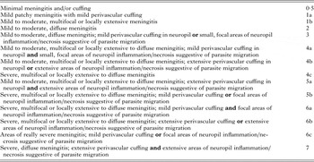
Snails were digested using artificial gastric juice to obtain infective third-stage larvae (L3) of A. mackerrasae 4 weeks after infection. Twenty-four outbred Swiss mice were randomly divided into two groups and infected by oral gavage with 30 third-stage larvae of either A. mackerrasae or A. cantonensis. The same number of uninfected control mice was also randomly allocated to each group. In addition, four tri-coloured guinea pigs were infected with 35 larvae using clear gelatin capsules directed into their oesophagus using a feline tablet applicator. The animals were monitored daily post infection for changes in behaviour indicative of infection and neurological changes. Four randomly selected mice from the infected and control groups were euthanized at 7, 14 and 21 days post infection (dpi). The infected guinea pigs were euthanized 16 dpi. Brain and spinal cord were removed and stored in 10% formalin for 72 h before histopathological sections were prepared from the tissues. Haematoxylin and eosin-stained paraffin sections were examined blindly using light microscopy in order to avoid bias in scoring. The histopathological lesions of mice and guinea pigs were graded based on a scoring system developed for this study based on (Baums et al. Reference Baums, Hermeyer, Leimbach, Adamek, Czerny, Horstgen-Schwark, Valentin-Weigand, Baumgartner and Steinhagen2013) in order to compare the severity of infection caused by the two species of Angiostrongylus (see Table 1).
Histopathology scoring system used in this study to compare lesions caused by A. mackerrasae with those caused by A. cantonensis in Swiss mice

Splenocyte culture and flow cytometry
Spleens were removed from each mouse and pressed through 70 µ m cell strainers (Falcon). Red blood cells were lysed using red blood cell lysing buffer (Sigma-Aldrich) and the remaining live leukocytes were enumerated using the trypan blue exclusion method. Splenic cultures were plated at 5 × 10⁵ cells per well and incubated at 37 °C in Iscove's Modified Dulbecco's Medium (IMDM) containing 10% fetal calf serum, penicillin streptomycin, 55 µ m betamercaptoethanol with phorbol myristate acetate (PMA) (10 ng mL−1) and Ionomycin (1 µg mL−1) in 5% CO2 flow. After 1 h, brefeldin A was added at 10 µg mL−1. Four hours later, cells were harvested and labelled for flow cytometry using anti-mouse CD3-BV510, CD4-Alexa Fluor700, CD8 PE-Cy7 and BD Horizon™ fixable viability stain 450 (all by BD biosciences). Cells were fixed and permeabilized using BD cytofix/Cytoperm buffer and stained for intracellular expression of cytokine using anti mouse interleukin (IL)-17 Alexa Fluor 647, IL-4 PE CF594 and interferon γ (IFNγ)-PerCp Cy5·5 (BD Biosciences). Fluorescence minus one control were also generated. Samples were collected on a BD Fortessa 4 Laser flow cytometer and the results were analysed using FACS DIVA software and GraphPad Prism version 6 using t-test and Wilcoxon rank sum tests to determine if any differences between the infected mice and control groups were statistically significant at a 95% level of confidence.
Serological comparison
Antigen preparation
Total soluble extract (TE) was obtained from harvested female worms that were macerated in liquid nitrogen and homogenized extraction buffer [phosphate-buffered saline (PBS; pH 7·4), 0·01% triton X-100 and proteases inhibitors kit (QIAGEN)]. The suspension was centrifuged at 12 000 g for 1 h at 4 °C, and the supernatants were used to derive the TE protein concentrations was determined by Bradford assay according to the instructions of the manufacturer. TE from male worms was produced as described above.
Sodium dodecyl sulphate-polyacrylamide gel electrophoresis (SDS–PAGE) and Western blots (WB)
One-dimensional electrophoresis of 4–12% polyacrylamide Bis-Tris gels with SDS–PAGE was used to resolve proteins of TE, which were then stained with Comassie Brilliant Blue. For the WB resolved 100 ng, 1 µg or 3 µg of TE were electro-transferred onto nitrocellulose membranes, and then blocked with 5% powdered milk for 2 h at room temperature. The membranes were then incubated for 2 h with different sera: (1) pool of human sera (1:200 dilution), prepared from three patients diagnosed with cerebral angiostrongyliasis; (2) rat sera (1:200 dilution); and (3) guinea pig sera (1:200 dilution). After three washes, the membranes were probed with a secondary peroxidase-conjugated anti-human IgG (1:5000; Abcam, Cambridge, UK), anti-Rat IgG (1:5000; Abcam, Cambridge, UK) or anti-guinea pig IgG (1:5000; Abcam, Cambridge, UK) for 2 h at room temperature. After three washes of PBS Tween 0·05%, Clarity Western ECL Blotting Substrate (Biorad) was added.
RESULTS
A. mackerrasae infections in mice
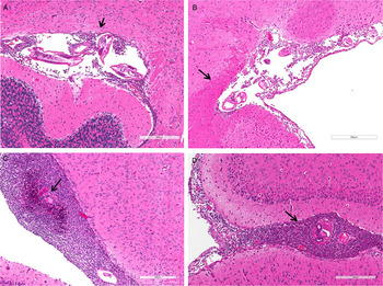
Grossly, hyperaemia was observed on the surface of the brain tissue in all infected mice. The first group of mice, euthanized 7 dpi, did not show any clinical neurological signs and the inflammatory reaction to the parasite was mild. Of the four mice in this group, two showed minimal meningitis and/or perivascular cuffing in the neuropil. Angiostrongylus larvae were present in the neuropil in two of the mice. The second group of four mice, euthanized 14 dpi, all showed moderate to marked diffuse eosinophilic meningitis with intralesional nematodes in three out of four of the mice (Fig. 3A). Multifocal, mild eosinophilic encephalitis was also observed in two out of four of these mice. In the group of mice euthanized 21 dpi, there was diffuse moderate to marked eosinophilic and granulomatous meningitis. Some nematodes appeared viable; however, degenerate parasites were also noted and were often associated with areas of granulomatous meningitis.
Pathological section of mice brain tissue infected with A. mackerrasae and A. cantonensis 2 weeks (A, B) and 3 weeks (C, D) post infection. Arrow points at larvae.

A. cantonensis infection in mice
Most of the infected animals showed variable degrees of pathological changes in the brain tissue. The mice euthanized on 7 dpi, showed mild patchy meningitis with mild perivascular cuffing. Small numbers of viable parasite were seen in focal area of the brain in one mouse. In the second group of mice euthanized on 14 dpi, eosinophilic meningitis was observed in all mice and moderate numbers (at least three per section) of parasites were present in multiple locations in the meninges (Fig. 3B). In the group of mice euthanized on 21 dpi, moderate to severe eosinophilic meningitis was observed in all mice (Table 2). Small numbers of viable parasites were noted in three out of four of the mice.
Histopathological scores of Swiss mice infected with the two species of Angiostrongylus

Comparing pathogenicity caused by the two Angiostrongylus species
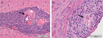
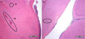
The group of 12 mice infected with A. cantonensis, showed brain lesions similar to those observed in A. mackerrasae-infected mice. The comparison between the two groups of mice infected with A. cantonensis and A. mackerrasae revealed that the two species cause similar lesions of similar severity in infected Swiss mice (Table 1; Figs 1 and 2). There were small points of difference however, including a more rapid development of meningitis in the A. cantonensis-infected mice. Moreover, the mice infected with A. mackerrasae developed meningitis of a more granulomatous nature, particularly at week 3 post infection, which was associated with an increased proportion of degenerate larvae in the A. mackerrasae-infected mice.
Pathological section of mice brain tissue infected with A. mackerrasae and A. cantonensis and 3 weeks post infection. A. cantonensis larvae is still viable 3 weeks post infection (A); Degenerated larvae of A. mackerrasae 3 weeks post infection (B).

Grade 7 infection with A. mackerrasae in mice 2 weeks post infection (A) A1: Perivascular cuffing, A2: Larvae migration track. Grade 4a infection with A. cantonensis in mice 2 weeks post infection; perivascular cuffing (B).

A. mackerrasae infections in guinea pigs
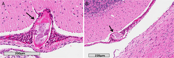
All infected guinea pigs also showed various degrees of pathological changes (scored between 1b to 4c) in the brain sections. Larvae of A. mackerrasae were present in the meninges of all four animals. Within the cerebral and cerebellar meninges and variably the nerve root meninges and stroma of the choroid plexus, there were multifocal, moderate to dense populations of eosinophils, lymphocytes and macrophages in all four animals (Fig. 4). As opposed to mice, no changes were observed in cerebral neuropil and the inflammation was limited to meninges.
Eosinophilic meningitis in infected guinea pigs with Angiostrongylus mackerrasae, 16 dpi. (A, B) Larvae of A. mackerrasae in cerebral meninges surrounded by inflammatory cells.

Flow cytometry analysis
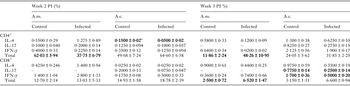
The results of flow cytometric analysis of splenocyte cultures from Angiostrongylus-infected mice are summarized in Table 3. Data values obtained from unstimulated media only control samples were subtracted from the values measured of PMA/Ionomycin re-stimulated samples to give the percentage values reported in Table 3.
Flow cytometry analysis of splenic T cells in mice infected with A. mackerrasae and A. cantonensis on 14 and 21 dpi

A.m., Angiostrongylus mackerrasae; A.c., Angiostrongylus cantonensis, PI, post infection.
a Significantly different values are bolded.
Comparison of the T cell counts and cytokines showed almost no significant differences between infected (both species) and control groups. Angiostrongylus cantonensis-infected mice showed decreased numbers of splenic T cells both at 2 weeks and 2 weeks post infection in comparison with the control group. Mice infected with A. mackerrasae showed decreased splenic T cells 2 weeks post infection but the number of cells increased after 3 weeks. In A. cantonensis-infected mice, significant differences included the decreased level of CD4 IL-4 (P = 0·049), 2 weeks post-infection, decreased expression of CD8 IL-17 (P = 0·041) 3 weeks post infection and the reduction of CD8 IFN-γ levels (P = 0·021) 3 weeks post infection in comparison to the uninfected control group. There was no significant cytokine variations observed between mice infected with A. mackerrasae and the control group. The percentage of stimulated CD4 T cells in A. mackerrasae-infected mice was significantly lower (P = 0·03) than the control group 2 weeks post infection. In addition, the percentage of stimulated CD4 and CD8 T cells were significantly higher in A. mackerrasae-infected mice 3 weeks post infection (P = 0·01 and 0·04, respectively).
SDS–PAGE and WBs
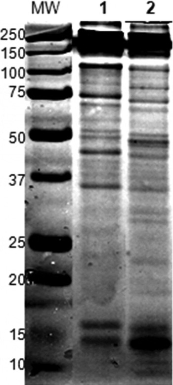
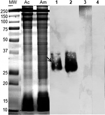
The TE from female A. mackerrasae, female A. cantonensis (Australian strain), male A. mackerrasae and female A. cantonensis (Brazilian strain) all showed similar recognition at 31 kDa band to the serum from a human infected with A. cantonensis from Brazil (positive control), serum from experimentally infected rats and guinea pigs (Figs 5 and 6).
SDS–PAGE of A. mackerrasae worms soluble total extract (TE). 1: A. mackerrasae female; 2: A. mackerrasae Male. MW: Molecular weight (kDa).

Identification of 31 kDa antigen produced by A. mackerrasae. Female worm soluble total extract (TE) from Australian A. cantonensis (Ac) and A. mackerrasae (Am) were resolved in 1DE gel and transferred on Western blot membranes. Lane 1: Ac-TE and Lane 2: Am-TE, probed with pool of positive controls for cerebral angiostrongyliasis (from Brazil); Lane 3: Ac-TE and Lane 4: Am-TE, probed with pool of normal human sera (100 ng TE per lane).

DISCUSSION
This research demonstrates for the first time that A. mackerrasae can cause severe meningitis in infected mice and guinea pigs, which is an indication that it may be pathogenic in humans, dogs and other animals. The lesions caused by A. mackerrasae in this study were consistent with those caused by A. cantonensis in humans (John and Martinez, Reference John and Martinez1975) as well as with lesions noted in previous work on guinea pigs (Perez et al. Reference Perez, Capron, Lastre, Venge, Khalife and Capron1989) and mice (Sugaya and Yoshimura, Reference Sugaya and Yoshimura1988) infected with A. cantonensis. This result was expected as molecular comparison of A. mackerrasae with A. cantonensis showed that the two species share highly similar genetic identity (Aghazadeh et al. Reference Aghazadeh, Traub, Mohandas, Aland, Reid, McCarthy and Jones2015b ). However, the result indicates that A. cantonensis endures for a longer time in the brain of accidental hosts than previously reported. Although A. mackerrasae is genetically very similar to A. cantonensis, it naturally has a different host choice (native Rattus spp.) and it caused a more severe immune response in mice in this study. This suggests that infection caused by A. mackerrasae could have a different clinical outcome in humans and other accidental hosts.
Our study showed minimal cytokine activity attributable to Angiostrongylus infection. The percentages of IL-17 and IFN-γ producing cells were lower in mice infected with A. cantonensis compared with uninfected mice in this study, consistent with the observations of Liu et al. (Reference Liu, Luo, Shen, Li, Ding and Chen2013). This was also observed with A. mackerrasae-infected mice but only 3 weeks post infection. IL-17 and IFN-γ both act as mediators in delayed type immune responses to extracellular pathogens and both are expected to be increased in parasitic eosinophilic meningitis. Liu et al. (Reference Liu, Luo, Shen, Li, Ding and Chen2013) speculated that the reduction of splenic IL-17 in A. cantonensis infection might be induced by high expression of IL-4 restricting IL-17 secretion. However, in this study IL-4 was not highly expressed in the infected mice. Sugaya et al. (Reference Sugaya, Aoki, Abe, Ishida and Yoshimura1997) also detected a peak of IL-4 in cerebrospinal fluid of BALB/c and C57BL/6 mice on 12–15 and 20 dpi by A. cantonensis, but they did not study the levels of IL-17. In the present study, IL-4 was decreased at both time points in A. cantonensis group but CD4 IL-4 was only increased on 14 dpi in A. mackerrasae group. It must be noted that A. mackerrasae-infected animals showed slightly less pathology at this time point, which could be attributable to this difference in IL-4 production although the percentage of cells producing it is very small.
Parasites are known to actively evade the host immune responses to ensure their ongoing survival and proliferation (Schmid-Hempel, Reference Schmid-Hempel2009). The subdued immune responses measured in this study may suggest that the parasites themselves are able to down-regulate or dampen the host immune response in some way, although the data collected cannot confirm this. Factors such as number of larvae used to induce infection, the time post infection, mouse strain and the involvement of brain blood barrier (the brain as an immune privileged site) could also be responsible for the variation of cytokine expression in different studies. It is therefore plausible to suggest that splenic T cells and expressed cytokines in this instance could not give a clear picture of the intensity of immune response to this parasite.
This study also reveals that both proteins of A. cantonensis and A. mackerrasae are recognized by positive sera of cerebral angiostrongyliasis patients. The 31 kDa band previously described (Morassutti et al. Reference Morassutti, Levert, Perelygin, da Silva, Wilkins and Graeff-Teixeira2012) from A. cantonensis was also observed from A. mackerrasae, indicating that the two species express a set of very similar proteins. Previously, Ben et al. (Reference Ben, Rodrigues, Agostini and Graeff-Teixeira2010) demonstrated the successful use of heterologous antigens in immune diagnosis of angiostrongyliasis (A. cantonensis and A. costaricensis), and Morassutti et al. (Reference Morassutti, Levert, Perelygin, da Silva, Wilkins and Graeff-Teixeira2012) corroborated these data showing that the 31 kDa antigen from A. cantonensis TE is recognized both by sera from patients with eosinophilic meningitis and abdominal angiostrongyliasis, indicating that both species of parasites produce very similar antigens although the pathology caused by each of these two species are different. Here we found that another species of Angiostrongylus, A. mackerrasae can also be recognized by sera from human cases of eosinophilic meningitis caused by A. cantonensis. The cross reactivity observed between A. cantonensis and A. mackerrasae was expected. However, further research is required to determine whether A. mackerrasae can potentially infect humans and other animals and cause a pathogenic effect. Investigating its ability to develop to patency in humans is also a priority.
ACKNOWLEDGEMENTS
The authors would like to thank Dr Rogan Lee from University of Sydney and Dr John Stanisic from Queensland Museum and Mr David McNeilly from QIMR Berghofer Medical Research Institute for their help and support during this study.
FINANCIAL SUPPORT
M. A. was supported by a University of Queensland Postgraduate Award. This research was supported by Peter and Mary Ellen Stone Memorial Fund, awarded to Malcolm Jones.
CONFLICT OF INTEREST
None.