Atherosclerosis is a complex systemic multifactorial inflammatory disease, which synergistically promote lesion development. Atherosclerosis leads to the development of coronary artery disease (CAD) and carotid disease, which are two of the most common causes of mortality and morbidity worldwide (Ref. Reference Altaf1). Atherosclerotic lesions progress over decades with periods of subclinical manifestations, until atheroma plaques develop. Plaque enlargement is associated with stenosis of the arterial lumen (Ref. Reference Vaina and Stefanadis2) with subsequent alteration of blood flow, and plaque rupture and/or plaque surface erosion (Ref. Reference Romero3), leading to thrombus formation, lumen occlusion and ultimately, ischemic events (Ref. Reference Adams and Celermajer4). Traditional risk factors include high density lipoprotein-cholesterol and total cholesterol content (dyslipidemia), systolic blood pressure (hypertension), age, gender, diabetes mellitus, cigarette smoking, obesity, and left ventricle hypertrophy. However, research into a combination of additional and as yet unknown factors might help us to better understand the onset of this disease.
Factors that determine progression towards vulnerable plaque are being carefully investigated, since the early detection of vulnerable plaque may represent a big step towards increasing the life span of the population (Refs Reference Cocker5, Reference Davies6, Reference Botnar7, Reference Sadeghi8, Reference Vengrenyuk9, Reference Canet-Soulas and Letourneur10). Until now, arterial luminal diameter as detected by classical imaging technologies has been the only diagnostic procedure, and restricted to later stages of disease. However, positive remodelling, which is associated with plaque rupture, is not necessarily associated with luminal changes.
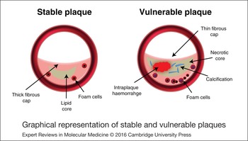
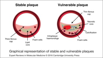
Oxidised low-density lipoproteins (oxLDL) play a major role during the onset of atherosclerosis by inducing the expression of several adhesion molecules at the cell surface of endothelial cells, promoting recruitment and adhesion, as well as internalisation of circulating monocytes from the intima layer, and internalisation to the media layer of the arteries (Refs Reference Canet-Soulas and Letourneur10, Reference Kanwar11, Reference Naghavi12). Histologically, atherosclerotic plaques can be differentiated into stable and vulnerable plaques, depending on whether they are stable or prone to rupture. Scavenger receptor-mediated uptake of ox-LDL by infiltrated monocytes causes them to differentiate into foam cells, producing significant amounts of proinflammatory cytokines and matrix metalloproteinases (MMPs), and leading to the formation of a fatty streak (Ref. Reference Li13). The lesions are composed of smooth muscle cells, cellular debris and a lipid-rich core covered by a fibrous cap (Refs Reference Majmudar14, Reference Millon15, Reference Pedersen16, Reference Ratib and Nkoulou17, Reference Rischpler, Nekolla and Beer18, Reference Bigalke19). Lesion progression continues with the formation of a necrotic core, mainly composed of apoptotic macrophages, smooth muscle cells, debris and a significant amount of free cholesterol coming from macrophages and erythrocyte membranes. The increase in free cholesterol is closely associated with lesion instability (Refs Reference Hatsukami20, Reference Jones21, Reference Xu22, Reference Cai23). Stable plaques show small necrotic cores and thick fibrous caps, whereas vulnerable plaques are composed of large necrotic cores and thin fibrous caps. The fibrous cap is mostly composed of smooth muscle cells, which produce extracellular matrix (ECM) components including collagen and elastin, hence contributing to the thickening of the fibrous cap. By contrast, those signals, which trigger smooth muscle and macrophage apoptosis will give rise to plaques with thinner fibrous caps, as shown in vulnerable atherosclerotic plaques (Fig. 1). Neoangiogenesis is another key signature of plaque vulnerability. Plaque vascularization coming from the vasa vasorum contributes a continuous influx of erythrocytes, lymphocytes and macrophages. Intraplaque vessels are immature, with a reduced number of smooth muscle cells; hence intraplaque haemorrhage also contributes to plaque rupture (Ref. Reference Heeneman24).
Graphical representation of stable and vulnerable plaques.
The thinner the fibrous cap is, the more prone it is to rupture. Therefore, new non-invasive imaging approaches providing information on plaque burden and composition are needed, in order to better identify population at risk (Refs Reference Botnar7, Reference Iqbal25, Reference Dweck26, Reference Mateo27). To this end, the combination of the most recent cutting edge imaging modalities – including hybrid positron emission tomography (PET)/magnetic resonance imaging (MRI) – and preclinical models of atherosclerotic disease, has resulted in a more synergistic handling of human atherosclerosis disease, leading us closer to an effective means of tackling it.
Preclinical models of atherosclerosis
Mouse models of atherosclerosis
The use of genetically modified mouse models is, for several reasons, the most common tool for the study of atherosclerosis (Refs Reference U-King-Im28, Reference Wasserman29). Despite the differences between human and murine atherosclerosis – including vessel architecture, plaque composition and even plaque stability – and thanks to the extensive investigation in mouse models of atherosclerosis, we have a deep understanding of the mechanisms involved in atherogenesis (Refs Reference Boshuizen30, Reference Getz and Reardon31).
LDL receptor (LDLR) deficient mice develop mild atherosclerosis, in which the severity of the lesions can be accelerated when mice are fed a high-cholesterol diet (Refs Reference Li32, Reference Ru33, Reference Rensing34, Reference Marquart35, Reference Tang36, Reference Teodoro37, Reference Awan38). Aortic lesions can progress beyond the foam-cell fatty-streak stage to the fibro-proliferative intermediate stage.
The apolipoprotein E (apoE)-deficient mouse model tends to accumulate significant amounts of LDL and very LDL (VLDL), and develop atheroma plaques, which are accelerated under a high cholesterol diet, in a manner similar to that which occurs in humans, including foam cell formation and migration of smooth muscle cells to the damaged tissue (Refs Reference Bigalke19, Reference Zhao39, Reference Andrés40, Reference Higashimori41, Reference Meir and Leitersdorf42). Atheroma plaques are mainly composed of a necrotic core surrounded by proliferating smooth muscle cells and ECM components (Refs Reference Zhao39, Reference Bot43, Reference Akhtar44, Reference Fernández-Hernando45). Double apoE/class B scavenger receptor mutated animals have allowed investigators to study coronary artery atherosclerosis, thus making this double mutant murine model a valuable tool for the study of CAD (Refs Reference Vaisman46, Reference Kitayama47, Reference Zhang48, Reference Zhang49, Reference Sosnovik and Caravan50).
Mutated variants of apoE, including apoE 3Leiden, apoE2 and several single point mutated forms of the apoE gene, have also been overexpressed in mice to recreate human atherosclerosis, in which new atheroma plaque reducing drugs are tested (Refs Reference Zadelaar51, Reference Lardenoye52, Reference Lalloyer53).
Besides all the benefits of using genetically modified animals – relatively low cost, elevated sample size and ease of handling – a major limitation still remains: vulnerable atheroma plaque formation is extremely infrequent in mice when compared with other species, including humans. The recapitulation steps, beginning with the formation of large necrotic cores, the thinning of the fibrous cap and plaque vascularisation, which lead, finally, to the disruption of human atheroma plaques, are still beyond the preclinical models available. Finding a better mouse model of human vulnerable plaque could represent a big step forward in cardiovascular research. In light of the above-mentioned limitations, advances have been made in the development of mouse models suitable for the study of plaque rupture, including the following: brachiocephalic plaque disruption in aged apoE null mice fed a high fat diet (Ref. Reference Johnson54); plaque rupture in the brachiocephalic artery of mice fed a high fat diet and treated with angiotensin II, which improves the efficacy of the model (Refs Reference Katsuki55, Reference Matoba, Sato and Egashira56, Reference Sato57); mice transfected with hematopoietic stem cells overexpressing MMP-9, or uPA in macrophages (Refs Reference Gough58, Reference Hu59); plaque disruption in apoE null mice with a collar placement on the carotid artery and stimulated with lipopolysacharide and phenylephryne injection in a cold environment (Ref. Reference Ma60).
As stated above, intensive efforts to find better models of human atherosclerosis have made possible the discovery of new genes involved in human atherosclerosis. However, a crucial aspect that still deserves thorough investigation is related to the formation of occlusive thrombosis after the disruption of the fibrous cap in humans, which in the few vulnerable atheroma plaque mouse models is still missing, since mouse PAI-1 levels in plasma are lower than in humans (Ref. Reference de Witt61).
Recently, thanks to a combination of non-invasive molecular imaging technologies, including the use of MRI (Ref. Reference Desai and Lima62), we can characterise the precise location, size and the shape of atherosclerotic lesions, which enables us to discriminate between fibrous and lipid components in plaques (Refs Reference Momiyama and Fayad63, Reference Yonemura64, Reference Blum and Nahir65, Reference Oikawa66). In combination with other non-invasive imaging technologies, mouse models of atherosclerosis today also serve to test novel contrast agents and to design and target specific molecules involved in several aspects of cardiovascular diseases, and to study the molecular mechanisms governing plaque instability (Refs Reference Raman67, Reference Slevin68, Reference Winter69, Reference Cheng70).
Rabbit models of atherosclerosis
Rabbit models offer distinct advantages over mouse models, since rabbit arteries are large enough for testing endovascular therapeutic devices and for the quantification of atherosclerotic aortic components by MRI, which accurately quantifies fibrotic and lipid components, allowing serial analysis of therapeutic strategies focused on atherosclerotic plaque stabilization (Refs Reference Abran71, Reference El-Sheakh72, Reference Fan73, Reference Hyafil74, Reference Fang75, Reference Kong76, Reference Millon77, Reference Shiomi, Koike and Ito78, Reference Tian79, Reference Yanni80, Reference Johnstone81).
Rabbits have been largely used to study the influence of lipid lowering (through diet or pharmacologically) on macrophage accumulation and inflammation (Refs Reference Shiomi, Koike and Ito78, Reference Shukla82, Reference Yang83, Reference Helft84, Reference Rival85, Reference Tijburg86). To this end, a new model – in which a combination of femoral injury and induced-knee arthritis in hyperlipidemic rabbits – has been developed, allowing us to study inflammation-associated atherosclerosis (Ref. Reference Largo87).
The absence of reliable models of vulnerable plaque has been recently overcome by using a rabbit model of vulnerable atherosclerotic plaque, consisting of vascular injury in rabbits fed a hyperlipidemic diet (Refs Reference Zhao88, Reference Phinikaridou89, Reference Shimizu90, Reference Abela91). The animals developed a lipid-rich core, plaque accumulation of macrophages, and a thin fibrous cap, similar to human vulnerable plaques. In addition, another model of rabbit vulnerable plaque consists of a LDLr-deficient rabbit model: the Watanabe heritable hyperlipidemic rabbit, which resembles human familial hypercholesterolemia and shows evidence of progressive disease of the aorta (Refs Reference Shiomi, Koike and Ito78, Reference Zhang92, Reference Ito and Shiomi93).
Porcine models of atherothrombosis
Swine may represent the best preclinical model for the study of human atherosclerosis: pigs exhibit similar vessel wall architecture, respond in the same way as humans to the exposure of significant amounts of oxLDL, develop atheroma lesions in response to insulin resistance and, as in humans, pigs develop vulnerable atheroma plaques (Refs Reference Mohler94, Reference Gerrity95).
Due to the size of their vessels and the facility with which coronary artery atherosclerosis in pigs may be studied, neointimal hyperplasia and neovascularisation of the vessel have been extensively evaluated in these animals (Ref. Reference Hamada96). Pigs are also excellent models for the development and validation of coronary imaging technologies and devices, including new state of the art imaging equipment (Ref. Reference Behler97) and new generations of drug-eluting stents (Refs Reference Behler97, Reference Deuse98, Reference Virmani, Kolodgie and Farb99).
Non-invasive molecular imaging of atherosclerosis
Molecular imaging can be defined as a non-invasive in-vivo procedure to target and/or to quantify specific molecules and/or molecular pathways. In recent years, advances in biomedical sciences have been boosted by the introduction of new non-invasive imaging technologies. MRI, PET and computerised tomography (CT) are now widely used to diagnose the onset and progression of several pathologies, including atherosclerosis (Refs Reference Botnar7, Reference Beller100, Reference Nörenberg101, Reference Phinikaridou102).
A great effort is being made to find specific targets to cover all aspects of atherosclerotic disease from the initial stages, even at subclinical levels, up to the point of plaque destabilisation and rupture. To this end, several laboratories have been focusing on inflammatory macrophages, but recently specific molecules – including trans-membrane proteins, enzymes, cytokines and others – have been used to detail and to quantify, with high level of precision, specific aspects of the disease (Refs Reference Misri, Saatchi and Häfeli103, Reference Chen104, Reference Klink105, Reference Mulder106).
MRI detection of atherosclerosis
MRI provides high spatial and temporal resolution in functional assays, and allows both qualitative and quantitative discrimination of the different elements of atheroma plaques. MRI has the potential to acquire multicontrast images of the same specimen, distinguishing between blood and tissue composition within the vessel wall, and the different components of atheroma plaque, ionizing radiation free. T1-, T2-, and proton density weighted imaging sequences enable us to visualise and quantify the extension of the necrotic core, calcification, intraplaque haemorrhage and the visualisation of the fibrous cap and the presence of plaque vascularisation (Refs Reference Yonemura64, Reference Phinikaridou89). Thus, MRI is chosen for the extensive assessment of human coronary, aortic and peripheral atheroma plaques (Refs Reference Yuan107, Reference Stuber108, Reference Worthley109, Reference Fayad110, Reference Corti111, Reference Yuan112, Reference Corti113, Reference Yuan114, Reference Kramer115, Reference Di Leo116).
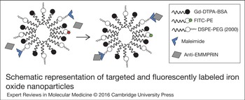
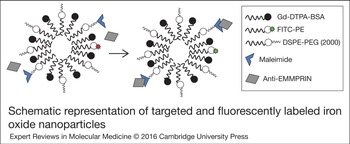
In combination with the use of targeted contrast agents, MRI has emerged as a powerful tool to evaluate atherosclerosis. Depending on the relaxation times (T1, T2), acquired images may reveal enhancement or inhibition of the signal, according to the probes used for the assay. T1 contrast agents incorporate Gadolinium (Gd) into micelles, immunomicelles with conjugated antibodies on the surface, and liposomes, to improve detection and sensitivity. By contrast, T2 contrast agents are usually coated with dextran, polyethylene glycol or polyvinyl alcohol, and contain an iron oxide superparamagnetic core (Refs Reference Makowski117, Reference Mulder118) (Fig. 2). According to the core size, they can be classified into ultrasmall superparamagnetic, iron-oxide (USPIO, 18–50 nm), or SPIO (100–200 nm) nanoparticles. Both particles exhibit a very large magnetic moment, leading to a quick signal loss in T2 images; thus, histological validation is highly recommended (Refs Reference Segers119, Reference Metz120, Reference Klug, Bauer and Bauer121, Reference Ma122).
Schematic representation of targeted and fluorescently labelled iron oxide nanoparticles.
USPIO nanoparticles are swallowed by macrophages in vivo, a feature that makes them an excellent tool for non-invasive intraplaque detection of inflammation by MRI both in mice, rabbits and pig models of cardiovascular disease (Refs Reference Millon77, Reference Sigovan123, Reference Bjerner124, Reference Sigovan125). Alternatively, the use of T2 nanoparticles containing cell specific conjugated antibodies or peptides is another strategy for plaque detection, to quantify extension and stage of the lesion, and recently for quantification of pharmacological therapeutic effect on plaque regression. An example of this is the recently reported use of USPIOs conjugated with anti-P-selectin antibodies expressed in cells activated during the early stages of atherogenesis, as a tool for non-invasive detection of early atheroma plaque formation (Ref. Reference Jacobin-Valat126). Likewise, the use of USPIO nanoparticles with conjugated antibodies against activated platelets, which recognise αIIbβ3 integrins, within atherosclerotic lesions (Ref. Reference Burtea127), provides information about advanced atherosclerosis, intraplaque haemorrhage and inflammation, in apoE deficient mice. Iron oxide based nanoparticles conjugated with two antibodies have also been successfully used to target immune cells within the lesion. Dual-ligand microparticles of iron oxide (MPIO), targeted to P-selectin and vascular cell adhesion molecule-1 (VCAM-1) (PV-MPIO) were efficient in atherosclerotic apoE null mice at visualising by MRI the distribution of plaque macrophages closely with the burden (Ref. Reference McAteer128). The effect of statin therapy on atherosclerosis was validated by using different USPIOs in atherosclerotic rabbits (Ref. Reference Millon77) and in humans (Ref. Reference Tang129).
Iron oxide based nanoparticles have some limitations. Internalisation and compartmentalisation of iron oxide particles within cells, including macrophages, cause MR signal loss because of the generation of local magnetic field inhomogeneities that induce shortening of T2 and T2 * relaxation times, which results in a signal deficit on magnetic resonance images, rendering dark signals (Ref. Reference Sigovan123). At the same time, other factors can also induce signal loss, causing ‘blooming artifacts’, and thereby generating loss of distinction of anatomical boundaries and distortion of normal tissues. Additionally, SPIOS suffer from partial-volume effects. They lose activity because of the limited resolution of the imaging system. This is one of the main reasons for which extensive investigations are focusing on generating SPIO-specific positive contrast, namely, to distinguish the signal surrounding SPIO nanoparticles from the dark signal derived from local field inhomogeneity (Ref. Reference Diwoky130).
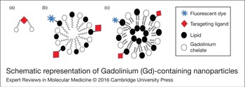
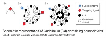
Gd-based T1 nanoprobes (chelates, micelles and liposomes, Fig. 3) offer the advantage of positive contrast as compared with iron oxide particles. In addition, their size facilitates endothelial crossing, offering a significant signal-to-noise ratio. However, and depending on the target, Gd-based nanoparticles are not adequate for detection of targets represented at low concentrations in living organisms. A strong correlation between macrophage density and MRI detection was found in a mouse model of atherosclerosis, by using a contrast agent consisting of Gd3+-loaded micelles targeted to the macrophage scavenger receptor (Ref. Reference Canet-Soulas and Letourneur10) and oxLDL in atheromatous plaques with MRI (Ref. Reference Kanwar11), the anti-inflammatory marker IL-10 (Ref. Reference Almer131), the adipose protein adiponectin (Ref. Reference Hansen132), or plaque rupture and thrombotic marker fibrin (Ref. Reference Andia133).
Schematic representation of Gadolinium (Gd)-containing nanoparticles. (a) Target ligand conjugated to Gd chelates. (b) Target ligand conjugated to Gd containing micelles. (c) Target ligand conjugated to Gd containing liposomes. Gd, Gadolinium.
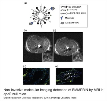
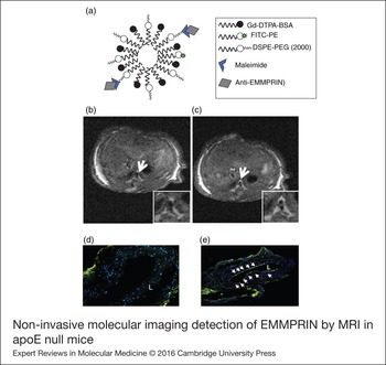
Gd-nanoprobes have been used to detect collagen, a major ECM component, which provides vessel tensile strength to the arteries (Refs Reference Chen134, Reference Spuentrup135), and elastin, the most abundant ECM component of the vessel wall (Ref. Reference Phinikaridou136). Immune foam cell expression of MMPs was also detected by Gd-based nanoparticles in mouse and rabbit models of atherosclerosis (Ref. Reference Hyafil137). MMPs are valuable markers of atherosclerosis, since they play a pivotal role in cell migration and plaque rupture. MMPs are mostly produced by foam cells, and therefore they are not suitable for detection of early atherosclerotic events. However, we found that ECM Metalloproteinase Inducer (EMMPRIN), a trans-membrane protein which induces the expression of several MMPs, is highly expressed at early stages of atherosclerosis in apoE null mice, as detected by immunoblot and by molecular imaging of apoE deficient mice with immunomicelles with conjugated anti-EMMPRIN antibodies (Fig. 4).
Non-invasive molecular imaging detection of EMMPRIN by MRI in apoE null mice. (a) Composition of immunomicelles conjugated with specific anti-EMMPRIN antibody. Eight-week old apoE atherosclerotic null mice were anesthetised and injected with Gd-containing immunomicelles conjugated with anti-EMMPRIN antibody. (b) T1 4.7T MRI scan of mice at time 0 h after injection. (c) T1 4.7T MRI scans of the same mice 2 h after injection, showing Gd enhancement in the aorta (white arrow). (d and e). Ex vivo tissue sections of paraffin embedded abdominal aortas of mice injected with Gd-containing immunomicelles conjugated with anti-EMMPRIN antibody, and isolated at time 0 h (d) and 2 h (e) after injection, and visualised by confocal microscopy. Arrows represent FITC positive cells containing nanoparticles, L, lumen. apoE, apolipoprotein E; EMMPRIN, extracellular matrix metalloproteinase inducer; Gd, Gadolinium; MRI, magnetic resonance imaging.
PET detection of atherosclerosis
PET imaging is based on the use of radiolabeled tracers, providing a highly sensitive method to quantify several metabolic events, including plaque composition, inflammation, calcification and more. Unfortunately, PET images must be overlapped with images taken using better spatial resolution equipment, like MRI, since PET resolution is below the minimum threshold required in order to translate results from bench to bedside.
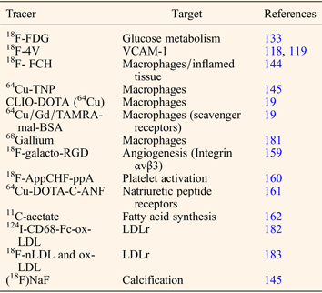
Radiolabeled fluorodeoxyglucose [(18F)FDG] is, among other tracers, the most common glucose analogue. (18F)FDG uptake by activated macrophages takes place in scenarios of high metabolic rates, as within atherosclerotic plaques (Refs Reference Pedersen16, Reference Millon77, Reference Zhao88, Reference Liu138, Reference Tawakol139, Reference Hag140, Reference Rosenbaum, Millon and Fayad141, Reference Calcagno142, Reference Rudd143, Reference Rudd144). In fact, a high correlation between (18F)FDG uptake and macrophage infiltration in atheroma plaque has made possible the use of (18F)FDG as a marker of vulnerable plaque (Refs Reference Chen and Dilsizian145, Reference Tawakol146). Accordingly, (18F)FDG has been used to detect important features of plaque disruption, including neovessel formation (Ref. Reference Calcagno142). In addition, (18F)FDG is an important tool for testing novel therapeutic strategies, (18F)FDG uptake in response to LXR agonist R211945 (Ref. Reference Vucic147), statin administration (Ref. Reference Davies148), the macrophage infiltration inhibitor probucol (Ref. Reference Ogawa149) and the anti-inflammatory properties of pioglitazone (Refs Reference Vucic150, Reference Lobatto151).
As shown above, (18F)FDG microPET has been extensively used to study the onset, progression and regression of atherosclerosis in mouse and rabbit models of disease. Besides, new tracers of atherosclerosis have been successfully implemented to analyse in more detail specific aspects of the disease. Such is the case of (11)C-PK11195, a PET radiotracer which labels a selective ligand of the translocator protein, which is highly expressed by activated macrophages in mice (Ref. Reference Laitinen152) and in human atherosclerosis (Ref. Reference Gaemperli153); taking this approach, via a direct and specific labelling of macrophages – rather than indirect glucose consumption, as detected by (18F)FDG – allows us to characterise and to measure inflammation in more detail. Similar approaches include the use of 18F-fluorocholine (18F-FCH), since choline is metabolised to phosphatidylcholine and incorporated into cell membranes of activated macrophages, as shown in hypercholesterolemic atherosclerotic apoE null mice (Ref. Reference Matter154). Activated macrophages are also traced with the multimodal agent 64Cu trireporter nanoparticle (64Cu-TNP), a dextranated and diethylenetriaminepentacetate (DTPA)-modified magnetofluorescent 20-nm nanoparticle, labelled with the tracer 64Cu to yield a PET, magnetic resonance and optically detectable multimodal imaging agent (Ref. Reference Nahrendorf155). Likewise, other multimodal tracers, for example the cross-linked dextran-coated iron oxide (CLIO)- 1,4,7,10-tetracarboxymethyl-1,4,7,10-tetraazacyclododecane (DOTA) (64Cu) probe or the 64Cu/Gd/TAMRA-mal-BSA tracer (Ref. Reference Bigalke19), were used in apoE null mice and in rats, respectively, to detect carotid atherosclerosis by targeting inflammatory macrophages.
Atherosclerosis is a systemic inflammatory disease, thus adhesion of circulating monocytes to the endothelial layer is a key stage of atherogenesis, in which VCAM, including the highly expressed VCAM-1, play a pivotal role (Ref. Reference Libby, DiCarli and Weissleder156). With the aim of studying early progression of atherosclerosis, a new radiotracer detecting VCAM-1 was synthesised, allowing non-invasive PET-imaging detection of early inflammation (Ref. Reference Nahrendorf157).
Additional radiotracers include 2-(2-nitro-1H-imidazol-1-yl)-N-(2,2,3,3,3-pentafluoropropyl) acetamide [(18F)EF5], a marker of hypoxia used to quantify plaque density, since large atherosclerotic plaques contain significant areas of hypoxia, as detected in atherosclerotic mice (Ref. Reference Silvola158). Plaque angiogenesis may be detected by targeting integrin aνβ3, a key component of the mechanism involved in the formation of new vessels, with the radiolabeled integrin binding RGD peptide radiotracer 18F-galacto-RGD (Ref. Reference Laitinen159). Activated platelets were detected in rabbit atherosclerotic lesions by using a radiotracer (18F-AppCHF-ppA), based on adenosine diphosphate-induced platelet aggregation (Ref. Reference Elmaleh160). In addition, rabbit atherosclerosis was also used to test inflammation with the C-type natriuretic factor-based tracer 64Cu-DOTA-C-ANF (Ref. Reference Liu161).
Other applications of PET in atherosclerosis include the quantification of plaque calcification through the use of (18F) sodium fluoride (Ref. Reference Chen and Dilsizian145), and fatty acid synthesis with (11)C-acetate (Ref. Reference Derlin162).
A summary of different PET tracers used to study atherosclerosis is depicted (Table 1).
Radiotracers to study atherosclerosis by PET

ox-LDL, oxidised low-density lipoproteins; PET, positron emission tomography; LDLr, low density lipoprotein receptor; VCAM-1, vascular cell adhesion molecule-1.
Simultaneous PET/MR technology
Three decades ago, professionals in clinical practice were faced with the momentous challenge of sequentially combining different imaging technologies in an attempt to improve the sensitivity and accuracy of diagnosis (Refs Reference Zerhouni163, Reference Skorton and Collins164). Today, multimodal imaging is a reality, and the combination of PET and CT offers unique sensitivity and anatomical information in one multimodal assay. Significant limitations of PET/CT are related to non-simultaneous data acquisition (Refs Reference Bailey165, Reference Delso, Ter Voert and Veit-Haibach166, Reference Disselhorst167). The CT scan is followed by PET acquisition. Given that CT scans take seconds, while PET images take longer, organ motion and different respiratory cycles may lead to significant artefacts (Ref. Reference Martinez-Möller168). Additionally, the radiation risk associated with CT scans, and the reduced soft tissue contrast as compared with MRI, are key concerns when applying this multimodal imaging technology (Refs Reference Martinez-Möller and Nekolla169, Reference Nappi and El Fakhri170).
PET/CT scans are traditionally used in preclinical and clinical practice to overcome poor spatial resolution exhibited by PET scans alone. In spite of the fact that it is still being studied extensively, the combination of MR with PET offers superb spatial resolution, thanks to the excellent sensitivity afforded by PET technology. In the next few years, great progress will be made in biomedical and nanoparticle research and multimodal probe design to be used in combination with this hybrid system. Simultaneous PET/MR imaging of atherosclerosis should be further studied, especially in combination with functional MR imaging techniques.
Four main challenges to combining both platforms still remain to be overcome. The first involves avoiding PET performance degradation. Photomultiplier tubes required for PET scanners are not compatible with the magnetic fields generated by MR. In this regard, several approaches have been taken, although only two of them with promising translation to clinical practice. Avalanche photodiode detectors detect gamma quanta at a 9.4 Tesla magnetic field (Refs Reference McCallum, Clowes and Welch171, Reference Maramraju172). More recently, several groups have implemented silicon photomultipliers as MR compatible PET detectors because of the fact that the timing resolution qualifies for time-of-flight PET detection mode (Ref. Reference Yoon173). With very promising results at the preclinical level, this new technological advance will be a great milestone achievement when implemented in human PET/MR scanners.
Second, there is a reduction in the quality of MRI images as a result of the radiofrequency generated by the PET insert. However, this problem has recently been overcome in a human study to assess the regional FDG uptake and transmurality of late Gd enhancement using a fully integrated whole body PET/MRI scanner in the sub-acute phase after myocardial infarction (Ref. Reference Rischpler174). Similarly, in lung disease and cancer, the performance of PET/MR versus PET/CT was also evaluated with no significant differences (Ref. Reference Schwenzer175), although the detection rate of small lung lesions is still inferior when using PET/CT (Ref. Reference Rauscher176).
Third, attenuation correction for PET/CT has already been established. However, extensive investigation in developing new PET/MR attenuation correction algorithms is crucial. Values are collected from the same CT images, in which attenuation coefficients corresponding to bone (apart from the rest of the tissues) are separated. However, magnetic resonance of bones is very similar to that of air and therefore requires improved attenuation sequences (Refs Reference Paulus, Tellmann and Quick177, Reference Mollet178).
Fourth, a main challenge in coming years will be the demand for tailored PET/MR multimodal tracers, which depict localisation of specific signals (tissue and/or molecular targets). More important, however, is the need to quantify the amount of specific particles associated with specific pathophysiological events at the molecular level, including atherosclerosis, with the possibility of non-invasively testing drug therapy and treatment. The upcoming years will be crucial for the implementation of this brand new technological achievement.
The first preclinical hybrid PET/MR scans on atherosclerotic mice, rabbits and pigs (Refs Reference Pedersen16, Reference Millon77) have shown very promising results for future clinical applications in humans, not just to follow up plaque progression, but most importantly for the new possibilities that the system offers. Since they provide concomitant anatomical, functional and chemical information about atherosclerotic plaques, these hybrid scans would therefore be useful in forecasting the outcome of plaque progression in terms of stability or vulnerability towards plaque disruption. In humans, assessment of carotid atherosclerotic plaques have recently been analysed (Ref. Reference Ripa179). Compared with PET/CT, hybrid PET/MR yields comparable (18F)FDG quantification and superior delineation of the vessel wall. However, these studies have been undertaken so far using single PET radiotracers. In the coming years when multimodal reagents are fully available, the real capabilities of this technology will come to light. To this end, rabbit atherosclerosis has been recently studied with a combination of USPIO nanoparticles and (18F)FDG, confirming the possibility of combining two functional imaging modalities in order to assess changes in the inflammation of atherosclerotic plaques by using hybrid PET/MR (Ref. Reference Millon77). In addition, the first study of rat atherosclerosis combining hybrid PET/MR with dual PET/MR nanoparticles was performed to image thrombus formation in a rat arterial thrombus model. In the study, the dual PET/MR probe was generated on the basis of EP-2104R – a well-established fibrin-targeted Gd-based MR contrast agent – by partial exchange of Gd for copper 64 (64Cu), demonstrating that fibrin-targeted dual PET/MR probe enables simultaneous, direct MR and PET imaging of thrombus (Ref. Reference Uppal180).
Outstanding research questions
Preclinical hybrid whole-body PET/MRI scanners are now a reality. In the coming years, great progress will be made in biomedical research thanks to different groups working around the world. Here we have outlined the enormous potential of hybrid PET/MR technology in cardiovascular research. When compared with CT, MR offers superior tissue resolution and functional imaging and is the best option to scan the population, thanks to the absence of ionizing radiation and the safety of the use of targeted MR contrast agents. Technically, PET motion compensation and image reconstruction are more accurate when combined with MR imaging, which in the end may offer better anatomical specificity and disease quantification. Hence, the coming years may represent a new era of PET/MRI; but first, three key issues must be addressed before its full implementation in clinical practice. First, clinically efficient attenuation correction algorithms are still under intense investigation, and worldwide standardised protocols are far from being a reality. Second, research into simultaneous multimodal reagents is now underway, and should be continued and intensified. Third, safety protocols should be investigated before transferring this cutting edge technology from bench to bedside.
Acknowledgements
Carlos Zaragoza designed the manuscript in its present form. Irene Cuadrado, Borja Castejón, Marta Saura, Ana María Martin, Irene Herruzo, Nikolaos Balatsos, Jose Luis Zamorano, and Carlos Zaragoza, drafted the manuscript in its final form. This work was supported by research grants from CZ: ‘Ministerio de Economía y Competitividad’ SAF 2011-28375, ‘Instituto de Salud Carlos III’ PI14/02022 (Plan Estatal de I+D+i 2013–2016) and co-financed by the European Development Regional Fund ‘A way to achieve Europe’ (ERDF), European COST action TD1007, and ‘Universidad Francisco de Vitoria 2014’