Introduction
Malpighian tubules are the chief excretory and osmoregulatory organs of insects, being highly specialized for organic solute transport, metabolism and detoxification (Dow, Reference Dow2009). They are homologous to the vertebrate nephron in its function of removing different chemical substances from the body fluids, and are actively associated with water and ion transport. They produce an isosmotic fluid termed as primary urine. The filtration of haemolymph and the production of primary urine take place by a secretory process involving the active transport of ions into the tubule lumen (Pannabecker, Reference Pannabecker1995). This filtered fluid or primary urine carries excretory waste products and toxic materials to the hindgut where this fluid is modified by selective absorption of useful substances. This selective modification of primary urine occurs in the ileum, rectum or even in the Malpighian tubules themselves (Maddrell, Reference Maddrell1971; Maddrell and Gardiner, Reference Maddrell and Gardiner1974; Maddrell, Reference Maddrell1981; Bradley, Reference Bradley1985; Chapman, Reference Chapman2008).
Recently, it has been observed that the Malpighian tubules of a Drosophila sp. (Diptera: Drosophilidae) play a critical role in immune responses (McGettigan et al., Reference McGettigan, McLennan, Broderick, Kean, Allan, Cabrero, Reguiski, Pollock, Gould and Davies2005) and these structures do not only function as a kidney but also as a liver and an autonomous immune system (Dow, Reference Dow2009). The Malpighian tubules are ideal for the investigations of physiological–morphological relationships in a fluid transporting tissue because of simple tubular shape and convenient size (Gaertner and Morris, Reference Gaertner and Morris1999). Thus, physiological functions of the Malpighian tubules are correlated with the ultrastructural features. This prompted us to study the ultrastructural details of the larval Malpighian tubules of a cyclorrhaphous dipteran, Sarcophaga ruficornis Fab. (Diptera: Sarcophagidae), which has received little attention.
Materials and methods
A culture of S. ruficornis was reared and maintained in the laboratory at 27 ± 1 °C and 75 ± 5% relative humidity. Fresh slices of goat's kidney and liver were used for larviposition and also for feeding the larvae. For transmission electron microscopy, third instar larvae were dissected in insect Ringer solution to remove Malpighian tubules, which were immediately fixed in 4% glutaraldehyde in 0.1 m sodium phosphate buffer, pH 6.8, at 4 °C for 1 h to accomplish primary fixation. Following primary fixation, the tubules were rinsed in 0.1 m sodium phosphate buffer for 10–20 min at room temperature and post-fixed for 45 min in a solution of 1% OsO4, prepared with 0.05 m sodium phosphate buffer and 0.1 m sucrose and dehydrated with ethanol series and embedded in Araldite®. Sections were cut on an ultramicrotome (Reichert ultracut E) and stained with 1% uranyl acetate and lead citrate, and viewed and photographed on a Philips CM 10 electron microscope at 100 KV.
For scanning electron microscopy (SEM), specimens were fixed in glutaraldehyde solution, as described above, to accomplish primary fixation. Then, the tubules were rinsed twice in phosphate buffer followed by treatment with 1% OsO4 for 1 h. Post-fixation of tubules was followed by rinsing two to three times with phosphate buffer solution and dehydrating with alcohol series. Specimens were then subjected to critical point drying and then viewed with an LEO 435 VP scanning electron microscope.
Results
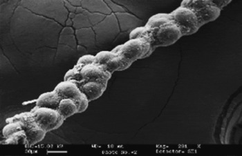
There are two pairs of Malpighian tubules, each pair connected to the alimentary canal by a common ureter opening at the junction of the midgut and hindgut. The SEM image reveals that the tubules have a beaded appearance and bulging outgrowths throughout their length (Fig. 1). The larval Malpighian tubule of S. ruficornis consists of two types of cells, namely principal and stellate cells.
SEM image of the Malpighian tubule of third instar larva of Sarcophaga ruficornis

Principal cells
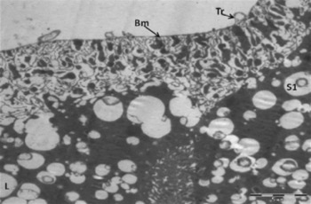
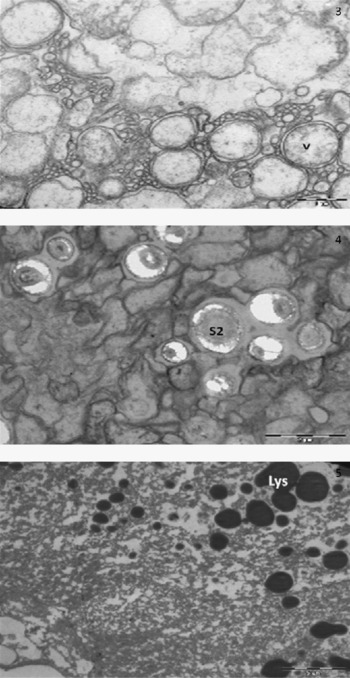
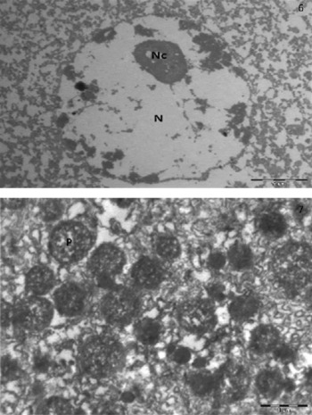
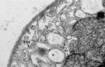
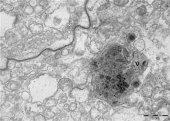
The outer surface of the epithelial cells is covered by a thin basement membrane and the tracheoles supplying the tubules are located on the outer surface of the basement membrane (Fig. 2). There are extensive basal membrane infoldings having mitochondria in cytoplasmic compartments. Multiple spherical cell inclusions are found in the central cytoplasm including clear vacuoles, lipid droplets, large vacuoles having double membranes, mineral concretions or spherocrystals (Figs 2–4) and lysosomes (Fig. 5). In the central cytoplasm, there is a large euchromatic nucleus with a nucleolus (Fig. 6). Several peroxisomes have also been observed in the central region (Fig. 7). The deep basal membrane infoldings are found to be associated with the endoplasmic reticulum, and there are several small and big vacuoles filled with granular secretory material (Fig. 8).
Cross-section of the Malpighian tubule showing tracheoles (Tr), basement membrane (Bm), lipids (L), spherocrystals (S1) and microvilli in the centre

Cross-sections of the Malpighian tubule showing the formation of large double-membrane vacuoles (V), large spherocrystals (S2) and lysosomes (Lys)

Cross-sections of the Malpighian tubule showing a large nucleus (N) with nuclear membrane and a nucleolus (Nc) and peroxisomes (P)

Cross-section of the Malpighian tubule showing basal membrane infoldings (Bmi) associated with endoplasmic reticulum (er) and vacuoles (V) filled with granular secretory material

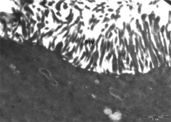
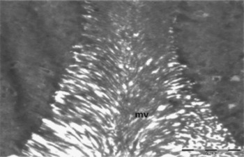
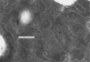
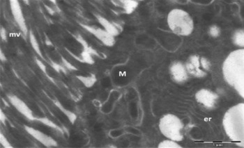
The apical surface of the epithelial cells shows the presence of luminal microvilli containing mitochondria running up to the tips of the microvilli (Figs 9 and 10). Mitochondria with prominent cristae are also found to be concentrated at the bases of luminal microvilli and the endoplasmic reticulum is clearly visible in the cytoplasm (Figs 11 and 12). Septate junctions separate the cells, and a large storage vacuole filled with granular secretory material is visible (Fig. 13).
Luminal surface with microvilli (mv) having mitochondria running up to the tips and also found in the apical region

Luminal microvilli (mv) from two inner cell surfaces interleaving at their tips

Mitochondria showing prominent cristae (cr)

Cross-section of the Malpighian tubule showing luminal microvilli (mv) with mitochondria (M) running up to the tips and also concentrated at their bases, and endoplasmic reticulum (er) in the central region

Cross-section of the Malpighian tubule showing septate junction

Stellate cells
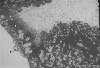
The tubular epithelium apart from the principal cells shows the presence of the stellate cells. The electron micrograph shows a stellate cell with a principal cell on either side. The stellate cell is thin, having a few cell inclusions and with many short luminal microvilli devoid of mitochondria. This fits with its proposed function of allowing the passive flow of water through the cell driven by the osmotic gradient set by the principal cells. The basal membrane infoldings are extremely reduced and peripheral tracheoles on the membrane are visible (Fig. 14).
Cross-section of the Malpighian tubule showing the stellate cell (S) with the principal cells (P) on either side

Discussion
The ultrastructure of the larval Malpighian tubules of S. ruficornis reveals all the characteristics of transporting epithelia involved in the secretion and reabsorption process leading to the formation of urine. Contrary to only one cell type found in the tubules of some insects such as Formica polyctena Förster (Garayoa et al., Reference Garayoa, Villaro, Montuenga and Sesma1992) and Solenopsis saevissima (Smith) (both Hymenoptera: Formicidae) (Arab and Caetano, Reference Arab and Caetano2002), the tubular epithelium consists of two types of cells, namely principal and stellate cells. Similar cell types have also been found in adult Calliphora erythrocephala Mg. (Diptera: Calliphoridae) (Berridge and Oschman, Reference Berridge and Oschman1969), larval C. erythrocephala (Alkassis and Schoeller-Raccaud, Reference Alkassis and Schoeller-Raccaud1984), Aedes taeniorhynchus (Wiedemann) (Diptera: Culicidae) (Bradley et al., Reference Bradley, Stuart and Satir1982), Chironomus tentans Fab. (Diptera: Chironomidae) (Jarial, Reference Jarial1988), Paragnetina media (Walker) (Plecoptera: Perlidae) (Kapoor, Reference Kapoor1994), and Drosophila hydei Sturtevant and D. melanogaster Meigen (both Diptera: Drosophilidae) (Wessing et al., Reference Wessing, Zierold and Polenz1999).
The principal cells are characterized by deep basal infoldings of basement membrane and extensive and well-developed closely packed microvilli towards the luminal surface. The mitochondria are found to be associated with the basal membrane infoldings and microvilli, running up to the tips of the microvilli and also concentrated in large numbers at the bases of luminal microvilli. These features point out that the principal cells are the major ion-transporting cells (Berridge and Oschman, Reference Berridge and Oschman1969; Sohal, Reference Sohal1974; Green, Reference Green1979; Bradley et al., Reference Bradley, Stuart and Satir1982; Alkassis and Schoeller–Raccaud, Reference Alkassis and Schoeller-Raccaud1984; Jarial, Reference Jarial1988) where the cell membrane is actively involved in the transport of ions (Nicolson, Reference Nicolson1993), and the basal membrane infoldings and microvilli are the sites where ion pumps are located (Maddrell, Reference Maddrell1977; Pannabecker et al., Reference Pannabecker, Aneshansley and Beyenbach1992). Berridge and Oschman (Reference Berridge and Oschman1969) have suggested that an osmotic gradient is established in the basal folds and the microvilli of Malpighian tubules as a result of the active transport of solutes followed by the passive transport of water from the haemolymph into the tubular lumen. The close and deep association of mitochondria with basal membrane infoldings suggests the involvement of solute pumps in creating an osmotic gradient and the transport of potassium from the haemolymph into the lumen against a concentration gradient for normal fluid formation (Maddrell, Reference Maddrell1971). Similarly, deep penetration of mitochondria into luminal microvilli is suggestive of the active transport site as the absence of mitochondria in the luminal microvilli of tubules of C. tentans is the principal cause for the lack of this function and the microvilli may perform some other functions such as sweeping biliverdin away from the tubular lumen (Jarial, Reference Jarial1988).
The cytoplasm of the principal cells contains endoplasmic reticulum, mineral concretions or spherocrystals, lipid droplets, lysosomes, clear vacuoles, vacuoles filled with secretory material and peroxisomes. One of the important functions of the principal cells is the storage of materials (Bradley et al., Reference Bradley, Stuart and Satir1982). The presence of clear vacuoles or vacuoles filled with secretory material is a characteristic feature of transporting principal cells. The clear vacuoles observed in these cells probably contain mineral concretions or spherocrystals (Garayoa et al., Reference Garayoa, Villaro, Montuenga and Sesma1992; Arab and Caetano, Reference Arab and Caetano2002). The spherocyrstals or mineral concretions are storage products of mineral and organic materials that could have been derived from the larval diet (Sohal and Lamb, Reference Sohal and Lamb1979; Wessing and Zierold, Reference Wessing and Zierold1992) and these may serve as storage excretion sites (Maddrell, Reference Maddrell1971, Reference Maddrell1977; Sohal, Reference Sohal1974; Sohal et al., Reference Sohal, Peters and Hall1976, Reference Sohal, Peters and Hall1977; Bradley, Reference Bradley1985).
The cytoplasm of the principal cells also contains lipid droplets and peroxisomes. The lipid droplets are found in the Malpighian tubule cells of many insects (Berridge and Oschman, Reference Berridge and Oschman1969; Green, Reference Green1979; Arab and Caetano, Reference Arab and Caetano2002) and may serve as nutrients and also play a role in processing lipid-soluble toxins and transporting them through the tubule system for excretion (Hazelton et al., Reference Hazelton, Townsend, Richter, Ritter, Felgenhauer and Spring2002). Peroxisomes have been observed in the tubule cells of several insects such as the glow-worm Arachnocampa luminosa (Diptera: Keroplatidae) (Green, Reference Green1979) and the fire ant S. saevissima (Hymenoptera: Formicidae) (Arab and Caetano, Reference Arab and Caetano2002). In D. melanogaster, abundant peroxisomes have been found in the adult Malpighian tubules and gut (Beard and Holtzman, Reference Beard and Holtzman1987). Peroxisomes are involved in the elimination of several toxic molecules besides being associated with lipid metabolism (Green, Reference Green1979; Alberts et al., Reference Alberts, Bray, Lewis, Raff, Roberts and Watson1994; Arab and Caetano, Reference Arab and Caetano2002). Peroxisomes play an important role in uric acid metabolism containing a number of enzymes related to nitrogen metabolism including amino acid oxidases, uricase and catalase (Hruban and Rechcigl, Reference Hruban and Rechcigl1969). In Periplaneta americana (Blattaria: Blattidae), peroxisomes found in the Malpighian tubules play an important role in uric acid metabolism, converting it into ammonia, which is the main excretory product (Wall et al., Reference Wall, Oschman and Schmidt1975). The larvae of flesh-eating flies such as Lucilia and Sarcophaga produce ammonia-rich excreta (Gillott, Reference Gillott2005), and the presence of peroxisomes in the larval tubule cells of Sarcophaga suggests their involvement in uric acid metabolism.
In tubule cells, abundant endoplasmic reticulum has been found to be in close proximity to basal membrane infoldings, suggesting their involvement in transcellular transport moving the majority of transported substances across the cytoplasm of the cell (Wessing and Eichelberg, Reference Wessing and Eichelberg1975).
Septate junctions separate the tubule cells. Such junctional complexes serve the purpose of intercellular adhesion and constitute a permeability barrier, restricting the paracellular diffusion (Berridge and Oschman, Reference Berridge and Oschman1972; Noirot-Timothee and Noirot, Reference Noirot-Timothée and Noirot1980; Bradley, Reference Bradley1985; Skaer et al., Reference Skaer, Le, Harrison and Maddrell1990). Such junctional complexes have also been observed in the Malpighian tubules of several insects such as Calliphora sp. (Berridge and Oschman, Reference Berridge and Oschman1969), P. americana (Wall et al., Reference Wall, Oschman and Schmidt1975), Rhodnius prolixus (Hemiptera: Reduviidae) (Skaer et al., Reference Skaer, Le, Harrison and Maddrell1990), F. polyctena (Garayoa et al., Reference Garayoa, Villaro, Montuenga and Sesma1992) and S. saevissima (Arab and Caetano, Reference Arab and Caetano2002).
The stellate cells found in the larval tubules of S. ruficornis are extremely thin and situated between the principal cells. These cells have short and wide basal infoldings and extremely short microvilli devoid of mitochondria, indicating that these are not involved in the active transport mechanism that requires expenditure of energy. These cells are analogous to type II cells of Calliphora (Diptera: Calliphoridae) larvae (Alkassis and Schoeller-Raccaud, Reference Alkassis and Schoeller-Raccaud1984), type II cells of Musca domestica L. (Diptera: Muscidae) (Sohal, Reference Sohal1974) and type I cells of Arachnocampa luminosa (Diptera: Keroplatidae) (Green, Reference Green1979). It has been suggested that the function of the stellate cells is reabsorptive and the cells absorb sodium ions from the lumen (Berridge and Oschman, Reference Berridge and Oschman1969). This has been subsequently confirmed by Wessing et al. (Reference Wessing, Zierold and Polenz1999) in D. hydei and D. melanogaster, where the sodium content in the stellate cells was significantly higher than that found in the principal cells.
The Malpighian tubules of S. ruficornis are richly supplied with tracheoles as found in the adult Calliphora (Berridge and Oschman, Reference Berridge and Oschman1969), F. polyctena (Garayoa et al., Reference Garayoa, Villaro, Montuenga and Sesma1992) and blowfly larvae (Alkassis and Schoeller-Raccaud, Reference Alkassis and Schoeller-Raccaud1984), whereas the absence of tracheal supply to the Malpighian tubules of C. tentans is related to the presence of haemoglobin in the haemolymph and the involvement of tubule cells in the uptake of biliverdin and haemoglobin from the haemolymph (Jarial, Reference Jarial1988).