INTRODUCTION
Prostate cancer is the second most common cancer in men worldwideReference Center, Jemal and Lortet-Tieulent 1 and, in the United Kingdom, radiotherapy is the most commonly used curative treatment for localised prostate cancer.Reference Jackson, Murthy and Dearnlaley 2
Currently, in radiotherapy planning, no account is made for body habitus when creating planning target volume (PTV) margins. Yet obese patients have been shown to have greater external set-up variation and tend to have larger prostates.Reference Formiguera and Cantón 3
Investigations looking at the relation of body mass index (BMI) and the extent of prostate motion have revealed conflicting results. A number of studies of varying design have been carried out and while some conclude that there is a definite increase in prostate motion with increased BMI,Reference Wong, Gao, Merrick, Uematsu, Wilson and Cheng 4 – Reference Piotrowski, Kaczmarek and Jodda 8 others state that there is no differenceReference Butler, Morris, Merrick, Kurko and Murray 9 – Reference Osei, Jiang, Barnett, Fleming and Panjwani 11 and some go as far as to suggest that, intrafractionally, an increase in patient weight may have a stabilising effect on the prostate.Reference Thompson, Gill and Thomas 12 Considering the rising trend in obesity prevalence and that obesity itself is an aetiological factor for prostate cancer,Reference Ma, Li and Giovannucci 13 this uncertainty requires clarification.
To date, it has not been possible to infer PTV margins based on BMI, and indeed it is unclear whether BMI is a useful measurement for examining prostate motion. Few studies have examined patient subcutaneous adipose tissue thickness (SAT) although Wong et al.Reference Wong, Gao, Merrick, Uematsu, Wilson and Cheng 4 , Reference Wong, Gao and Merrick 5 have shown it to correlate with prostate shifts in the left–right direction, as well as patient weight.
Image-guided radiotherapy (IGRT) using implanted gold fiducial markers is the gold standard treatment technique for prostate radiotherapyReference Smyth, McCallum, Pearson and Lawrence 14 – Reference Zelefsky, Kollmeier and Cox 17 as it does not have the dose associated with cone-beam computed tomography (CBCT), it is not open to variation in prostate delineation by practitionersReference Barney, Lee, Handrahan, Welsh, Cook and Sause 18 and has the potential for reducing PTV margins and thus enable greater dose escalation and reduce organ at risk (OAR) toxicity.Reference Poulsen, Muren and Hoyer 19 – Reference van Haaren, Bel, Hofman, van Vulpen, Kotte and van der Heide 21
However, fiducial markers are invasive and expensive and it is unclear whether some patients would benefit more from IGRT than others. Some studies have highlighted a possible link between obesity and biochemical failure of patients post radiotherapy of the prostate,Reference Bujold, Craig, Jaffray and Dawson 15 , Reference King, Spiotto and Kapp 22 – Reference Ly, Reddy, Klein and Ciezki 24 suggesting a greater extent of uncertainty in daily prostate position.
The aim of this study was to examine the relationship between patient BMI and the extent of interfractional prostate movement during a course of radical radiotherapy and hence determine patient-specific PTV margins based on patient BMI. We also sought to determine whether it was possible to identify certain patient characteristics that would suggest a greater benefit from the use of implanted prostate fiducial markers.
MATERIALS AND METHODS
All patients treated with radical prostate IGRT between June 2013 and August 2013 using fiducial markers were included in the study. Patients were identified from the patient management system. Written informed consent was obtained from all participants. The study was approved by the NHS Trust Research and Development Committee.
Data
Patient height and mass, daily IGRT correction shift measurements in the superior/inferior, anterior/posterior and right/left directions and anterior SAT (measured from computed tomography data at level of pubic symphysis) were collected. Patients were grouped into two groups: obese (BMI≥30) and not obese (BMI<30) (World Health Organisation obesity definitions). Using the mean daily correction measurements for each patient, it was possible, first, to see if there was a difference in shifts between the two groups, and then to calculate a mean isocentre shift in each direction for each patient group and hence examine possible PTV margins based on patient BMI.
Planning and treatment
Patients had three gold fiducial markers placed into the prostate in the right base, left midgland and right apex under rectal ultrasound guidance, 2 weeks before CT simulation. They were instructed to follow a high-fibre diet from 1 week before CT simulation and throughout the course of their treatment. On the day of the scan, patients were instructed to empty their bowel and bladder, drink 500 ml of water and wait 45 minutes for the bladder to refill. Patients were positioned supine with their feet in indexed foot stocks and a foam rest under the knees. A CT scan was performed using 3-mm axial slices with the OARs (bladder, rectum, bowel, penile bulb) and clinical target volume (CTV) contoured on the CT dataset. The anterior SAT at the level of the pubic symphysis (skin surface to rectus abdominis) was also measured on the CT dataset. A PTV was then expanded from the CTV using 0·5 cm margins in all directions and standard dynamic arc radiotherapy treatment planning was carried out. Treatments were delivered to the PTV in 200 cGy/fraction to a total dose of 7,400 cGy.
Daily alignment on the treatment couch using skin markers and a laser coordination system was carried out. Orthogonal kilovoltage images were obtained and online image match using the fiducial markers was performed to triangulate the anatomic isocentre. Isocentre deviation was calculated and recorded in the anterior/posterior, superior/inferior and left/right directions and the treatment couch was then shifted to account for these deviations. Online image matching was carried out by two trained radiographers.
Data analysis
Descriptive statistics were used for patient mass, BMI, SAT and corrective shifts. The Pearson’s product-moment correlation coefficient for shifts and BMI, SAT and patient mass, respectively, were calculated using a two-tailed level of significance. Linear regression analysis with the mean shift and BMI, SAT and patient mass was carried out for each dimension of displacement. In order to compare the mean shifts for the two groups (BMI≥30 versus BMI<30), a two-tailed independent t-test was carried out for the three shift dimensions. A significance level of p<0·05 was used. All data were analysed using SPSS 18 (SPSS Inc., Chicago, IL, USA).
RESULTS
The sample comprised of 38 patients who received dynamic arc IGRT to the prostate. Median mass was 88·6 kg (range 66·2–137·0). The median BMI was 28·4 kg/m2 (range 21·4–44·7) and median SAT was 4·3 cm (range 1·0–8·7).
Table 1 shows the mean fiducial marker-based corrective shifts for the whole sample. The greatest shift was in the anterior/posterior direction (mean 3·4 mm; range 1·4–8·6), followed by the superior/inferior direction (mean 3·1 mm; range 1·0–5·9) and the left/right direction (mean 1·8 mm; range 0·5–4·4).
The mean and SD for the mean fiducial marker-based corrective shifts for n=38 patients receiving dynamic arc image-guided radiotherapy to the prostate

There was no significant correlation between any of the variables of patient BMI, SAT or mass and the mean corrective shifts in any direction (Table 2).
Results of Pearson’s product-moment correlation coefficient analysis for the relationship between mean shifts and the variables of body mass index (BMI), subcutaneous adipose tissue thickness (SAT) and mass for the sample (n=38) of patients receiving dynamic arc image-guided radiotherapy to the prostate

Note: A two-tailed level of significance was used.
Abbreviations: r, result of the Pearson’s product-moment correlation coefficient analysis; ρ, level of significance.
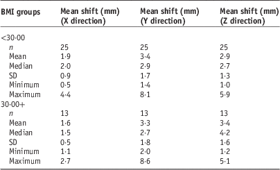
Table 3 shows the characteristics of the IGRT corrective shifts for the obese (n=13) versus non-obese (n=25) patients. Mean corrective shifts were similar for both groups in all directions, with the greatest correction in the Y and Z axes.
Mean corrective shifts in the X (left/right), Y (anterior/posterior) and Z (superior/inferior) directions for prostate image-guided radiotherapy patients with a body mass index (BMI)<30 kg/m2 and BMI≥30 kg/m2
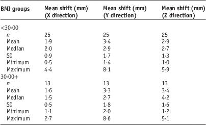
There was no significant difference in mean corrective shifts between the two groups in any direction (X shift: ρ=0·320; Y shift: ρ=0·839; Z shift: ρ=0·325). Linear regression analysis of BMI, SAT and patient mass to mean corrective shifts is shown in Table 4. It can be seen in Table 4 that the R 2 values are all very low. The greatest values can be seen for SAT and the extent of corrective shifts, with the highest of these being in the superior/inferior direction.
The R 2 values for the linear regression analysis of patient body mass index (BMI), subcutaneous adipose tissue thickness (SAT) and mass, with the extent of mean corrective shifts in patients receiving radical image-guided radiotherapy to the prostate (n=38)

Abbreviation: ρ, level of significance.
DISCUSSION
This study showed no difference in mean prostate shifts in any direction between obese and non-obese patients, and while the results from this study also show no relation between BMI, weight, SAT and interfraction prostate movement, they raise some interesting questions. It is possible that patient BMI does have an effect on the extent of prostate motion, but that the effect is being occluded by other patient variables, one of which being variation in rectal volume. Few studies into the effects of BMI on prostate motion have involved any sort of patient bowel preparation and according to Stasi et al.Reference Stasi, Munoz and Fiorino 25 the rectal volume can vary with an average random fluctuation of 4·4 mm over a course of treatment. Although patients in our study were instructed to follow a high-fibre diet throughout the course of their treatment in order to achieve a consistency of bowel transit and volume, others have concluded that a high-fibre diet does not improve the consistency of rectal filling.Reference Faithfull 26 – Reference Jotwani, Surendran, Chilukuri, Ramamohan, Ibrahim and Shivakumar 28
The use of daily CBCT to visualise the extent of rectal filling, as well as PTV position, has been employed as an IGRT method in some departments and it would certainly be interesting to use CBCT to examine whether there is a correlation between patient size and the extent of variation in rectal volume between fractions.
Another possible reason why we found no significant difference between the two groups and their respective prostate movement could be due to movement of the fiducial markers themselves within the prostate which can be on average 1–2 mm between fractions.Reference Delouya, Carrier, Beliveau-Nadeau, Donath and Taussky 29 Given the mean shifts for the sample in this study were between 1·8 and 3·4 mm, it could potentially impact on the reliability of the data. It would seem prudent in future investigations to examine whether there are any patient variables, such as mass, BMI or SAT, that influence the extent of fiducial marker migration.
The fact that no correlation between anterior SAT and interfraction prostate movement was found could be linked to the way in which SAT was measured. Although all SAT measurements were carried out by one individual in order to minimise interobserver error, measurements were only taken on a single CT slice and hence may not be entirely representative of patient SAT. Lateral SAT may also have been a useful measurement but was not taken here. However, we employed a comparable technique to that carried out by Wong et al.,Reference Wong, Gao, Merrick, Uematsu, Wilson and Cheng 4 where a correlation was observed. While it therefore remains unclear as to whether there is a link between SAT and interfraction prostate movement, it is possible that more SAT does not so much lead to a greater extent of prostate movement as increased random set-up error as a result of increased mobility of external alignment marks.
We have demonstrated that, regardless of patient BMI, the extent of interfraction prostate movement varied widely, with mean shifts for some patients as high as 8 mm in the anterior/posterior direction. Without the use of fiducial marker-based IGRT there is a significant risk of underdosing the PTV while overdosing the surrounding tissue.Reference Millender, Aubin, Pouliot, Shinohara and Roach 6 Therefore, all patients receiving radical prostate radiotherapy should be given gold fiducial markers. Indeed the National Radiotherapy Advisory Group 30 report (2007) states that IGRT is the gold standard for prostate radiotherapy and IGRT using fiducial markers is more accurate than IGRT with CBCT.Reference Lazos, Mourad and Hauerstock 31
Without the use of fiducial markers and without a consensus on either the effect of BMI on prostate movement or an effective bowel preparation, it would seem that the best way to ensure that the prostate is in the same place each day is to immobilise the prostate itself. This could be achieved by using a rectal balloon catheter that is inserted into the rectum and then inflated to a set volume, effectively pinning the prostate to the pubic symphysis, ensuring the rectal volume is more or less consistent throughout the course of the radiotherapy treatment.Reference Wachter, Gerstner and Dorner 32 , Reference Smeenk, Teh, Butler, van Lin and Kaanders 33 Rectal balloon catheters may represent a viable and cheaper alternative to fiducial markers for early stage disease prostate radiotherapy patients. However, in a study looking at the use of rectal balloon catheters and IMRT for prostate cancer Teh et al.Reference Teh, Woo and Mai 34 concluded that the most favourable outcomes were for patients with rectal balloon catheters and fiducial markers.
Given the overall mean interfraction prostatic shifts, and considering the extent to which fiducial markers may migrate within the prostate, the current margins of 5 mm in all directions from CTV to PTV seem sensible, but in the absence of fiducial marker-based image-guided treatment, these margins should be increased in the anterior/posterior and superior/inferior directions to 7 mm. An increase in the posterior margin would, however, lead to an increase in the volume of rectum receiving a high dose and hence greater rectal toxicity.Reference Huang, Catton, Jezioranski, Bayley, Rose and Rosewall 35 , Reference van der Laan, van den Bergh, Schilstra, Vlasman, Meertens and Langendijk 36 In an effort to increase treatment accuracy and reduce PTV margins, the use of fiducial markers should be encouraged.
Study limitations
It is not expected that patient BMI would change dramatically during the course of radiotherapy treatment, but a significant loss or gain in weight could affect the quality of the data. Future studies should measure BMI at regular intervals throughout treatment so that prostate shifts could be related to a current BMI and the effects of any change in patient mass would be minimised.
CONCLUSION
BMI is not a useful indicator of the potential extent of interfraction prostate motion, nor is patient mass or SAT. Therefore current PTV margins should not be reduced. Infact, without the use of fiducial markers, margins should be increased in the anterior/posterior directions and superior/inferior directions to around 7 mm to ensure that the CTV is not missed at treatment.
Gold fiducial markers should be introduced as standard in the delivery of prostate radiotherapy to increase treatment accuracy.
Acknowledgements
None.
Financial support
This research received no specific grant from any funding agency, commercial or not-for-profit sectors.
Conflicts of Interest
None.