Implications
The continuous decline in milk yield during lactation is attributed to the decline in number of mammary epithelial cells (MECs) as a result of apoptosis. Insulin-like growth factor-binding protein-5 (IGFBP-5) is highly upregulated during involution. IGFBP-5 binds IGF-I and IGF-II in the mammary gland and regulates their bioavailability. Mostly, indigenous lactating animals in the Indian subcontinent are low yielders with poor persistency of lactation. We have observed comparatively higher expression of IGFBP-5 both at the transcript level (in MECs) and the protein level (in milk) in late lactation and in short lactating animals. Therefore, expression pattern of IGFBP-5 may be explored as an indicator for lactation function.
Introduction
The insulin-like growth factor-binding proteins (IGFBPs) are a family of six related proteins (IGFBP-1, -2, -3, -4, -5, -6) involved in regulating the effects and the bioavailability of the IGFs such as IGF-I and IGF-II. IGFBPs have been reported to be involved in growth and differentiation of cells and tissues (Zapf, Reference Zapf1995). IGFBP-5 is a 29-kDa glycoprotein, which is secreted by a wide variety of cell types during different growth conditions in vitro (Firth and Baxter, Reference Firth and Baxter2002). In vivo, IGFBP-5 is expressed in many cell types and is overexpressed during the differentiation of neurons (Cheng et al., Reference Cheng, Shy and Feldman1999), osteoblasts (Thrailkill et al., Reference Thrailkill, Quarles, Nagase, Suzuki, Serra and Fowlkes1995), myoblasts (Xu et al., Reference Xu, Li, Zhao, Maures, Yin and Duan2004) and the forebrain of adult rat (Stenvers et al., Reference Stenvers, Zimmermann, Gallagher and Lund1994). In addition to the presumed IGF-dependent effects, IGF-independent effects of IGFBP-5 have also been suggested. Senescence in human dermal fibroblasts (Yoon et al., Reference Yoon, Kim, Kim, Song, Kim, Kim, Baek, Kim and Kim2004) and endothelial cells (Hampel et al., Reference Hampel, Fortschegger, Ressler, Chang, Unterluggauer, Breitwieser, Sommergruber, Fitzky, Lepperdinger and Jansen-Durr2006) is also associated with increased IGFBP-5 expression. IGFBP-5-induced senescence is associated with induction of the tumor suppressor p53 (Kim et al., Reference Kim, Seu, Baek, Kim, Kim, Kim and Kim2007). IGFBP-5 has also been shown to induce migration of macrophages (Yasuoka et al., Reference Yasuoka, Hsu, Ruiz, Steinman, Zhou, Choi and Feghali-Bostwick2009), an important component of the inflammatory response evident in fibrotic tissues. IGFBP-5 also binds components of the extracellular matrix and the cell membrane (Andress, Reference Andress1995; Butt and Williams, Reference Butt and Williams2001).
IGFBP-5 has been reported to be expressed in mammary tissues of mammals (Tonner et al., Reference Tonner, Allan, Shkreta, Webster, Whitelaw and Flint2000; Allar and Wood, Reference Allar and Wood2004). Primary bovine mammary epithelial cells (MECs) secrete IGFBP-2, -3, -4, -5 in culture, which are identical to those present in bovine milk (Gibson et al., Reference Gibson, Staley and Baumrucker1999). IGFBP-5 is also overexpressed during mammary gland involution. There is an increase in IGFBP-5 protein concentration, which has been observed in rat milk 48 h after removal of the suckling young (Tonner et al., Reference Tonner, Barber, Travers, Logan and Flint1997). In vivo, increased expression of IGFBP-5 has been observed in tissues undergoing apoptosis, such as the involuting mammary gland (Tonner et al., Reference Tonner, Barber, Allan, Beattie, Webster, Whitelaw and Flint2002). In particular, the mammary gland has been used as an ideal model for cell biology studies, because most of its development occurs after birth and undergoes repeated phases of growth, differentiation and apoptosis that are coordinated by multiple growth factors and steroid or peptide hormones (Hovey et al., Reference Hovey, Trott and Vonderhaar2002). The milk production and lactation persistency in farm animals are governed by several signaling pathways and molecules. IGFBP-5 has been reported to bind IGF-I and IGF-II with high affinity and modulate the biological actions of IGFs in the mammary gland. Involution of the mammary gland takes place when the animal ceases to lactate. During involution, remodeling of the mammary gland takes place by apoptosis of mammary alveolar tissue due to the action of various pro-apoptotic factors. IGFBP-5 is highly upregulated during this stage and has been associated with apoptosis of secretory MECs (Tonner et al., Reference Tonner, Allan, Shkreta, Webster, Whitelaw and Flint2000). During lactation, the continuous decline in milk production is attributed to the decline in number of MECs, and at the end of lactation the mammary gland undergoes involution, which corresponds to loss of MECs (Yart et al., Reference Yart, Lolliviera, Marneta and Dessauge2014). Lactation in farm animals such as in cows and buffaloes follows a sigmoid pattern representing early, mid and late lactation. Persistency of lactation in lactating cows is important for optimal milk yield. Mostly, indigenous lactating animals in the Indian subcontinent are low yielders with poor persistency of lactation (Dhara et al., Reference Dhara, Ray and Sinha2006). An undefined population of cows does not complete full lactation length leading to a phenomenon called short lactation. We hypothesize that high level of expression of pro-apoptotic factors like IGFBP-5 in alveolar epithelial cells will cause apoptosis of the mammary gland and result in decreased milk yield. To evaluate our proposed hypothesis, we have performed comparative expression profiling of IGFBP-5 during lactation and have also compared their levels between Bos indicus (Sahiwal) and Bubalus bubalis (Water buffaloes). To the best of our knowledge, this is the first report on profiling of IGFBP-5 throughout lactation in cows (B. indicus) and water buffaloes (B. bubalis).
Material and methods
Collection of milk samples
Milk samples were collected from lactating animals (Sahiwal cows and Murrah buffaloes), which were maintained in the dairy herd of the National Dairy Research Institute (NDRI), Karnal, India. The animals were healthy without any incidence of mastitis. Milk samples (100 to 200 ml) were collected randomly (six cows and six buffaloes) from a pool of 100 animals (60 cows and 40 buffaloes) at different time points such as days 0 (colostrum), 50, 100, 150, 200, 250 and 300 of lactation. The sampling represents the entire lactation length and can be broadly divided into groups such as early (0 to 100 days), mid (100 to 200 days) and late lactation (200 to 300 days). Apart from these normal lactating points, we collected mammary secretion from the selected animals 4 to 6 days after cessation of milking at the normal end of their lactation period (290 to 305 days), which represented the milk samples from involution stage. Samples were also collected from those animals having a history of short lactation and were producing <2 l of milk at around 100 days of lactation. Milk samples were also randomly collected from six animals from each breed, which were diagnosed with clinical mastitis.
Isolation of MECs from milk and preparation of whey
The MECs were isolated from the milk samples of all the selected animals as described by Boutinaud et al. (Reference Boutinaud, Ben Chedly, Delamaire and Guinard-Flament2008) and Janjanam et al. (Reference Janjanam, Jamwal, Singh, Kumar, Panigrahi, Hariprasad, Jena, Anand, Kumar, Kaushik, Dang, Mukesh, Mishra, Srinivasan, Reddy and Mohanty2013) with minor modifications. In brief, 2 to 4 l of fresh milk samples of Sahiwal cows were cooled to 4°C and centrifuged (15 min, 1500×g) in several 250 ml centrifuge bottles at 4°C to obtain a pellet of milk somatic cells. Fat layer was removed with a spatula and the remaining skim milk was discarded. The pellet of milk somatic cells was washed twice (10 min, 1000×g) with 100 ml of cold phosphate buffered saline (PBS) and the final pellet was pooled and re-suspended in PBS containing 0.1% BSA to give a final cell count of (5×10⁶) cells/ml. MECs were isolated by immuno-magnetic separation technique using Dynabeads coated with Pan Mouse IgG (Dynal Biotech, Invitrogen, Carlsbad, CA, USA) that cross-reacted with the monoclonal antibody against cytokeratin 18 (Clone KS-B17.2, Catalogue No. C1399; Sigma, St Louis, USA). The final cell pellet was stored directly at −80°C in 1 ml of TRIzol® reagent (Invitrogen) for RNA preparation.
For the preparation of whey, milk samples were centrifuged at 1500×g for 20 min to remove fat. After centrifugation, the milk fat formed a uniform layer that was removed by a spatula. The defatted milk was centrifuged at 50 000×g in a high-speed refrigerated centrifuge (Sorvall RC6+) for 2 h (Gibson et al., Reference Gibson, Staley and Baumrucker1999). After centrifugation, casein was separated as a loose pellet at the bottom of tube. The milk whey was separated from casein and was transferred very carefully to a fresh microcentrifuge tube without disturbing the casein pellet. Whey was stored at −80°C until use.
Isolation of RNA and reverse-transcriptase PCR (RT-PCR) of IGFBP-5 in MECs
RNA was isolated from MECs by following the TRIzol method with suitable modification. In brief, 5×10⁶ cells were used for RNA isolation. MECs were washed twice with diethylpyrocarbonate-treated PBS (DEPC; Sigma, USA), centrifuged at 600×g for 10 min at 4°C and re-suspended in 10 ml DEPC-PBS. One milliliter of TRIzol® reagent (Invitrogen) was added to 200 µl cell suspension. The mixture was vortexed well for homogeneous mixing. Chloroform (0.2 ml) was added to the cell lysate, mixed for 15 s followed by incubation for 3 to 5 min at room temperature. The sample was then centrifuged at 12 000×g for 15 min at 4°C. The aqueous phase was separated from the organic phase very carefully into a fresh microcentrifuge tube. The RNA was precipitated from the aqueous phase adding 0.5 ml isopropanol followed by incubation at room temperature for 10 min and centrifugation at 12 000×g for 10 min at 4°C. The precipitated RNA pellet was washed once with 1 ml of 75% ethanol followed by centrifugation at 7500×g for 5 min at 4°C. RNA pellet was air-dried for 10 min. The isolated RNA was dissolved in 20 µl RNase-free water and incubated for 10 min at 55°C to 60°C and stored at −80°C. The integrity of the isolated RNA was checked by electrophoresis in 1.5% agarose (Sigma) gel.
For designing primers, the IGFBP-5 gene sequence of cattle was retrieved from GenBank (www.ncbi.nlm.nih.gov). Primer3 software was used for designing the primer for RT-PCR. Primers were custom synthesized from Sigma Chemicals Co., Bangalore, India. The sequences of the primers used in the study are shown in Supplementary Table S1. The isolated RNA was reverse-transcribed using Revert Aid First Strand cDNA Synthesis Kit (Thermo Scientific, Waltham, MA, USA). Double-stranded cDNA was prepared by adding DreamTaq Green PCR Master Mix (2X, Thermo Scientific) to the first-strand cDNA mix following the manufacturer’s protocol. GAPDH was used as internal control. Then PCR product was run in agarose gel to observe the expression of IGFBP-5.
Real-time PCR
Real-time PCR of the transcripts of IGFBP-5 was carried out on different days of lactation using LC-480 light cycler (Roche, Mannheim, Germany). The level of IGFBP-5 transcript on the 100th day in lactation was used as the calibrator. The reaction mixture comprised of 10 µl reaction volume containing 5 µl of SYBR Green master-mix (SYBR Green, Roche), 0.2 µl of 10 µM of each primer and 2×diluted c-DNA. Thermal cycling conditions consisted of initial denaturation at 95°C for 5 min, followed by 40 cycles of 15 s at 95°C, 15 s at 58°C and 15 s at 72°C followed by 95°C for 10 s. All the primer pairs used were confirmed for their PCR efficiency, and specific products were checked by melt curve analysis and for the appropriateness of size by 2% agarose gel electrophoresis. Two housekeeping genes ribosomal protein S9 (RPS 9) and RPS 23 were used as internal control (Yadav et al., Reference Yadav, Singh, Mukesh, Kataria, Yadav, Mohanty and Mishra2012). The expression data were normalized to the expression of mean of the above two housekeeping genes. The experiment was carried out in three biological and two technical replicates, respectively.
ELISA of IGFBP-5 in whey
Experimental design
The standard curve for IGFBP-5 was prepared by using the kit components that resulted in a negative parabolic curve. The standard curve was linearized by logarithmic transformation of concentration in µg/ml and optical density, which showed higher accuracy. Samples were appropriately diluted to bring the concentration of IGFBP-5 within the standard curve range. All the samples were loaded in the ELISA plate including the standard to limit the error. ELISA of IGFBP-5 protein in milk for each group (n=6) was performed in duplicate at different time points during lactation. ELISA was performed by using Bovine-specific IGFBP-5 ELISA Kit (Catalogue No.: E2659Bo; Uscn Life Science Inc., Wuhan, China). The kit was based on sandwich enzyme immunoassay principle for in vitro quantitative measurement of bovine IGFBP-5 in serum, plasma and other biological fluids. The manufacturer’s protocol was strictly followed with the whey samples in our study.
Statistical analysis
Between-group comparisons among cattle and buffaloes were carried out using two-way ANOVA using SigmaPlot 11.0 software (Chicago, IL, USA). The data sets were analyzed to see the effect of species and days of lactation on IGFBP-5 concentration by two-way ANOVA, using balanced design of 2×7. The levels of the first factor (species) were cattle and buffalo. The levels of the second factor were days of lactation (0, 50, 100, 150, 200, 250, 300 days). The dependent variable was IGFBP-5 concentration measured in milk whey. All comparisons were done by the Holm-Sidak test. Between-group comparisons during lactation days were carried out using one-way ANOVA for analyzing the real-time PCR data. Differences were considered to be statistically significant at P<0.05. Data were presented as mean±s.e.m. All graphics were generated with Microsoft office Excel 2007.
Results
Detection of IGFBP-5 transcripts and quantitative real-time PCR
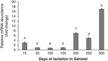
Milk and MECs indicate the physiological health of the mammary gland. IGFBP-5 was observed to be expressed in MECs by RT-PCR (data not shown). The relative mRNA abundance of IGFBP-5, as illustrated in Figure 1, was found to be significantly (P<0.05) higher on day 15 of lactation compared with 50 to 150 days of lactation in Sahiwal cows. There was no significant difference (P<0.05) in the transcript level during early and mid-lactation (50 to 150 days). However, comparatively higher levels of IGFBP-5 mRNA expression were observed at around 300 days followed by 200 and 250 days, respectively (P<0.05).
mRNA expression profile of insulin-like growth factor binding protein-5 (IGFBP-5) measured by real-time PCR in mammary epithelial cells (MECs) of Sahiwal cows. Bars with different superscripts differ significantly (P<0.05).

ELISA of IGFBP-5
Milk concentration of IGFBP-5 during lactation and mammary gland involution
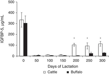
In colostrum and late lactation milk, the IGFBP-5 level was observed to be higher in comparison with early stage of lactation (50 to 150 days) in both cows and buffaloes. Interestingly, very high level of IGFBP-5 was observed in both species, which was significantly (P<0.05) higher than other days of lactation (Figure 2). We observed no significant variation in the concentration of IGFBP-5 protein (5 to 11 μg/ml) during early phase of lactation (days 50 to 150). However, the concentration of IGFBP-5 (93 to 113 μg/ml) varied significantly (P<0.05) during the late phase of lactation (days 200 to 300) in cows. In buffaloes, no significant difference (P<0.05) was observed in IGFBP-5 concentrations during 50 to 200 days (5 to 8 µg/ml). However, IGFBP-5 concentration (25 to 30 µg/ml) differed significantly in samples collected during the late lactation period (250 and 300 days). As lactation proceeded from early to late lactation stage, progressively increased concentrations of IGFBP-5 were observed in cattle as well as in buffaloes. Buffaloes showed comparatively lower concentrations of IGFBP-5 protein than cows on days 200, 250 and 300. Low level of IGFBP-5 concentration was maintained until 150 and 200 days in cows and buffaloes, respectively (Figure 2). The concentration of IGFBP-5 was very high during involution, and they measured 2315±190 and 1142±175 µg/ml in cows and buffaloes, respectively. Highest concentration of IGFBP-5 was observed during involution followed by colostrum, late phase and early phase of lactation in both the species.
Comparison of insulin-like growth factor binding protein-5 (IGFBP-5) concentration in milk throughout lactation in Sahiwal cattle and Murrah buffaloes (n=6). Asterisk (*) shows significant difference (P<0.05) between IGFBP-5 levels of cattle and buffaloes at different time points throughout lactation.

Milk concentrations of IGFBP-5 in short lactating and mastitis diagnosed animals
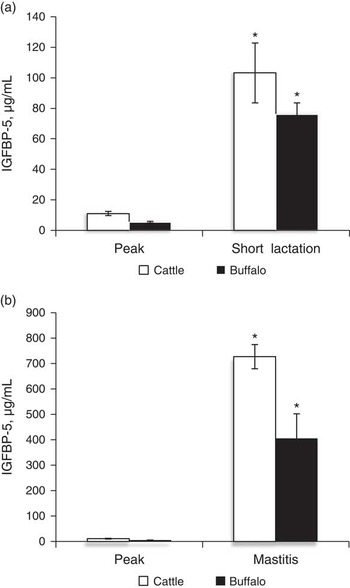
Short lactating animals do not follow the complete lactation duration and stop yielding milk quite early in lactation. This phenomenon is exhibited by an undefined population of animals. We have performed relative comparison of the level of IGFBP-5 in milk of short lactating cows and buffaloes at around 100 days in lactation. In both species, there was significant (P<0.05) increase in concentrations of IGFBP-5 in comparison with normal lactating cows and buffaloes at their peak lactation (Figure 3a). Both cows and buffaloes diagnosed with mastitis showed higher (P<0.05) IGFBP-5 concentrations in milk in comparison with its concentration in milk of normal animals during peak of lactation (Figure 3b).
Comparison of insulin-like growth factor binding protein-5 (IGFBP-5) concentration in milk between (a) normal (peak: 100 days) and short lactating Sahiwal cows and Murrah buffaloes, (b) normal and mastitic milk of Sahiwal cows and Murrah buffaloes (n=6). Bars marked with an asterisk differ significantly from the other group (P<0.001).

Discussion
The present investigation was undertaken to measure the concentration of IGFBP-5 in milk of lactating cows and buffaloes during different time points of lactation. IGFBP-5 is a physiological regulator of MECs survival and synthetic activity in the mammary gland, which is secreted into milk (Marshman et al., Reference Marshman, Green, Flint, White, Streuli and Westwood2003). Plath-Gabler et al. (Reference Plath-Gabler, Gabler, Sinowatz, Berisha and Schams2001) detected expression of mRNAs of all the six IGFBPs (1 to 6) in bovine mammary gland tissue. The expression of IGFBP-3 and -5 were significantly higher in comparison with all the other IGFBPs. In our study, we detected the expression of IGFBP-5 using RT-PCR in milk somatic cells and MECs. IGFBP-5 has been reported to be expressed in MECs in previous studies (Tonner et al., Reference Tonner, Barber, Allan, Beattie, Webster, Whitelaw and Flint2002). IGFBP-5 has an established role in the control of milk yield through regulation of free IGF molecules in the mammary gland (Clemmons, Reference Clemmons1998).
Expression profile of IGFBP-5 through lactation in cattle and buffaloes
The effective length and lactation yield in cattle and buffaloes are dependent on the balance between proliferation and apoptosis of MECs, which in turn are regulated by signaling molecules. One such molecule in this complex network is IGFBP-5, which suppresses IGF-1-mediated survival of MECs, and thus causes enhanced apoptosis at the time of involution (Marshman et al., Reference Marshman, Green, Flint, White, Streuli and Westwood2003). Expression analysis of IGFBP-5 throughout lactation at various time points is important, as it enables us to understand its role on lactation persistency and milk yield. IGFBP-5 was expressed at relatively higher levels in the colostrum in both the species. In bovines, IGFs and IGFBPs have been reported to be highly concentrated in the prepartum secretion and colostrum and were found to be very low in mature milk (Blum and Baumrucker, Reference Blum and Baumrucker2002). This may be because during the colostrum period, there is involvement of IGF-dependent functions for normal growth of mammary systems. A comparatively higher concentration of IGFBP-5 during the colostrum period in present study may be due to the accumulation of IGFBP-5 in the mammary gland as a result of pre-parturient accumulation of milk ensuing parturition. A similar observation was reported in bovines by Gibson et al. (Reference Gibson, Staley and Baumrucker1999). It has also been suggested that, although IGFBP-5 appear in milk, leakage or bio-transport of IGFBP-5 from blood to milk may have happened during the pre-colostrum period leading to an increase in its concentration (Gibson et al., Reference Gibson, Staley and Baumrucker1999). Vega et al. (Reference Vega, Gibson, Skaar, Hadsell and Baumrucker1991) have reported that high concentrations of IGFBP-5 during the colostrum period may be due to the accumulation of IGF and IGFBP in the pre-colostral milk, 2 to 4 weeks prepartum. As lactation progressed, the level of IGFBP-5 plummeted drastically on day 50 and remained lower in the early phase of lactation till 150 days in cattle and 200 days in buffaloes. However, once the animal entered the late stage of lactation, 200 to 300 days in cows and 250 to 300 days in buffaloes, the concentration of IGFBP-5 was comparatively very high. The level of IGFBP-5 kept on increasing from early to late lactation in cattle as well as in buffaloes. It was observed that its expression at the involution stage was ~200 times higher than that during peak lactation, which indicates that IGFBP-5 is a pro-apoptotic molecule, and, therefore, is expressed during mammary gland involution. The concentration of IGFBP-5 increased sharply during the involution (Allan et al., Reference Allan, Beattie and Flint2004) stage that coincided with the process of apoptosis. A large increase in the production of IGFBP-5 by the mammary gland of rat during involution exerts an apoptotic effect by abrogating the survival effects of IGF-I (Tonner et al., Reference Tonner, Barber, Travers, Logan and Flint1997). In addition, it has been reported that IGFBP-5 is present in high concentrations (∼60 μg/ml milk) in the involuting rat mammary gland (Tonner et al., Reference Tonner, Allan, Shkreta, Webster, Whitelaw and Flint2000). Involution stage is a period when remodeling of the mammary gland takes place due to programmed cell death of MECs, and IGFBP-5 acts as regulatory molecule, which mediates apoptosis in the mammary gland by interacting with IGF-I (Tonner et al., Reference Tonner, Quarrie, Travers, Barber, Logan and Wilde1995). Although involution in cows is characterized by extensive cell turnover, which includes apoptosis, the concurrent gestation of cows prevents the total loss of alveolar structure unlike rodents (Capuco and Akers, Reference Capuco and Akers1999). Tight junctions between epithelial cells also deteriorate during these early stages of the dry period in cows and this calls into question the source of IGFBP-5 in the mammary secretion sampled. To make sure that IGFBP-5 synthesis took place in the mammary gland, we clearly observed very high level expression of IGFBP-5 transcripts in milk somatic cells of cows during the involution period (data not shown). However, perfusion of IGFBP-5 from blood into mammary secretion during involution cannot be ruled out, as has been reported elsewhere (Gibson et al., Reference Gibson, Staley and Baumrucker1999). IGFBP-5 has an independent role in inducing extracellular matrix remodeling through plasminogen activator inhibitor-I (Tonner et al., Reference Tonner, Allan, Shkreta, Webster, Whitelaw and Flint2000), apart from suppressing the survival action of IGFs. The concentration of IGFBP-5 increases leading to cell death as shown in the involution phase. Because of the pro-apoptotic nature of IGFBP-5, this protein may be playing an important role in apoptosis of MECs during late lactation, resulting in decreased milk synthesis by MECs. In our study, we have observed a sharp decline of IGFBP-5 concentration in early lactation and with gradual increase during late lactation. Baumrucker and Erondu (Reference Baumrucker and Erondu2000) have reported similar profile of IGFBP-5 in terms of absorbance from its western ligand blotting experiment in milk of Holstein cows.
Real-time PCR
Real-time PCR performed for IGFBP-5 in MECs showed similar pattern with that of ELISA results in Sahiwal cows. Relative mRNA abundance of IGFBP-5 in MECs on immediate early phase (15th day of lactation) was significantly higher than the transcript level during 50 to 150 days of lactation. Although we have not analyzed the level of transcripts in MECs during the colostrum period, its level is expected to be very high because the transcript level during different time points in lactation coincides with ELISA data. Highest level of IGFBP-5 expression was observed during late lactation (~300 days), indicating that there is accelerated loss of MECs due to apoptosis. In a similar study, relative expression profiling of IGFBP-5 transcripts in mouse mammary gland tissue exhibited high level expression during the involution stage and no difference was observed between day 18 of pregnancy and 1st and 10th day of lactation (Boutinaud et al., Reference Boutinaud, Shand, Park, Phillips, Beattie, Flint and Allan2004). We also tried to measure the level of transcripts in mastitic cows. However, we failed to isolate MECs from mastitic cows. MECs from buffaloes were not isolated due to non-availability of species-specific antibody.
IGFBP-5 expression profile in relation to lactation curve
As the lactation begins, the milk production rises constantly till it reaches a plateau followed by a decline in the milk yield. In the ascending period of the lactation curve, the MECs are active in synthesis and secretion of milk constituents. IGF-I is an important regulatory molecule, which influences lactation and milk yield (Allan et al., Reference Allan, Beattie and Flint2004). IGFBP-5 has been reported to interact with IGF-I, which indirectly affects milk yield. Very low concentrations of IGFBP-5 during early lactation suggest that more number of IGF-I molecules will be available during the early and peak lactation, whose expression influences milk yield (Ewton et al., Reference Ewton, Coolican, Mohan, Chernausek and Florini1998). On correlation of the expression profile of IGFBP-5 with the lactation curve, both exhibit an inverse pattern (figure not shown). In the early part of lactation, the concentration of IGFBP-5 is less and the milk yield is high. As the lactation progressed, the concentration of IGFBP-5 increased gradually and at the same time milk production decreased gradually. The present findings suggest that IGFBP-5 may have an important role in lactation persistency.
Comparative expression analysis of IGFBP-5 in cattle and buffaloes
In colostrum and the early part of lactation, the concentration of IGFBP-5 was almost similar in cattle and buffaloes. However, there was a sharp decline in its concentration in the early part of lactation in comparison with the colostrum period. In cattle and buffaloes, the level of IGFBP-5 remained lower up to 150 days, whereas in buffaloes the lower level was maintained till 200 days, following which we observed increased concentrations after 250 days. The trend of IGFBP-5 in buffaloes was more or less similar compared with cattle; however, the concentration of IGFBP-5 differed in both species. A low level of IGFBP-5 concentration that persists up to 200 days of lactation in buffaloes indicates that the onset mammary gland remodeling in Sahiwal cows starts earlier than in buffaloes. Low level of IGFBP-5 in Murrah buffaloes suggests that there will be availability of more free IGF-1. This may prevent remodeling of the mammary system making it more persistent in milk yield. This may be one of the possible explanations as to why Murrah buffaloes are comparatively better persistent milk yielders in comparison with Sahiwal cows.
IGFBP-5 level in short lactating and mastitic animals
To understand the possible role of IGFBP-5 in short lactating animals, concentration of IGFBP-5 was measured in samples from the above-mentioned animals. These animals were hardly able to reach up to 100 days of lactation, leading to drastic reduction in milk yield and cessation of lactation. When the IGFBP-5 was measured in milk of short lactating animals, its concentration was found to be 9.32 and 14.1 times higher than normal lactating cows and buffaloes in their peak yield, respectively. The concentration of IGFBP-5 in short lactating animals during peak lactation was within the normal range (data not shown), as observed in normal lactating cows and buffaloes. Reduction in milk yield in short lactating animals concomitant with increased level of IGFBP-5 suggests accelerated apoptosis of milk secreting cells. Thus, increase of IGFBP-5 at the early stage of lactation could be an indicator of short lactation.
Expression of IGFBP-5 protein was also measured in milk of animals suffering from clinical mastitis. The expressions of IGFBP-5 were 65.8- and 75.3-fold higher in mastitis in comparison with peak lactation in cattle and buffaloes, respectively. Higher value of IGFBP-5 in mastitis may be due to severe inflammation leading to apoptosis. This may also be due to leakage of systemic IGFBP-5 from blood to milk as a result of increased vascular permeability.
Conclusion
Expression profiling of IGFBP-5 throughout lactation revealed higher expression in the colostrum, which subsequently declined during early lactation. It was clearly indicated that there is a gradual increase in the level of IGFBP-5 in the mammary gland from early to late lactation, and a sharp increase took place during the involution process. This observation suggests the role of IGFBP-5 as a regulator of mammary gland involution in addition to the role of other regulatory molecules. We observed a significantly higher expression level of IGFBP-5 protein in mastitic milk. The concentration of IGFBP-5 was comparatively lower in buffaloes than in cows during lactation. Short lactating animals showed significantly higher concentration of IGFBP-5 at the protein level in comparison with normal lactating animals during peak yield, suggesting its possible role in lactation persistency.
Acknowledgments
The authors acknowledge their sincere thanks for the financial support from Niche Area of Excellence (NAE) on buffalo production and reproduction genomics and National Agriculture Innovation Project (NAIP), Indian Council of Agricultural Research, Government of India. S.K.M. is thankful to the Director, National Dairy Research Institute, Karnal, for granting him fellowship.
Supplementary materials
To view supplementary materials for this article, please visit http://dx.doi.org/10.1017/S1751731114002985