Introduction
The volumetric-modulated arc therapy (VMAT) technique was developed by Otto and has become an effective form of rotational intensity-modulated radiotherapy (IMRT), allowing the delivery of complex dose distributions in a single arc.Reference Otto1 Usually, these treatments are delivered by modulating continuously the shape of the radiation beam and dose rate at the maximum gantry speed, in order to reduce the treatment time. When the maximum dose rate is reached, the gantry speed is modulated. To ensure proper administration of treatments, the linac must be in optimal conditions both mechanically and dosimetrically. Possible sources of error affecting the dose delivery of VMAT treatment are as follows:Reference Oliver, Gagne, Bush, Zavgorodni, Ansbacher and Beckham2 multileaf collimator (MLC) calibration leaves speed, acceleration and deceleration of the gantry, unexpected dose rate fluctuations, incorrect calibration of the collimator rotation, etc. As VMAT treatments involve a collimator rotation ranging between 15° and 45°,Reference Clivio, Fogliata and Franzetti-Pellanda3–Reference McGrath, Matuazak and Yan7 it is important to assess the consequences of having an error in the collimator angle. Thus, the objective of this work was to evaluate the dosimetric errors associated with the effect of the collimator angle error in prostate and head and neck (H&N) VMAT treatment plans.
Materials and Methods
Our treatment units consist of two VARIAN 2300 iX in mirror, equipped with a Millennium 120 MLC, including 60 pairs of leaves with 5-mm projection at the isocentre for the 40 central pairs and 10-mm projection for the 20 external pairs. The TPS used for planning was Eclipse version 10·0 (Varian Medical Systems, Palo Alto, CA, USA), and the dose calculation was performed with the AAA algorithm (Analytical Anisotropic Algorithm) and 2·5-mm grid size. At present, more than 50% of our patients, including H&N, prostate and central nervous system cancers, are treated using VMAT. Patient simulations are carried out in a dedicated Computed Tomography (CT) SIEMENS Sensation Open (manufactured by SIEMENS Medical, Henkestrasse, Germany), using 2-mm slice thickness for planning purposes.
A total of four patients, previously treated at our centre with different anatomical localisations, have been investigated in this study. The cases under study were as follows: (1) low-risk (LR) prostate, unique planning target volume (PTV) PTV-T (7000 cGy in 28 sessions); (2) high-risk (HR) prostate, including prostate PTV-T1 (7000 cGy in 28 sessions), seminal vesicles PTV-T2 (6020 cGy in 28 sessions) and pelvic nodal chains PTV-N (5040 cGy in 28 sessions); (3) H&N including PTV-H (6600 cGy) corresponding to the macroscopic tumour, PTV-I (6000 cGy) corresponding to intermediate risk nodal area and PTV-L (5400 cGy in 30 sessions) corresponding to the low-risk nodal area; and (4) holocranial prophylaxis treatment with protection of the hippocampus, with a unique PTV (2500 cGy in 10 sessions). All treatments were performed using VMAT with a single arc of 358°, collimator rotation of 30° and maximum dose rate of 600 Monitor Units (MU)/min. For treatments involving several PTVs at different dose levels, a simultaneously integrated boost technique was used to minimise the number of sessions (H&N and HR prostate cases). All the treatments were planned following our clinical protocols,Reference Salinas, Serna and Iglesias8 prescribing dose to the median (D 50) of the higher dose PTV and covering at least 98% of all the PTVs with 95% of the prescription dose (V95%>98%). In addition, no more than 5% of the volume should exceed 105% of the prescription dose (V105%<5%) for the higher dose PTV. Before the start of the treatment, a patient-specific quality control was carried outReference Serna, Puchades and Mata9, Reference Mata Colodro, Serna Berná and Puchades Puchades10 consisting of the following three procedures: portal dosimetry, PTW Array-729 and an independent monitor unit calculation with PTW-Diamond software.
For each case, a set of six systematic collimator angle errors was introduced, consisting of ±0·5°, ±1° and ±1·5°, performing new dose calculations with the original fluences. The selection of angles was made according to the following: ±0·5° corresponding to our linac specifications, ±1·0° as recommended by the international protocols for IMRT treatmentsReference Klein, Hanley and Bayouth11 and ±1·5° for checking larger deviations. In order to analyse the dosimetric differences among the modified and the original plans, three parameters were extracted from the dose–volume histogram – that is, the dose covering 50%, 98% and the maximum dose covering 2% – of the PTV volumes (D 50%, D 98% and D 2%, respectively) and the homogeneity index12 according to
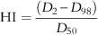 $${\rm HI}={{(D_{2} {\minus}D_{{98}} )} \over {D_{{50}} }}$$
The lower the value of HI, the better the plan is, in such a way that an increase of this index indicates degradation of the quality of the PTV’s dose distribution. An HI of zero indicates that the absorbed dose distribution is almost homogeneous.12 Likewise, for the organs at risk (OAR), the variation of the mean and the maximum dose were evaluated, considering the maximum dose for each OAR as the maximum dose covering 0·1 cc (D 0·1 cc) to spinal cord and brainstem and 2% of volume (D 2%) for the rest of OARs.12
All HI values, for each collimator position, were compared with the position of the collimator, which was assumed as the correct one, showing the relative differences concerning to that.
Results
A search for related works was carried out; however, none was found in the literature during the time of this study. In spite of the limited number of cases analysed, we think that these are representatives of the range of PTV lengths, corresponding to (1) long PTV (high-risk prostate case), where greater dose indices variations have been found, (2) intermediate PTV (H&N) and (3) small PTVs (low-risk prostate and holocranial cases) with no significant dosimetric deviations.
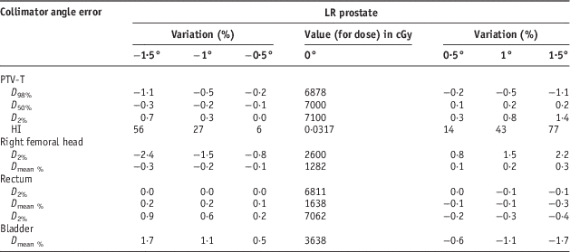
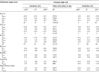
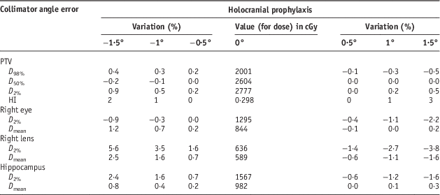
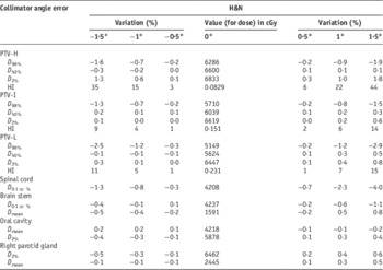
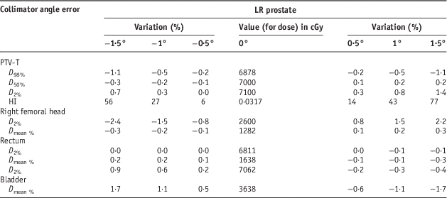
A summary table (Tables 1–4) is presented for each case, where the central column shows the dose in cGy corresponding to the original plan without error. The rest of the columns shows the percentage difference with respect to that plan. For OARs that are even and with bilateral symmetry (parotid glands, eyes, lenses and femoral heads), only the right side ones are presented, showing an opposite behaviour to the corresponding symmetrical OAR.
Results for the low-risk (LR) prostate case
Results for the high-risk (HR) prostate case

Results for holocranial prophylaxis

Results for the head and neck case (H&N)

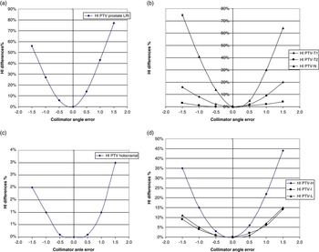
In Figures 1a–1d, the variation of the HI for each PTV is plotted as a function of collimation error for the cases studied. All the cases show a similar parabolic behaviour.
(a) Change in the Homogeneity Index for PTV in the LR prostate case; (b) change in the Homogeneity Index for PTVs in the HR prostate case; (c) change in the Homogeneity Index for PTV in the Holocranial case; and (d) change in the Homogeneity Index for PTVs in the head and neck case. Note: The units for the collimador angle error are in degrees. Abbreviations: PTV, planning target volume; LR, low risk.

Discussion
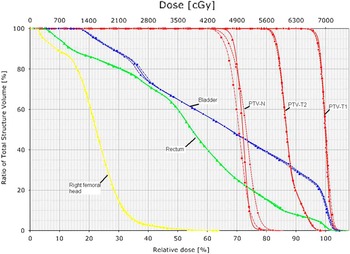
As shown in Tables 1–4 and in Figures 1a–1d, the collimator error angle has a noticeable impact on the dose distribution of the PTVs. A degradation of the dose distribution was observed for all the cases. This effect becomes more important as the PTV extends farther away from the isocentre. Among the four cases analysed, the PTV-N of the HR prostate case was the largest one. In this case, the worsening of the dose distribution was remarkable with a maximum relative variation of 75% of the HI and 1·8% decrease of the D 50 for an error of 1·5° in the collimator angle. Moreover, the isodose covering 98% of the PTV decreased by 4% and the maximum dose varied by 5%. In contrast with the PTVs, the dosimetric parameters of the majority of OARs did not present critical variations. This affirmation can be observed in Figure 2 where dose–volume histograms for PTV-T1, PTV-T2, PTV-N, bladder, rectum and right femoral head are plotted, showing the quality lost as the collimator angle error increased. For a collimator angle error in the range of ±1° (AAPM recommendations),Reference Klein, Hanley and Bayouth11 a maximum median dose variation of 0·1%, 0·0% and 1·2% for PTV-T1, PTV-T2 and PTV-N, respectively, covering dose was obtained.
Dose–volume histograms of the femoral right head, rectum, bladder, PTV-N, PTV-T2 and PTV T1 in the prostate HR treatment: triangles for original plan, squares for +1·5° and circles for a −1·5° collimator angle error. Abbreviations: PTV, planning target volume; HR, high risk.

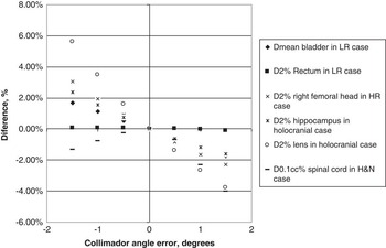
However, when the PTVs are smaller and the isocentre is centred on them, as in the LR prostate case (Table 1 and Figure 1a) and the holocranial case (Table 3 and Figure 1c), the loss of quality and the D 50 are less sensible to the collimator angle errors. In these cases, the HI and the D 50 are almost constant in the range of collimator errors analysed in this study. For instance, the median doses of the PTVs in the low-risk prostate and the holocranial cases have a maximum variation of 0·3% and 0·2%, respectively, when the collimator angle error war 1·5°. The most significant variations were observed for the lenses in the holocranial case, with a maximum dose increase of 5% (Figure 3). The variations for the OARs in the low-risk prostate case were almost negligible.
Plot of some organs at risk (OARs) as a function of collimator angle error.

The H&N case can be considered as an intermediate case (Table 4 and Figure 1d). The PTVs and OARs, including spinal cord and brainstem, presented variations of <4% for all of them. In this particular case, for the spinal cord, this variation represents 0·9 Gy and 1·7 Gy for a systematic collimator angle error of 1·0° and 1·5°, respectively (Figure 3). These variations in the OARs, without being very significant, must be taken into account in assessing possible clinical risks.
Conclusions
The evaluation of the dosimetric errors associated with the effect of the collimator angle error in prostate and H&N VMAT treatment plans has been studied. It has been observed that when the PTVs are away from the isocentre, they might have significant dosimetric discrepancies between the calculated and the planned values, even when fulfilling the widely accepted tolerance of ±1°. Therefore, it seems reasonable to restrict the tolerances to a lower value. In addition, on selecting the position of the isocentre, one should be cautious, and whenever possible one must choose a position close to the geometrical centre of the PTVs. We have not found important variations regarding the dose parameters of OARs, even if they are away from the PTVs, with the exception of the lens and the spinal cord with differences of 5% and 4%, respectively.
Acknowledgements
None.
Conflicts of Interest
None.
Ethical Standards
No animals were used in this study.