Introduction
Cumulus cells (CCs) are a subpopulation of granulosa cells that surround the oocyte. Cumulus cells have been considered as playing an important role in oocyte maturation by keeping the germ cell under meiotic arrest, inducing meiotic resumption and supporting cytoplasmic maturation (Warriach & Chohan, Reference Warriach and Chohan2004). It is known that the CCs and the oocyte are functionally and physically connected (Suzuki et al., Reference Suzuki, Jeong and Yang2000) and due to a sophisticated network of bidirectional communication, which is mediated by gap junctions (Gandolfi et al., Reference Gandolfi, Brevini, Cillo and Antonini2005), CCs confer developmental competence to the oocyte (Bogliolo et al., Reference Bogliolo, Ariu, Fois, Rosati, Zedola, Leoni, Succu, Pau and Ledda2007) by way of transfer of nutrients to the internal compartments. Owing to the fact that adequate rates of in vitro maturation (IVM) of oocytes derived from ovariohysterectomized bitches are low, measures that are more specific are required in the process of identifying factors presumably favouring or interfering with the communication between the CCs and the oocyte during culture. It is believed that the lack of suitability of the oocyte or the CCs influences subsequent development of the cumulus–oocyte complex (COC).
In humans, the oocyte morphology has recently been viewed as a prognostic factor in the developmental potential of an embryo (Ebner et al., Reference Ebner, Moser and Tews2006). The integrity of the COC phenotype is regarded not only as an indicator of its viability, but also as a potential marker of the ability for transcription of genes involved in maturation and early embryonic development.
Morphological assessment of COCs has been used in research that involves IVM of dog oocytes for the purpose of selecting high quality oocytes and estimating their viability. Thickness of CCs is one of the morphological targets used in this process (Lee et al., Reference Lee, Seo, Yin, Cho, Lee, Kim, Cho and Kon2007); because it has been shown that only oocytes with two or more layers of attached CCs continue to develop in vitro (Nickson et al., Reference Nickson, Boyd, Eckersall, Ferguson, Harvey and Renton1993). Cumulus cell expansion and/or in vitro mucification has been interpreted as an indicator of oocyte maturation (Trumpan, Reference Trumpan2001).
By far, most studies have tracked the progression of meiosis by evaluating the pattern of nuclear chromatin decondensation in oocytes selected for in vitro culture (De Los Reyes et al., Reference De Los Reyes, de Lange, Miranda, Palominos and Barros2005; Bolamba et al., Reference Bolamba, Russ, Harper, Sandler and Durrant2006; Santos et al., Reference Santos, Rodrigues and Rodrigues2006). However, to our knowledge, there are no in vitro matured dog COC reports that have examined the relationship between the morphology of CCs, the nuclear status of oocytes and the profile of circulating progesterone concentrations of ovary donors. Therefore, the aim of this study was to evaluate the hallmark features of CCs and those from nuclear chromatin configurations in in vitro matured COCs by establishing, when appropriate, a relationship between them. Moreover, because developmental competence of in vitro canine oocytes has been attributed to ovary donor reproductive status (Otoi et al., Reference Otoi, Ooka, Muramaki, Karja and Suzuki2001; Willingham-Rocky et al., Reference Willingham-Rocky, Hinrichs, Westhusin and Kraemer2003; Hossein et al., Reference Hossein, Kim, Jang, Oh, Koo, Kim, Kang, Lee and Hwang2007), we evaluated the influence of the in vivo female endocrine environment on the viability of COCs matured in vitro. Furthermore, due to the reported high levels of degeneration of in vitro matured COCs (Rodrigues & Rodrigues, Reference Rodrigues and Rodrigues2003; Lee et al., Reference Lee, Seo, Yin, Cho, Lee, Kim, Cho and Kon2007), we hypothesized that morphological features of CCs, including the signs of apoptosis, could be used in COCs as an intrinsic predictor of oocyte viability in vitro.
Materials and Methods
Ovaries and oocyte retrieval
Ovaries were retrieved from two prepubertal (6 months) and 14 adult canine females (Canis familiaris) ranging from 1 to 13 years of age (mean age was 3.7 years). Purebred (n = 8) and crossbred (n = 8) bitches had their ovaries subjectively categorized by identification of macroscopic external ovarian tissue morphology as follows: (i) without follicles or luteal tissue (n = 4); (ii) with one or more visible follicles (n = 3); and (iii) with one or more pronounced corpora lutea (n = 9). Furthermore, females were grouped according to their serum progesterone (SP) concentrations as follows: (a) bitches with serum progesterone levels between 0 ng/ml and 1 ng/ml (n = 4); (b) bitches with serum progesterone levels between 1.1 ng/ml and 2.5 ng/ml (n = 3); (c) bitches with serum progesterone levels between 2.6 ng/ml and 5 ng/ml (n = 5); and (d) bitches with serum progesterone levels higher than 5 ng/ml (n = 4). Serum concentrations of progesterone were measured by chemiluminescence (Nobel RIE). Ovaries were obtained by routine and elective ovariohysterectomy (n = 14), after Caesarean section operation (n = 1), or after pyometra surgery (n = 1), following anesthaesia at the Veterinary Hospital of Federal University of Rio Grande do Sul (UFRGS) (Av. Bento Gonçalves, 9090/Bairro Agronomia–Porto Alegre/RS; Brazil); Zoonoses Control Centre of Porto Alegre (Estrada Bérico José Bernardes, 3489–Porto Alegre/RS; Brazil) and at local private clinics. The study followed the guidelines for ethical conduct in the care and use of animals instituted by COBEA (Colégio Brasileiro de Experimentação Animal).
The females' ovaries were brought to the laboratory in modified phosphate-buffered saline (PBS) solution at ambient temperature within 1 h of removal. The COCs were recovered by slicing of ovarian tissue at 37 °C in PBS supplemented with 1% fetal calf serum (FCS) (Nutricell). Cumulus–oocyte complexes were recovered and morphologically classified by the following grades: (i) darkly pigmented ooplasm surrounded by one or more layers of CCs; or (ii) lightly pigmented ooplasm with incomplete layers of CCs (Hewitt et al., Reference Hewitt, Watson and England1998). A total of 266 COCs were morphologically classified as Grades 1 and 2 and were matured in vitro. The medium used in the maintenance of the COCs before maturation was TCM 199 with 25 mM HEPES (Sigma), supplemented with 2.38 mM NaHCO3 (Sigma), 50 μg/ml gentamicin (Sigma), 22 μg/ml pyruvic acid and 1 mg/ml BSA (Sigma).
In vitro maturation
The COCs were cultured in 100 μl drops (four drops per dish) under mineral oil (Sigma°) at 37 °C in a humidified atmosphere with 5% CO2 in air (high oxygen tension) for 48 h. Oocytes were exposed to the following medium: TCM 199 with 2.2 mg/ml sodium bicarbonate + Earle's salts + l-glutamine (Gibco), 10% fetal bovine serum (Nutricell), 50 μg/ml gentamicin (Sigma), 22 μg/ml pyruvic acid (Sigma), 20 μg/ml estradiol (Sigma), 0.5 μg/ml FSH (Foltropin-V, Vetrepharm, Inc.), 0.03 IU/ml hCG (Profasi HP, Serono) and a final concentration of 1 μg/ml human somatotropin (hST; Lilly).
Cumulus cell expansion
The oocytes were evaluated for expansion on a four-point scale as previously reported (Ball et al., Reference Ball, Ax, First, Mahash, Muldoon, Saxana and Sadler1980). Briefly, complexes were rated as follows: (i) D1 (compact, unexpanded complexes); (ii) D2 (outer CCs slightly expanded); (iii) D3 (outer and inner CCs slightly expanded); and (iv) D4 (fully expanded complexes).
Removal, identification and quantification of cumulus cell types
Removal of CCs was performed by repeated passage of the oocytes through a fine-bore, fire-polished glass pipette in modified PBS. The CC population was pipetted onto a microscope slide, stained with 1 mg/ml propidium iodide (PI) (Sigma) and squashed with a coverslip. After that, the coverslip was sealed with clear nail polish. Cumulus cells were evaluated among the PI-stained cells by counting 100 cells at each group of subjectively estimated cumulus expansion degree (D1, D2, D3 and D4) in various randomly selected cumulus mass fields by using a contrast phase microscope (Olympus BX41) at magnification of ×1000.
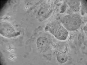
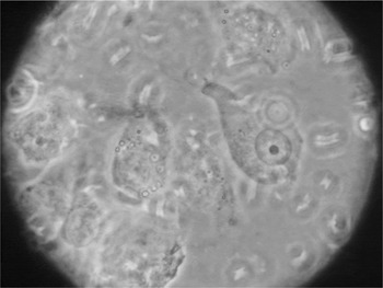
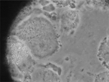
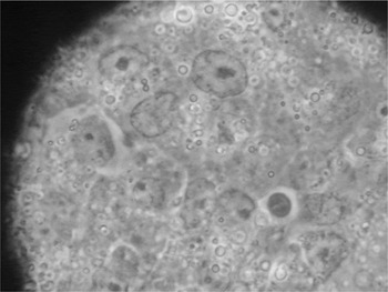
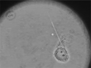
Cumulus cells were classified into morphological types (C1, C2, C3), following the description from Calado et al. (Reference Calado, Rocha, Colaço and Sousa2003) in which type C1 cells had small and very dense nuclei, type C2 cells had intermediate size and dense nuclei and type C3 cells presented large and lightly stained nuclei (Figs. 1–3). Features of apoptosis in CCs were observed and identified as follows: (a) marginated chromatin; (b) pyknotic appearance; (c) multiple nuclear fragments; and (d) apoptotic bodies (Yang & Rajamahendran, Reference Yang and Rajamahendran2000) (Figs. 1, 4 and 5).
Photo of canine cumulus cells (CCs) after 48 h IVM. Note two C2 type cells (top right), a C1 type cell (bottom right) and a cumulus cell with three nuclear fragments (propidium iodide stain; contrast phase microscope; oil immersion) (×1000) (Rodrigues, 2008).

Cumulus cell (CC) type C1. Note the small dense nucleus (propidium iodide stain; contrast phase microscope; oil immersion) (×1000) (Rodrigues, 2008).

Cumulus cell (CC) type C3. Note the large and lightly stained nucleus (propidium iodide stain; contrast phase microscope; oil immersion) (×1000) (Rodrigues, 2008).

Feature of apoptosis in cumulus cells (CCs) of canine oocyte matured in vitro: apoptotic body. Note the distinct round dark structure (bottom right) (×1000) (Rodrigues, 2008).

A fragmented canine cumulus cell (CC) after 48 h IVM (top left). Note the four nuclear fragments (propidium iodide stain; contrast phase microscope; oil immersion) (×1000) (Rodrigues, 2008).

Photos were taken with the aid of a digital Still Camera (Sony) and made with the camera lens resting over the microscope eyepiece. Different images sizes were obtained by means of the zoom function in the camera.
Chromatin evaluation
The chromatin configuration was characterized according to Brück et al. (Reference Brück, Bézard, Baltsen, Synnestvedt, Couty, Greve and Duchamp2000). Nuclear maturation stages were: (a) germinal vesicle (GV): finely granulated nucleoplasm with filamentous chromatin configuration; (b) germinal vesicle breakdown (GVBD): condensed filamentous chromatin within finely granulated nucleoplasm; (c) metaphase I (MI): condensed chromosomes oriented in a metaphase plate; (d) metaphase II (MII): two sets of condensed chromosomes of which one set with orientation in a metaphase plate, while the second (polar body) more or less randomly oriented in a cluster; and (e) others: no chromatin or small spots of dense chromatin. Chromatin configuration in oocytes after IVM was evaluated by Hoechst 33342 (10 μg/ml) staining as previously described (Rodrigues et al., Reference Rodrigues, dos Santos and Rodrigues2004).
Statistical analysis
Data were analyzed using chi-squared analysis with adjusted residual. Differences were considered to be significant at probability p < 0.05. Fisher's exact test was used to assess differences in oocyte configurations after in vitro maturation.
Results
Qualitative findings
Under the contrast phase microscope, the phenotype of CCs among the expansion grades and various concentrations of progesterone from bitches used in this study, were distributed into three cell types: C1, C2 and C3. Apoptotic cells were those belonging to the same recorded types, showing additional features of cell death, among which the most representative was fragmentation of the CC nucleus.
Quantitative findings
Statistically significant results were found among the different types of CCs with respect to serum progesterone level or degree of expansion. These results are presented in Tables 1–3. In Table 2, the total number of CCs varied among the progesterone concentrations due to the lack of one, or more than one, cell type (C1, C2, C3 or apoptotic cell) in the observed degrees of expansion. In Table 3, the total number of CCs were not evenly distributed between the three degrees of expansion due to the lack of presence of one or more degrees within the experimental routines.
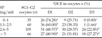
Influence of serum progesterone (SP) level on degree of cumulus expansion (DCE) of canine oocytes in vitro matured.

Values with different superscripts in the same column (ab, cd) differ significantly (chi-squared analysis with adjusted residual) (p< 0.001).
*D1: degree 1 cumulus expansion; D2: degree 2 cumulus expansion; D3: degree 3 cumulus expansion.
#G1, G2: grade 1 and 2 oocytes
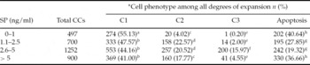
Patterns in cumulus cells (CCs) in COCs of bitches at various serum progesterone (SP) concentrations.

Values with different superscripts (ab, cd, ef, gh) within a column, differ significantly (chi-squared analysis with adjusted residual) (p < 0.05).
*C1: cumulus cell type 1; C2: cumulus cell type 2; C3: cumulus cell type 3.
Cumulus cell patterns in relation to the degree of expansion in cumulus mass.

Values with different superscripts (ab, cd, ef, gh) within a column, differ significantly (chi-squared analysis with adjusted residual) (p < 0.05).
*D1: degree 1 cumulus expansion; D2: degree 2 cumulus expansion; D3: degree 3 cumulus expansion.
Chromatin configuration did not yield statistical differences in relation to the same parameters. Data for chromatin configuration are presented in Tables 4 and 5. The quantitative findings are described in detail below.
Influence of degree of expansion on nuclear chromatin configuration of canine oocytes matured in vitro.

GV: germinal vesicle; GVBD: germinal vesicle breakdown; MI: metaphase I; MII: metaphase II; Others: degenerated or unclassified.
*D1: degree 1 cumulus expansion; D2: degree 2 cumulus expansion; D3: degree 3 cumulus expansion.
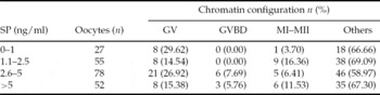
Influence of female serum progesterone (SP) concentration on oocyte chromatin configuration.

GV: germinal vesicle; GVBD: germinal vesicle breakdown; MI: metaphase I; MII: metaphase II; Others: degenerated or unclassified.
Cumulus cell expansion in relation to progesterone concentrations
Table 1 shows the relationship between degree of CC expansion and different serum progesterone levels of ovarian donors. The most frequent degree of cumulus expansion seen in oocytes derived from bitches at serum progesterone levels between 0 ng/ml and 1 ng/ml was for category 1. Frequencies in this category were 74.28% (26/35) and these rates were statistically different from the remaining progesterone levels in the same category of expansion. For expansion category 3, frequencies were 1.66% at serum progesterone levels between 1.1 ng/ml to 2.5 ng/ml. Statistically, this rate was associated to a number lower than expected for that category. A negative adjusted residual (−3.5) confirmed a high statistical significance (p < 0.001). In contrast, compared to the low progesterone levels, cumulus expansion category 3 showed enhanced rates at serum levels between 2.6 ng/ml and 5 ng/ml (22.85%) and at serum levels higher than 5 ng/ml (27.27%). None of the oocytes matured in modified TCM 199 for 48 h showed CCs expansion in category 4.
Patterns in cumulus cells in relation to serum progesterone concentrations
The results presented in Table 2 show the predominance of CC type C1 (55.13%) in COCs of bitches at serum progesterone levels between 0–1 ng/ml. Type C2 displayed higher numbers than those expected for progesterone levels between 1.1–2.5 ng/ml. In addition, normal CCs of types C2 and C3, which were presented with rates of 20.52% and 15.9% respectively at serum progesterone levels between 2.6–5 ng/ml, showed a significant difference between the observed and expected frequencies, tested using the adjusted standardized residuals of chi-squared test (p < 0.05). Apoptosis, mostly recognized in this study by fragmentation of CC nuclei, was high, independent of bitch progesterone serum concentrations. However, observed frequencies of fragmentation at serum progesterone levels from 0–1 ng/ml (40.64%) and at serum progesterone levels above 5 ng/ml (36.6%) were numerically higher and therefore considered statistically significant (p < 0.05), when evaluating the remaining concentrations of progesterone for the same parameter.
Patterns in cumulus cells in relation to degree of expansion
The phenotype of cells in cumulus mass of expanded oocytes is shown in Table 3. There was a prevalence of CC type C1 (49.57%) at D1 degree of expansion. Significantly, more CCs with this phenotype were seen, when compared to expanded degrees D2 and D3 (p < 0.05). Numbers of apoptotic cells were statistically less than expected in D1 expanded COCs, while in D2 expanded COCs a positive adjusted residual (+3.6) endorsed the significance of apoptotic pattern (32.58%) at this expansion grade (p < 0.05).
Chromatin configuration
Due to loss during mechanical displacement of CCs, numbers of oocytes were less (n = 212), when evaluating chromatin configuration in the oocytes:
• Chromatin configuration in relation to degree of expansion – there was no correlation between nuclear chromatin configuration and cumulus expansion in COCs after 48 h of in vitro maturation (p = 0.622) (Table 4).
• Chromatin configuration in relation to serum progesterone concentrations – the results summarized in Table 5 do not show any significant correlation between the circulating progesterone profile of ovary donors and the nuclear chromatin configuration in COCs matured in vitro (p = 0.087).
Discussion
Mammalian ovaries contain a distinguishable population of oocytes, which consists of growing, medium-sized and fully grown germ cells. From the three listed populations, only the fully grown oocytes resume maturation when they are released from follicles and cultured in vitro (Mrazek & Fulka, Reference Mrazek and Fulka2003).
In bitches, various oocytes selected for in vitro culture are collected from atretic follicles during the slicing procedure. Late atretic and early to mid-atretic follicles can be found at estrus and diestrus phases of the cycle respectively (Willingham-Rocky et al., Reference Willingham-Rocky, Hinrichs, Westhusin and Kraemer2003). Although apparently healthy oocytes can be retrieved from atretic follicles (McNatty et al., Reference McNatty, Heath, Henderson, Lun, Hurst, Ellis, Montgomery, Morrison and Thurley1984), this contingent of germ cells may account for the observed sensitivity to degeneration of in vitro matured COCs (Rodrigues & Rodrigues, Reference Rodrigues and Rodrigues2003), because granulosa cells of an atretic follicle have a reduced synthesis of DNA (Greenwald, Reference Greenwald1989) and more numbers of apoptotic cells (Van Soom et al., Reference Van Soom, Vandaele, Goossens, de Kruif and Peelman2007). It is noteworthy that oocytes from atretic follicles undergo meiosis-like changes (germinal vesicle breakdown), which are followed by oocyte fragmentation and disruption of the oocyte–cumulus connection (Tsafriri & Braw, Reference Tsafriri and Braw1984).
Cumulus–oocyte complexes expand under the influence of gonadotropins during in vitro maturation (Yooko et al., Reference Yokoo, Miyahayashi, Nagamura, Kimura, Sasada and Sato2002) and expansion in vitro has been interpreted as a marker of oocyte maturation (Trumpan, Reference Trumpan2001). Nevertheless, large expanded and/or mucified canine CCs, after in vitro maturation, are not necessarily those achieving meiosis (Rodrigues & Rodrigues, Reference Rodrigues and Rodrigues2006). Similar observations have been made by other researchers. Srsen et al. (Reference Srsen, Kalous, Nagyova, Sutovsky, King and Motlik1998) reported on expressive cumulus expansion without correspondent meiosis in in vitro matured blue fox oocytes (Alopex lagopus). Bolamba et al. (Reference Bolamba, Russ, Harper, Sandler and Durrant2006) attributed the in vitro expansion of CCs to the synergistic action of steroids and gonadotropins in medium. Lee et al. (Reference Lee, Seo, Yin, Cho, Lee, Kim, Cho and Kon2007) reported on the presence of high numbers of FSH receptors in CCs of canine oocytes. Supplementation of media with FSH is thought to reduce the incidence of apoptosis in CCs (Hussein et al. Reference Hussein, Froiland, Amato, Thompson and Gilchrist2005). Consistent to what was reported by De Los Reyes et al. (Reference De Los Reyes, de Lange, Miranda, Palominos and Barros2005), in the present study, the addition of gonadotropin in medium contributed to a great proportion of mucification of CCs in the COC. Mucification, which is the presence of mucus, was observed in COCs after 48 h of maturation. Nevertheless, morphological appearance (grade) of the oocyte–corona–cumulus complex has been observed not to be correlated with nuclear maturity, fertilization, or early embryo cleavage (Rattanachaiyanont et al., Reference Rattanachaiyanont, Leader and Leveille1999). Recently, we demonstrated that the degree of expansion showed no correlation with cleavage of dog oocytes matured in vitro and fertilized in vitro (Rodrigues et al., Reference Rodrigues, dos Santos and Rodrigues2007). While somewhat surprising, mucified complexes may not express factors that are essential in communicating with the oocyte (Rodrigues et al., Reference Rodrigues, dos Santos and Rodrigues2006). Therefore, mucification may be only an artifactural expression of an artificially imposed environment.
Currently, there are limited data available about the consequences involving cumulus quality and the fate of the canine oocyte in vitro. For this reason, we decided to investigate the paradox of expansion without meiosis. For instance, a series of studies performed by Calado et al. (Reference Calado, Rocha, Colaço and Sousa2001; Reference Calado, Rocha, Colaço and Sousa2003), identified a heterogeneous CC population inside the cumulus mass from COCs aspirated from small antral follicles during the various phases of the bovine estrous cycle. The three observed cell types (C1, C2 and C3) diverged qualitatively and quantitatively at distinct stages of the bovine ovarian cycle. The changing proportions of cell populations in COCs were assumed by the authors to be an explanatory basis for the physiological differences seen in vitro. Consistent with the findings from Calado and collaborators (2003), we also verified a progressive differentiation in CC types (from C1 to C3) of canine COCs, correlated with the in vivo endocrine environment of ovary donors at the time of ovary removal. From our observations, C1 cell type was the most prevalently healthy cell in the cumulus mass. Whereas COCs with grade 3 expansion showed increased numbers of C3 type cells, C1 type cells were the most prevalent in oocytes with cumulus expansion 1. The prevalence of C1 type in COCs with expansion 1 and levels of circulating progesterone between 0–1 ng/ml may reflect the in vivo endocrine physiology of bitches, where proliferative changes are not expected until follicular secretion increases at proestrus phase. With the increase in serum progesterone levels above 1.1 ng/ml, a transition of cell type C1 into C2 and C3 could be observed by us. Thus, phenotype in cumulus mass changed and C3 was numerically enhanced when progesterone levels achieved values between 2.6–5 ng/ml, which are near the concentrations estimated at the time of the luteinizing hormone surge (LH), i.e., 2–4 ng/ml (Feldmann & Nelson, Reference Feldman and Nelson1997). Intriguingly, in this study, C3 prevalence did not persist at hormone levels higher than 5 ng/ml, when type C3, the one with higher functional capacity in CCs of bovine COCs (Calado et al., Reference Calado, Rocha, Colaço and Sousa2003), had been expected to dominate. The reason for this behaviour is not clear. In the present study, C3 type may have escaped observation at the rate normally associated with further advancement of the in vivo endocrine milieu, due to species dependent differences or due to in vitro developmental specificities in canine COCs. In addition, it might be possible that inclusion of abnormal C3 types in the apoptotic group explains for itself the low numbers of C3 at enhanced (>5 ng/ml) serum progesterone concentrations. Furthermore, we cannot exclude the possibility that cell differentiation in cumulus mass is modulated by the process of cumulus expansion. In that case, an expressive prevalence of C3 would only be expected to be associated with more advanced levels of cumulus expansion, like for example expansion grade 4, which was not observed in the present study. It is important to notice that, in the unique estrus cycle of bitches, no expansion of CCs happens in COCs before fertilization (Van Soom et al., Reference Van Soom, Tanghe, de Pauw, Maes and de Kruif2002).
The numerical distribution of CC types within the cumulus mass was observed as a whole, because previous studies (Calado et al., Reference Calado, Rocha, Colaço and Sousa2003) demonstrated that similar frequencies of cell types (C1, C2 and C3) can be found as much in the closest as in the farthest zones from the oocyte. The cell population in the cumulus was pooled from distinct oocytes. The CCs were retrieved from various regions of the complexes and therefore thought of as being representative of the mass population. Nevertheless, Hussein et al. (Reference Hussein, Froiland, Amato, Thompson and Gilchrist2005) verified that the incidence of apoptosis is the lowest in the inner most layer of CCs and gets higher with increasing distance from the oocyte. Indeed, in the present study, apoptosis exhibited higher percentages in COCs at enhanced degrees of expansion (D2 and D3). Therefore, it seems plausible to state that the expressive fragmentation rates observed in cumulus mass in our study may correlate with the population muster evaluated and could possibly have been represented by the cells mostly localized at the oocyte's periphery zone.
The findings of Lopes et al. (Reference Lopes, Sousa, Luvoni and Rocha2007), reporting a low rate (less than 15%) of apoptosis in CCs from immature canine grade 1 COCs, with no differences in rates between bitches at diestrus and anestrus, seem not to be true when canine oocytes are submitted to in vitro maturation.
In this study, apoptosis of CCS, mostly represented by nuclear fragmentation, was a common feature among COCs matured in vitro. This phenotype was seen independent of bitch serum progesterone levels or degree of CC expansion after 48 h of culture. However, numbers of apoptotic cells were predominant at the lowest and at the highest measured progesterone serum levels of ovary donors, an observation partially similar to the findings from Van Cruchten et al. (Reference Van Cruchten, Van den Broeck, Duchateau and Simoens2003), who reported about the relationship between endometrial apoptosis and high progesterone concentrations in dogs.
In our opinion, canine CC apoptosis follows a time-dependent path. This observation was made recently in our laboratory by verifying the viability of CCs and its relationship with nuclear morphology of oocytes from bitches at various moments of culture (Rodriguez et al., Reference Rodriguez, Arruda, Cavalcante, Silva, Rodrigues, Rodrigues, England, Concannon and Schäfer-Somi2008). Variability among immature canine complexes at various stages of the estrous cycle was emphasized by Köhle et al. (Reference Köhle, Woehl-Wenigerkind, Sinowatz and Braun2005), who reported about increased percentages of cumulus apoptosis at metestrus stage, in contrast to those observed during proestrus and estrus stages. These findings are in agreement with the results herein. Apoptotic features were lower in number at serum progesterone levels between 1.1 ng/ml and 2.5 ng/ml and from 2.6 to 5 ng/ml, which are the corresponding hormone concentrations seen at the follicular phase of the cycle. It appears therefore that preovulatory to ovulatory circulating progesterone levels may sustain the integrity of cells in cumulus mass in a more appropriate manner. Despite these observations, in the present study, regardless of in vivo progesterone profile of the ovary donor, canine COCs after 48 h of in vitro culture, showed enhanced rates of CCs apoptosis and additionally, featured chromatin degeneration or arrest. Oocyte removal from the follicular environment may artificially induce CCs apoptosis (Van Soom et al., Reference Van Soom, Vandaele, Goossens, de Kruif and Peelman2007). However, Dunning et al. (Reference Dunning, Lane, Brown, Yeo, Robker and Russell2007) offered evidence-based results that alteration of the composition of the COC matrix occurs during in vitro maturation of human oocytes, which contributes to poor competence.
By comparing the final chromatin configuration with the expansion of in vitro matured oocytes, we verified that most of these oocytes were degenerated. As stated before, nuclear maturation in the bitch is negatively correlated with the degree of the cumulus expansion (Songsasen et al., Reference Songsasen, Yu and Leibo2002) and expansion of CCs, except for the corona radiata, does not depend on the intercellular communication with oocytes (Funahashy & Day, Reference Funahashy and Day1993). These findings reinforce our belief, that in vitro expansion of cumulus in COCs from dogs might be not a legitimate, but an artificial phenomenon instead.
Considerable evidence appears to support the idea that the degree of apoptosis in CCs is negatively correlated with oocyte competence (Yuan et al., Reference Yuan, Van Soom, Leroy, Dewulf, Van Zeveren, de Kruif and Peelman2005). Apoptosis involves a meticulous series of biochemical events and is characterized by morphologic changes including DNA fragmentation, pyknosis of cell nuclei, formation of apoptotic bodies and death (Kerr et al., Reference Kerr, Wyllie and Currie1972; Perez et al., Reference Perez, Tao and Tilly1999). Recent studies have shown that there is often a continuum of apoptosis and necrosis following cell injuries (Zong & Thompson, Reference Zong and Thompson2006). Due to low specificity of current methods used for apoptosis detection, such as electron microscopy, fluorescence morphological staining, or TUNEL (terminal deoxynucleotidyl transferase-mediated dUTP nick end-labeling) assay, it has being difficult to differentiate the in vitro apoptotic process from necrosis (Fabian et al., Reference Fabian, Koppel and Maddox-Hyttel2005). Apoptosis in CCs may be one of the factors responsible for the inhibition of meiotic competence in the present study. However, this observation does not exclude the possibility that the oocyte itself might have been responsible for the degeneration rates herein observed.
Adenosine triphosphate (ATP) depletion, a situation compatible with the reduction in energy levels of oocytes in vitro cultured, may be listed among the possible reasons for the results of degeneration in COCs herein, since ATP is necessary for several functions, including maintenance of homeostasis and regulation of cell survival (St John, Reference St John2002). Therefore, apoptosis of the CCs and oocyte might be a reasonable explanation for the usually high degeneration levels observed in canine COCs matured in vitro (Rodrigues & Rodrigues, Reference Rodrigues and Rodrigues2003), due to apoptosis being energy dependent (Zong & Thompson, Reference Zong and Thompson2006).
Nevertheless it should be kept in mind that, spontaneous apoptosis occurs in COCs during suboptimal culture (Ikeda et al., Reference Ikeda, Imai and Yamada2003; Yuan et al., Reference Yuan, Van Soom, Leroy, Dewulf, Van Zeveren, de Kruif and Peelman2005) and specific factors or components in medium may be expected to interfere (positively or negatively) with the extent either of cell death or survival. While insufficient to enhance meiosis to the MII stage (Silva et al., manuscript in preparation), supplying media with glucose, the predominant energy substrate used for dog oocytes (Songsasen et al., Reference Songsasen, Spindler and Wildt2005), has an antiapoptotic effect on CCs as observed by ourselves recently (Silva et al., Reference Silva, Rodriguez, Cavalcante, Rodrigues, Rodrigues, England, Concannon and Schäfer-Somi2008). On the other hand, a negative effect on the structure of mitochondria was reported when bovine serum was included in the maturation medium for bovine oocytes (Abe & Hoshi, Reference Abe and Hoshi2003). In our study, the presence of bovine serum in medium should, therefore, be considered among the detrimental factors resulting in COC degeneration. Nevertheless, it is unknown to which extent our findings might have been influenced by the presence of protein (bovine serum) in the maturation medium or by meiosis-inhibiting factors present in TCM 199 (for example purines and purine precursors) (Roberts et al., Reference Roberts, Frank and Hardy2002).
This study has yielded the following major findings: (a) advanced cumulus expansion in canine COCs in vitro is predominant at bitch serum progesterone levels > 2.5 ng/ml; (b) CC apoptosis in vitro relates both to low (0–1 ng/ml) and to high (>5 ng/ml) in vivo serum progesterone levels; (c) distinct cell types (C1, C2 and C3) are observed in cumulus mass from dog oocytes matured in vitro and influenced by female's circulating progesterone concentration; and (d) oocyte nuclear maturation is influenced neither by in vivo serum progesterone values nor by the degree of expansion in CCs.
Currently, lack of knowledge about the real rate of viability of COCs and its influence on in vitro developmental potential makes the relationship between CC integrity and in vitro maturation of oocytes worthy of further investigation.
Acknowledgements
The authors thank the Veterinary Hospital of Universidade Federal do Rio Grande do Sul (UFRGS), the Zoonoses Control Centre of Porto Alegre and the local private clinics (Arca de Noé and Clinica Veterinária do Vale; São Leopoldo, RS, Brazil) for generously providing bitch ovaries. We acknowledge Mr Hratch Kalaidjian's valuable contributions in proofreading and English language assistance. In addition, we thank Prof. Jandyra Fachel (UFRGS) for statistical assistance.