Introduction
The suborder Phacopina is a common and globally distributed group of trilobites found in the Devonian sediments (Chlupáč, Reference Chlupáč1975; Crônier and François, Reference Crônier and François2014). Unlike the compound (holochroal) eyes of most trilobites, the eyes of the Phacopina are schizochroal, with surfaces that consist of numerous small lenses segmented by interlensar cuticles (sclerae); each lens is covered by a separated cornea (Clarkson, Reference Clarkson1975; Clarkson et al., Reference Clarkson, Levi-Setti and Horvath2006). These lenses, in addition to providing members of the Phacopina with a more efficient visual system (Clarkson and Levi-Setti, Reference Clarkson and Levi-Setti1975; Clarkson et al., Reference Clarkson, Levi-Setti and Horvath2006; Schoenemann, Reference Schoenemann2021), are used in systematic taxonomy, according to their number and arrangement (e.g., Clarkson, Reference Clarkson1966, Reference Clarkson1969; Eldredge, Reference Eldredge1972; Zhou and Campbell, Reference Zhou and Campbell1990; McKellar and Chatterton, Reference McKellar and Chatterton2009; Ghobadi Pour et al., Reference Ghobadi Pour, Popov, Omrani and Omrani2018). However, quantitative study of eye lenses in some members of the Phacopina has shown that the lens numbers of the same species were not stable; their numbers could change with individual ontogeny and evolution (Thomas, Reference Thomas1998; Crônier and Clarkson, Reference Crônier and Clarkson2001), and even members of the same species living in different environments could show changes in the number of eye lenses (Crônier et al., Reference Crônier, Feist and Auffray2004). Therefore, the emphasis on eye lens numbers in the taxonomic study of the Phacopina should be reconsidered. Quantitative study of eye lens numbers will provide a new understanding of the changes to the visual system during ontogeny and evolution, as well as the influence of environmental factors on the visual system of the Phacopina (Clarkson and Tripp, Reference Clarkson and Tripp1982; Crônier et al., Reference Crônier, Budil, Fatka and Laibl2015).
Omegops Struve, Reference Struve1976, one of the last phacopid trilobites, is found only in the Famennian and persists to the Devonian–Carboniferous boundary in many sections (Xiang, Reference Xiang, Ji, Wei, Wang, Wang, Sheng, Wang, Hou, Xiang, Feng and Fu1989; Brauckmann et al., Reference Brauckmann, Chlupáč and Feist1992; Feist et al., Reference Feist, Cornée, Corradini, Hartenfels, Aretz and Girard2021). Although Omegops has been found in Europe (Richter and Richter, Reference Richter and Richter1933; Struve, Reference Struve1976; Weber, Reference Weber2000), North Africa (Alberti, Reference Alberti1972; Belka et al., Reference Belka, Klug, Kaufmann, Korn, Döring, Feist and Wendt1999), Kazakhstan (Struve, Reference Struve1976), Iran (Feist et al., Reference Feist, Yazdi and Becker2003; Ghobadi Pour et al., Reference Ghobadi Pour, Popov, Omrani and Omrani2018), Afghanistan (Farsan, Reference Farsan1998), and China (Xiang, Reference Xiang1981, Reference Xiang, Ji, Wei, Wang, Wang, Sheng, Wang, Hou, Xiang, Feng and Fu1989; Yuan and Xiang, Reference Yuan and Xiang1998), the number of specimens reported in the past was small, and the eyes of some specimens were not completely preserved. Therefore, it was difficult to carry out quantitative study on the eye lenses, and it was unclear whether the eye lenses of this genus were as diverse as those of other members of the Phacopina (Ghobadi Pour et al., Reference Ghobadi Pour, Popov, Omrani and Omrani2018).
In the Devonian, western Junggar belonged to the Kazakhstan Block in the Northern Hemisphere. The marine deposits are rich in trilobite fossils in this area, but most of these trilobites have been listed only briefly in the Geological Survey Reports from the 1960s to the 1980s or simply described in the Paleontological Atlas of Northwest China (Xinjiang part) (Zhang, Reference Zhang1983). Only a few species have been systematically studied (Xiang, Reference Xiang1981; Guo, Reference Guo1989; Yuan and Xiang, Reference Yuan and Xiang1998; Crônier and Waters, Reference Crônier and Waters2022), including Omegops from the Hongguleleng Formation in western Junggar, Xinjiang. Omegops in the Hongguleleng Formation has low disparity but high abundance. I collected nearly 200 specimens with preserved eyes from this formation to conduct quantitative analysis on the numbers and distribution of their eye lenses and evaluate the possible causes for the diversity of these eye lenses.
Geological setting and materials
The Upper Devonian marine deposits in western Junggar include mainly three sedimentary types: marine and continental transitional facies (Tielieketi and Taketai formations), shallow marine facies (Hongguleleng Formation), and flysch facies (Taerbahatai Formation) (Gong and Zong, Reference Gong and Zong2015). Among these, the Hongguleleng Formation has abundant fossils and can be divided into three lithologic members (Hou et al., Reference Hou, Lane, Waters, Maples and Yang1993). The lower member comprises thin-bedded bioclastic limestones, shelly limestones, argillaceous limestones, calcareous siltstones, and shales. The middle member is composed of grayish-green and grayish-purple pyroclastic rocks and a few sandy limestones. The upper member is mostly grayish yellow calcareous clastic rocks with a few bioclastic limestones. The formation is Famennian in age (Ma et al., Reference Ma, Zhang, Zong, Zhang and Lü2017; Zong et al., Reference Zong, Wang, Fan, Song, Zhang, Shen and Gong2020). Trilobites are located mainly within the upper part of the lower member and the middle member, and a few trilobites extend near the Devonian/Carboniferous boundary (Suttner et al., Reference Suttner, Kido, Chen, Mawson and Waters2014; Zong and Gong, Reference Zong and Gong2017; Zong et al., Reference Zong, Wang, Fan, Song, Zhang, Shen and Gong2020).
Xiang (Reference Xiang1981) systematically described phacopid trilobites from the Hongguleleng Formation for the first time, but he did not document the specific horizon of these fossils. Judging from the preservation of trilobites, they seem to have been collected from the upper part of the lower member to the middle member of the revised Hongguleleng Formation (Zong et al., Reference Zong, Wang, Fan, Song, Zhang, Shen and Gong2020). Yuan and Xiang (Reference Yuan and Xiang1998) described the other two species of phacopids from the Hongguleleng Formation and registered their horizon as the upper part of the Hongguleleng Formation. It should be noted that in the early stratigraphic classification of the Hongguleleng Formation (e.g., Xu et al., Reference Xu, Cai, Liao and Lu1990; Zhao and Wang, Reference Zhao and Wang1990), the lower part was a set of continental sandy conglomerates (now incorporated into the underlying Zhulumute Formation), and the upper part was the marine beds (roughly equivalent to the revised Honguleleng Formation), and Yuan and Xiang (Reference Yuan and Xiang1998) probably used the old stratigraphic division scheme. This inference can be drawn from their field specimen numbers of trilobites (i.e., AEJ 484, AEJ 460, and AEJ 563); all three numbers appear in the description of the Hongguleleng Formation measured by Xu et al. (Reference Xu, Cai, Liao and Lu1990). These three fossil horizons are located in the limestones from the upper part of the lower member, rather than the upper member/part of the revised Hongguleleng Formation (Zong et al., Reference Zong, Wang, Fan, Song, Zhang, Shen and Gong2020). Their age corresponds roughly with the upper Palmatolepis rhomboidea to Palmatolepis marginifera conodont biozones of the Famennian (Suttner et al., Reference Suttner, Kido, Chen, Mawson and Waters2014). More recently, Crônier and Waters (Reference Crônier and Waters2022) described the phacopids of the Hongguleleng Formation that were collected from the Bulongguoer Reservoir section (type section of the Hongguleleng Formation) and the Aomage locality by Waters in 1995 and 2005, and they believed these phacopids occurred in the Palmatolepis crepida conodont biozone. However, the stratigraphic data they provided were not consistent with the conodont biozones. They suggested that Bulongguoer Reservoir section exposes the marly beds of the lower part of the Hongguleleng Formation and dark bryozoan-rich shales and coarse crinoidal grainstones from the upper part of the Hongguleleng Formation. Their studied trilobites were collected from bed 5 of this section. The “marly beds” of the Hongguleleng Formation were first reported in the stratigraphic study of the Hongguleleng Formation by Waters and his partners (see Hou et al., Reference Hou, Lane, Waters, Maples and Yang1993), equivalent to bed 4 of their measured Bulongguoer Reservoir section. Bed 4 and the overlying bed 5 are located in the upper part of the lower member of the revised Hongguleleng Formation, corresponding to Unit 2 of the Hongguleleng Formation divided by Suttner et al. (Reference Suttner, Kido, Chen, Mawson and Waters2014). The age is roughly equivalent to the upper P. rhomboidea to P. marginifera conodont biozones rather than the P. crepida biozone (Suttner et al., Reference Suttner, Kido, Chen, Mawson and Waters2014). Moreover, the fossil horizon labeled by Crônier and Waters (Reference Crônier and Waters2022) in figure 2 of their paper is a set of silty shales with a few limestone interlayers, equivalent to bed 3 of Hou et al. (Reference Hou, Lane, Waters, Maples and Yang1993) or the upper part of Unit 1 of Suttner et al. (Reference Suttner, Kido, Chen, Mawson and Waters2014); this horizon is trilobite free.
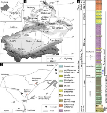
The specimens described as two new species (Omegops honggulelengensis and O. xiangi) in this study were collected from the bioclastic limestones and argillaceous limestones of the upper part of the lower member of the Hongguleleng Formation in the Buninuer and Bulongguoer sections, northern Hoxtolgay (Fig. 1). The stratigraphic extents of the two species are not exactly the same. Specifically, Omegops honggulelengensis also occurred in the middle member to top of this formation (from middle to top of the Famennian), but O. xiangi existed only in the upper part of lower member (middle Famennian) (Fig. 1.3). In addition to trilobites, abundant brachiopods, crinoids, corals, and bryozoans, smaller numbers of gastropods, bivalves, and cephalopods also occurred in the upper part of the lower member, deposited in a distal storm lithofacies sedimentary environment (Fan and Gong, Reference Fan and Gong2016). All trilobites were preserved three-dimensionally. Apart from some enrolled and articulated exoskeletons, which represent corpses, most trilobites were scattered sclerites. Some thoraces articulated with pygidia, but with cephala nearby, are identified here as exuviae (Zong et al., Reference Zong, Fan and Gong2016; Zong and Gong, Reference Zong and Gong2017). In other cases, the cephala, thoraces, and pygidia were separated from each other, including some isolated thoracic segments. This preservation indicates that the corpses and exuviae of trilobites might have been carried by seawater before they were ultimately buried.
(1, 2) Fossil locality maps of the Late Devonian Omegops in western Junggar, Xinjiang. (3) Stratigraphic column of the Hongguleleng Formation, stratigraphic distribution of Omegops, and the horizon of samples collected in this study (blue arrow). Conodont biozones after Suttner et al. (Reference Suttner, Kido, Chen, Mawson and Waters2014) and Zhang et al. (Reference Zhang, Over and Gong2021).

A total of 170 isolated cephala, 15 articulated cephala and thoraces, two enrolled exoskeletons, and nine exuviae consisting of articulated thoracopyga and isolated cephala were collected in this study. Among 196 cephala, 73 specimens had two intact schizochroal eyes, 62 specimens had only an intact left eye, and the other 61 specimens had only an intact right eye. All specimens were measured using vernier calipers or ImageJ software (Schneider et al., Reference Schneider, Rasband and Eliceiri2012), and the fossils were coated for photography with magnesium oxide; all photographs were captured using a Nikon D5100 camera with a Micro-Nikkor 55 mm F3.5 lens. The morphological terminology follows Whittington and Kelly (Reference Whittington, Kelly and Kaesler1997).
Repository and institutional abbreviation
BGEG = State Key Laboratory of Biogeology and Environmental Geology, China University of Geosciences (Wuhan).
Systematic paleontology
Order Phacopida Salter, Reference Salter1864
Superfamily Phacopoidea Hawle and Corda, Reference Hawle and Corda1847
Family Phacopidae Hawle and Corda, Reference Hawle and Corda1847
Subfamily Phacopinae Hawle and Corda, Reference Hawle and Corda1847
Genus Omegops Struve, Reference Struve1976
Type species
Calymene accipitrina Phillips, Reference Phillips1841
Omegops honggulelengensis new species
Figures 2
- Reference Xiang1981
Phacops (Omegops) accipitrinus mobilis Xiang, p. 186, pl. 2, figs. 1–10.
- Reference Zhang1983
Phacops accipitrinus mobilis; Zhang, p. 547, pl. 182, figs. 15–16.
- Reference Yuan and Xiang1998
Phacops (Phacops) circumspectans tuberculosus Yuan and Xiang, p. 29, pl. 1, fig. 3.
- Reference Yuan and Xiang1998
Phacops (Omegops) cornelius (Richter and Richter); Yuan and Xiang, p. 33, pl. 1, fig. 2.
- Reference Zong and Gong2017
Omegops cornelius (Richter and Richter); Zong and Gong, p. 159, fig. 1A–C.
- Reference Zong, Wang, Fan, Song, Zhang, Shen and Gong2020
Omegops sp.; Zong et al., p. 2470, fig. 7a–h.
- Reference Crônier and Waters2022
?Houseops cf. olonbulagensis Crônier in Crônier and Waters, fig. 6a–c.
- Reference Crônier and Waters2022
Undetermined phacopid; Crônier and Waters, fig. 6d.
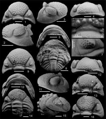
Omegops honggulelengensis n. sp. from the Upper Devonian Fammenian Hongguleleng Formation in western Junggar, NW China. (1–7) Cephalon (BGEG–HB–59): (1) dorsal view; (2) left side view; (3) right side view; (4) anterior view; (5) ventral view; (6, 7) close-ups of the intercalating ring (6) and postocular pad (7). (8) The exuvia consist of enrolled thoracopygon and isolated cephalon (BGEG–HB–55), ventral view of cephalon, and dorsal view of thoracopygon. (9–12) Cephalon (BGEG–HB–03): (9) dorsal view; (10) ventral view; (11) left side; (12) anterior view. (13) Incomplete cephalon (BGEG–HB–178), dorsal view; part of the shell was detached, revealing impressions of S2 and S3 (white arrows). (14, 15) Enrolled cephalon and thorax (BGEG–HB–05): (14) dorsal view; (15) right side view. (16) Cephalon (BGEG–HB–02), dorsal view. Specimen BGEG–HB–178 was collected from the Bulongguoer section; the other specimens were taken from the Buninuer section. Scale bars = 5 mm unless otherwise specified.

Holotype
Nearly complete cephalon, BGEG–HB–59 (Fig. 2.1– 2.7), from the upper part of the lower member of the Hongguleleng Formation (middle Famennian), Buninuer section, northern Hoxtolgay, western Junggar, Xinjiang, China.
Diagnosis
Species of Omegops with a broadly and evenly rounded anterior margin of the glabella. Glabellar axial furrows almost straight, posteriorly and medially divergent at 60–70°. The median part of the intercalating ring is barely visible, replaced by a pair of tubercles. Eyes moderately high with 45–49 lenses arranged in 15 dorsoventral files and with up to four lenses in one file. Occipital ring, postocular pad, and posterior border of cephalon are smooth. Thorax composed of 11 smooth thoracic segments. Pygidium with five pleural ribs, pygidial axis up to 85% of pygidial length, tapering posteriorly, with eight axial rings plus a short terminal piece. The surface of the pygidium is smooth.
Occurrence
Upper part of lower member to top of the Hongguleleng Formation (middle to top of the Famennian), Buninuer and Bulongguoer sections, northern Hoxtolgay, western Junggar, Xinjiang, China.
Description
Cephalon transverse, semicircular, ratio of length to width is 1:1.65–1.80 (Fig. 3.1; Supplemental file S1). Glabella subtrapezoid, slightly curved in front, and extending to the cephalic border (Fig. 2.1, 2.2, 2.4, 2.9, 2.11, 2.12); maximum glabellar width at the anterior termination of axial furrows. The width of cephalon is 1.60–1.75 times the maximum glabella width, while the latter is slightly larger than the length of the cephalon (Fig. 3.2; Supplemental file S1). The surface of the glabella is covered by irregularly arranged coarse tubercles that are larger and sparser in the glabella middle and rear and gradually become dense and smaller forward and to both sides (Fig. 2.1, 2.9, 2.16). S1 transverse, incised laterally, broadening and shallowing adaxially, and slightly upward sloping. S2 and S3 absent on the dorsal glabellar surface, but present faint impressions on the ventral side (Fig. 2.13); slightly arched, not connected in the middle part of glabella. Intercalating ring, transverse, weakly defined with a pair of smooth lateral tubercles (Fig. 2.6, 2.9, 2.16); the median part is barely visible, replaced by a pair of medium-sized tubercles and a few smaller tubercles (Fig. 2.6, 2.9). Occipital ring high, smooth, strongly vaulted transversely, slightly broadening adaxially (Fig. 2.9, 2.16). Posterior border curved posteriorly adaxially in dorsal view. Posterior border furrow gradually fading abaxially, curving along the rounded genal angles (Fig. 2.2, 2.11). Anterior border narrow, ridge-like, delineated by the narrow and shallow border furrow (Fig. 2.4, 2.11, 2.12). Axial furrows deep and broad, divergent at about 60–70°, almost straight posteriorly and medially. Palpebral furrows moderately deep, subparallel, very gently curved outward (Fig. 2.1). Palpebral lobes long, crescent in outline, distinctly raised above the palpebral area, with the outer rim of faint densely spaced fine tubercles. Palpebral area of fixigena moderately broad, covered by a few fine to medium-size tubercles (Fig. 2.1). Eyes large, kidney-shaped. Visual surface steeply falling down laterally, mostly consisting of 45–49 lenses arranged in 15 dorsoventral files and up to four lenses in one file; the most common pattern is 3/4/4/4/4/4/4/4/3/4/3/3/2/2/1. Postocular area of the fixigena slightly inflated, narrow adaxially widening abaxially with a smooth postocular pad (Fig. 2.7). Vincular furrow evenly deep, evenly curved. Middle part of postvincular doublure (Feist et al., Reference Feist, Mahboubi and Girard2016) almost flat, slightly inward curved on both sides, about 2.5 times as wide as the vincular furrow, ornamented with faint terrace lines and fine tubercles (Fig. 2.5, 2.10).
(1, 2) Omegops honggulelengensis n. sp.: (1) length versus width of cephalon; (2) length of cephalon versus maximum width of glabella. (3, 4) Omegops xiangi n. sp.: (3) length versus width of cephalon; (4) length of cephalon versus maximum width of glabella. From the Hongguleleng Formation in western Junggar, NW China.

Thorax composed of 11 smooth segments. Thoracic axial rings strongly arched, lacking lateral lobes (Fig. 2.14, 2.15), up to 36% as wide as whole segment in anterior segments and down to 32% in posterior segments. Anterior bands of thoracic pleurae narrow (exs. = exsagittal). Pleural furrow weakly defined abaxially, becoming prominent at the transition to a fulcrum, then gradually fading adaxially (Fig. 2.14). Posterior bands up to two times as wide as anterior bands.
Pygidium short, smooth, semielliptical; the ratio of length to width is 1:1.7–1.9. Pygidial axis conical, tapering posteriorly at 15–17°, almost 85% as long as pygidium and about 33% as wide as maximum pygidial width; eight axial rings plus a small terminal piece, separated by deep, almost transverse axial furrows. Axial furrows narrow and deep, moderately shallowing posteriorly. Pleural field gently vaulted, with five pleural ribs evenly bent posteriorly adaxially (Fig. 2.8). Three anterior pairs of the pleural furrows deep, posterior fourth pair significantly shallower. Pygidial border weakly defined without border furrow (Fig. 2.8).
Etymology
After Hongguleleng Formation, which yielded the holotype of O. honggulelengensis.
Materials
In addition to the holotype, assigned specimens including an exuvia consist of articulated thoracopygon and isolated cephalon (BGEG–HB–55), an articulated cephalon and thorax (BGEG–HB–05), and three cephala (BGEG–HB–03, BGEG–HB–178, BGEG–HB–02).
Remarks
This species was originally identified as a subspecies of Omegops accipitrinus, i.e., Omegops accipitrinus mobilis (=Phacops (Omegops) accipitrinus mobilis), by Xiang (Reference Xiang1981). However, many of its characteristics are not consistent with the Omegops accipitrinus group, specifically, the postocular pad of Omegops acc. accipitrinus (Phillips, Reference Phillips1841) is ornamented with eight large tubercles, the glabellar axial furrows are posteriorly and medially divergent at 45–60°, the visual surface consists of 56–70 lenses, and the pleural field has six pleural ribs (Struve, Reference Struve1976; Ghobadi Pour et al., Reference Ghobadi Pour, Popov, Omrani and Omrani2018). In Xinjiang specimens, the postocular pad is smooth (Fig. 2.7), the glabellar axial furrows are posteriorly and medially divergent at 60–70°, the visual surface consists of 45–49 lenses, and the pleural field has five pleural ribs (Fig. 2.8). In addition, the latter has a longer pygidial axis (85% versus 75% as long as pygidium). These characteristics distinguish it from other subspecies of Omegops accipitrinus (i.e., Omegops acc. maretiolensis (Richter and Richter, Reference Richter and Richter1933), Omegops acc. bergicus (Drevermann, Reference Drevermann1902), and Omegops acc. insolatus Struve, Reference Struve1976). Omegops honggulelengensis shows certain similarity to the Afghan Omegops paiensis Farsan, Reference Farsan1998 in cephalic and pygidial morphology, but the latter has 86 lenses on the visual surface with up to seven lenses in one file, which is obviously different from Xinjiang specimens. Although there is intraspecific variability in the number of eye lenses in some phacopid taxa (Crônier and Clarkson, Reference Crônier and Clarkson2001; Crônier et al., Reference Crônier, Feist and Auffray2004, Reference Crônier, Budil, Fatka and Laibl2015), among 86 specimens of Omegops honggulelengensis preserved with visual surface, the largest number of lenses is only 53, which is far fewer than the number of lenses in Omegops paiensis.
Omegops honggulelengensis can also be distinguished from Omegops cornelius (Richter and Richter, Reference Richter and Richter1933) by its smooth postocular pad, fewer eye lenses (45–49 versus 62), and five (not six) pleural ribs. Ghobadi Pour et al. (Reference Ghobadi Pour, Popov, Omrani and Omrani2018) named Omegops tilabadensis on the basis of the late Famennian specimens from northern Iran; O. honggulelengensis is very similar to it in terms of cephalic shape, number of eye lenses, and number of pleural ribs. The differences include the axial glabellar furrows diverge at an angle of about 60–70° (versus 40–45°), weak S2 and S3 impressions on the ventral side of the glabella, and a slightly shorter pygidial axis in O. honggulelengensis. In addition, the postocular pad is ornamented with eight tubercles, and the lateral lobes of the intercalating ring are ornamented with one pair of tubercles in Iranian specimens. However, the postocular pad and lateral lobes of O. honggulelengensis are smooth, and a pair of medium-sized tubercles and some smaller tubercles are arranged in the median part of the intercalating ring (Fig. 2.6). There are also differences between these two species in other ornamentations on the exoskeleton, i.e., the occipital ring, thoracic segments, pygidial axis, and pleural ribs of the Iranian specimens are covered with varisized tubercles, while these parts of the Xinjiang specimens are smooth (Fig. 2.1, 2.8, 2.14, 2.15). Xinjiang specimens have some similarity with Omegops acc. accipitrinus and Omegops acc. maretiolensis from Afghanistan reported by Farsan (Reference Farsan1998) in terms of the cephalic morphology, the number of eye lenses, and the number of pleural ribs. However, the latter two clearly do not belong to the Omegops accipitrinus group according to their number of eye lenses and pleural ribs, and Ghobadi Pour et al. (Reference Ghobadi Pour, Popov, Omrani and Omrani2018) questionably assigned them to Omegops tilabadensis.
Yuan and Xiang (Reference Yuan and Xiang1998) established a new subspecies of Phacops circumspectans, i.e., Phacops circumspectans tuberculosus, on the basis of an incomplete cephalon from the same horizon in the same region. This specimen is very similar to O. honggulelengensis in terms of cephalic and glabellar outlines, eye shape, and the decoration of the cephalon; the eye lens number is also within the variation range of O. honggulelengensis. The main difference lies in the fact that Yuan and Xiang (Reference Yuan and Xiang1998) believed that the former has a significant intercalating ring (especially the strongly convex median part). I reexamined the holotype of Phacops circumspectans tuberculosus and found that there was an extruded rupture from the posterior part of the glabella to the occipital ring. I concluded that the so-called convex median part was an artifact formed by the extruded part of the posterior part of the glabella. Hence, I classify this specimen as O. honggulelengensis here. In addition, Yuan and Xiang (Reference Yuan and Xiang1998) described an enrolled cephalon and thorax and an isolated weathered pygidium from the same horizon and ascribed them to Omegops cornelius. This pygidium has five pleural ribs that should not be ascribed to O. cornelius (Ghobadi Pour et al., Reference Ghobadi Pour, Popov, Omrani and Omrani2018); the first eight of the nine pygidial axes are clearer, but the ninth pygidial axis is poorly preserved, so it is difficult to exclude the artifact caused by weathering. The shape of this pygidium is very similar to the shape of my specimens, and I also assign it to O. honggulelengensis.
Crônier and Waters (Reference Crônier and Waters2022) described one fragmented cephalon and one mostly exfoliated thoracopygon from the same horizon (upper P. rhomboidea to P. marginifera conodont biozones, rather than the P. crepida biozone) at Aomage locality (very close to Buninuer section) and identified them as ?Houseops cf. H. olonbulagensis and undetermined phacopid, respectively. The ratio of length to width of the cephalon and morphology of the intercalating ring of the former and the number of pleural ribs and the smooth thoracopygon of the latter are very similar to O. honggulelengensis, and I hereby classify them as such.
Omegops xiangi new species
Figure 4
- Reference Yuan and Xiang1998
Phacops (Omegops) cornelius (Richter and Richter); Yuan and Xiang, p. 33, pl. 1, fig. 1.
- Reference Weber2000
Omegops sp.; Weber, p. 543, unfigured.
- Reference Zong, Fan and Gong2016
Omegops cornelius (Richter and Richter); Zong et al., p. 4, fig. 2 A–L.
- Reference Crônier and Waters2022
Clarksonops junggariensis Crônier; Crônier and Waters, fig. 3a–s.
Holotype
An exuvia consisting of articulated thoracopygon and isolated cephalon, BGEG–HB–04 (Fig. 4.1–4.4), from the upper part of the lower member of the Hongguleleng Formation (middle Famennian), Bulongguoer section, northern Hoxtolgay, western Junggar, Xinjiang, China.
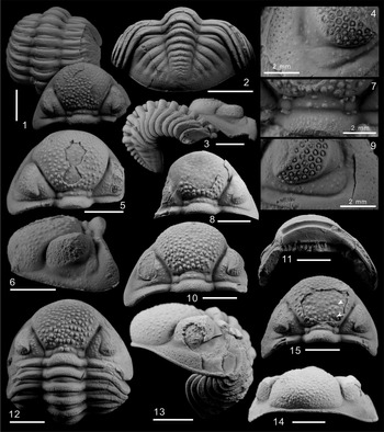
Omegops xiangi n. sp. from the Upper Devonian Famennian Hongguleleng Formation in western Junggar, NW China. (1–4) The exuvia consist of enrolled thoracopygon and isolated cephalon (BGEG–HB–04): (1, 3) dorsal and side views of exuvia; (2) dorsal view of pygidium and some thoracic segments; (4) close-up of left postocular pad. (5–7) Cephalon (BGEG–HB–49): (5) dorsal view; (6) side view; (7) close-up of intercalating ring. (8, 9) Incomplete cephalon (BGEG–HB–111): (8) dorsal view; (9) close-up of right postocular pad. (10, 11) Cephalon (BGEG–HB–32): (10) dorsal view; (11) ventral view. (12) Enrolled cephalon and thorax (BGEG–HB–68), dorsal view. (13, 14) Enrolled cephalon and thorax (BGEG–HB–16): (13) side view; (14) anterior view of cephalon. (15) Cephalon (BGEG–HB–86), dorsal view, part of the shell was detached, revealing impressions of S2 and S3 (white arrows). Specimen BGEG–HB–32 was collected from the Buninuer section; the other specimens were taken from the Bulongguoer section. Scale bars = 5 mm unless otherwise specified.

Diagnosis
Species of Omegops with a broadly and evenly rounded anterior margin of the glabella. Glabellar axial furrows almost straight, posteriorly and medially divergent at 45–55°. Intercalating ring is composed of the low median part and a pair of lateral lobes, and a pair of medium to large tubercles located on the median part. One medium-sized tubercle in the middle of S1. Occipital ring, postocular pad, and posterior border of cephalon are covered by fine tubercles. Eyes moderately high with 54–61 large lenses arranged in up to 15 dorsoventral files and with up to five lenses in one file. Thorax is composed of 11 segments ornamented with fine tubercles. Pygidium with six pleural ribs, pygidial axis up to 85% of pygidial length, tapering posteriorly, with eight axial rings plus a short terminal piece. The surface of pygidial axis and pleural ribs ornamented with fine tubercles.
Occurrence
Upper part of the lower member of the Hongguleleng Formation (middle Famennian), Buninuer and Bulongguoer sections, northern Hoxtolgay, western Junggar, Xinjiang, China.
Description
Cephalon transverse, semicircular, ratio of length to width is 1:1.65–1.75 (Fig. 3.3; Supplemental file S2). Glabella subtrapezoid, moderately vaulted, slightly curved in front, and extending to the cephalic border, maximum glabellar width at the anterior termination of axial furrows (Fig. 4.1, 4.5, 4.6, 4.10). The width of the cephalon is 1.71–1.81 times the maximum width of the glabella; the latter is slightly smaller than the length of the cephalon (Fig. 3.4; Supplemental file S2). The surface of the glabella is covered by irregularly arranged tubercles, which are composed of larger tubercles, and a few medium tubercles fill in the space among the larger ones (Fig. 4.1, 4.10, 4.12). S1 transverse, incised laterally, broadening and shallowing adaxially. S2 and S3 absent on the dorsal glabellar surface, but present clear impressions on the ventral side, straight but not connected in the middle part of glabella (Fig. 4.15). Intercalating ring, narrow, weakly defined with a pair of smooth lateral lobes and the low and flat median part (Fig. 4.1, 4.10); the median part ornamented with a pair of medium to large tubercles and a few smaller tubercles. A medium-sized tubercle near the intercalating ring in the middle of S1 (Fig. 4.7, 4.12). Occipital ring high, ornamented with dense fine tubercles (Fig. 4.1, 4.12). Posterior border transverse in the middle and curved posteriorly adaxially in dorsal view. Posterior border furrow narrow and deep, curving along the rounded genal angles (Fig. 4.6, 4.9). Anterior border narrow, ridge-like, delineated by the narrow and shallow border furrow (Fig. 4.13, 4.14). Axial furrows deep and broad, divergent at about 45–55°, straight posteriorly and medially. Palpebral furrows broad and shallow, very gently curved outward. Palpebral lobes crescentic in outline, distinctly raised above the palpebral area, with the outer rim of faint densely spaced fine tubercles. Palpebral area of fixigena moderately broad, slightly obliquely extends to posterior border furrow, covered by a few fine to medium tubercles (Fig. 4.1, 4.10, 4.12). Eyes large, kidney-shaped. Visual surface steeply inclined laterally, mostly consisting of 54–61 lenses arranged in 15 dorsoventral files, and up to five lenses in one file; the most common pattern is 4/5/4/5/4/5/4/4/4/4/3/3/3/3/2. Postocular area of the fixigena slightly inflated, narrow adaxially, widening abaxially with a distinct postocular pad covered with several small tubercles (Fig. 4.4, 4.9). Posterior border ornamented with dense fine tubercles (Fig. 4.6). Vincular furrow deep and broad, evenly curved. The middle part of postvincular doublure almost flat, slightly inward curved on both sides, about 2.5 times as wide as the vincular furrow, ornamented with faint terrace lines and fine tubercles (Fig. 4.11).
Thorax of 11 segments. Thoracic axial rings strongly arched, lacking lateral lobes (Fig. 4.1, 4.3, 4.12), up to 37% as wide as whole segment in anterior segments and down to 31% in posterior segments; ornamented by dense small tubercles (Fig. 4.1, 4.12). Anterior bands of thoracic pleurae narrow (exs.), smooth. Pleural furrow straight, slightly bend backward abaxially, becoming prominent at the transition to a fulcrum (Fig. 4.12). Posterior bands up to 2.5 times as wide as anterior bands, ornamented with fine tubercles, reducing adaxially and increasing abaxially (Fig. 4.12, 4.13).
Pygidium transverse, semioval; the ratio of length to width is 1:1.6–1.8. Pygidial axis conical, tapering posteriorly at 13–15°, almost 85% as long as pygidium and about 30% as wide as maximum pygidial width; eight axial rings plus a small terminal piece, separated by slightly arched axial furrows. Axial rings covered with small tubercles. Axial furrows wide and deep, moderately shallowing posteriorly (Fig. 4.2). Pleural field gently vaulted, with six pleural ribs evenly bent posteriorly adaxially. Middle part of the pleural ribs covered with small to medium tubercles gradually fading outward and inward (Fig. 4.2). Three anterior pairs of the pleural furrows deep, posterior two pairs significantly shallower. Pygidial border smooth, weakly defined without border furrow.
Etymology
For Chinese paleontologist Liwen Xiang, who first studied the trilobites from the Hongguleleng Formation.
Materials
In addition to the holotype, assigned specimens including two articulated cephala and thoraces (BGEG–HB–16, BGEG–HB–68) and four cephala (BGEG–HB–32, BGEG–HB–49, BGEG–HB–86, BGEG–HB–111).
Remarks
This species was originally classified as Omegops cornelius by Yuan and Xiang (Reference Yuan and Xiang1998) on the basis of an enrolled cephalon and thorax. More well-preserved specimens were obtained in this study, and Weber (Reference Weber2000) provided some key information on the cephalon based on a topotype of Omegops cornelius (the holotype and paratype specimens were lost in World War II), allowing the Xinjiang specimens to be compared in greater detail with Omegops cornelius. Although there are some similarities between them (Yuan and Xiang, Reference Yuan and Xiang1998), the differences are also obvious, reflected mainly in that the glabella of Xinjiang specimens is more convex, and the highest point is located in the middle of the glabella while the highest point of Omegops cornelius is located in the front of the intercalating ring (Weber, Reference Weber2000). The cephala of Xinjiang specimens are wider (length-to-width ratio is 1:1.65–1.75 versus 1:1.55–1.6), while the cephalon of Omegops cornelius is longer and narrower. The glabella of Xinjiang specimens are narrower (tr. = transverse) taking the width of the cephalon as reference (1:1.71–1.81 versus 1:1.5), and the maximum width of the glabella is slightly smaller than the cephalic length in Xinjiang specimens, while the maximum width of the glabella in Omegops cornelius is larger than the cephalic length (Weber, Reference Weber2000). Another difference is that the axial glabellar furrows diverge at an angle of about 45–55° in Xinjiang specimens versus 45° in Omegops cornelius. In addition, the postocular pad of Omegops cornelius is club-like and expands outward, with dense tubercles on its surface that contact each other abaxially (Weber, Reference Weber2000), while the Xinjiang specimens have a narrower, cudgel-shaped postocular pad with sparse tubercles on its surface that do not contact each other (Fig. 4.4, 4.9). The ornamentation of the intercalating ring is also different; in addition to two tubercles in the median part like Omegops cornelius, Xinjiang specimens have a medium tubercle near the intercalating ring in the middle of S1 (Fig. 4.7). Weber (Reference Weber2000) provided the arranged pattern of eye lens of the topotype of Omegops cornelius; it has 62 lenses on the visual surface arranged in 15 dorsoventral files and with up to five lenses in one file, i.e., 4/5/5/5/4/5/4/5/4/5/4/4/3/3/2. Although the eye lens numbers of Xinjiang specimens are similar, this arrangement is not seen in my collection of 110 specimens with well-preserved visual surface (Supplemental files S2, S4). On the basis of these differences, I believe that the Xinjiang specimens should not be attributed to Omegops cornelius.
Omegops xiangi and Omegops accipitrinus accipitrinus are very similar in terms of axial glabellar furrows divergent angle, the eye lens number and arrangement, and the number of pleural ribs. According to the revised and measured data by Richter and Richter (Reference Richter and Richter1933), the latter has wider cephalon and glabella, and the maximum width of the glabella is larger than the cephalic length, while the glabella of Omegops xiangi is more convex forward, and the maximum width is slightly less than the cephalic length (Fig. 3.4). In addition, the intercalating ring of Omegops acc. accipitrinus is weakly defined with only a pair of lateral lobes. However, in the Xinjiang specimens, the median part of the intercalating ring is low and flat, and in addition to a pair of tubercles on the median part, there is a tubercle in the middle of S1 on the anterior side of the intercalating ring, which is arranged in a triangle with the two tubercles on the median part (Fig. 4.7, 4.12). In terms of pygidial morphology, the pygidium of Omegops acc. accipitrinus is wider (tr.) (width-to-length ratio is 1.9–2.16:1 versus 1.6–1.8:1). There is also a difference in the decorations of the postocular pad, which is smooth adaxially in Omegops acc. accipitrinus and ornamented with several large tubercles abaxially, but for Xinjiang specimens, the surface of the postocular pad is covered by several sparse and evenly distributed small tubercles. The ornamentation of the postocular pad of Xinjiang specimens is similar to Omegops accipitrinus insolatus, but there are distinct differences in the glabellar width, the convex degree of the frontal glabellar lobe, and the shape of the intercalating ring and its decorations, which can also distinguish the Xinjiang specimens from other subspecies of Omegops accipitrinus.
Omegops xiangi can be easily distinguished from Afghan Omegops paiensis according to the number and arrangement of eye lenses and the number of pleural ribs (Farsan, Reference Farsan1998). It is obviously different from Omegops tilabadensis from Iran in terms of the shape of the intercalating ring and its decorations, the ornamentation of the postocular pad, and the axial glabellar furrows divergent angle (Ghobadi Pour et al., Reference Ghobadi Pour, Popov, Omrani and Omrani2018). Compared with Omegops honggulelengensis of the same area, the main differences are as follows: the glabella of the latter is slightly wider and the frontal glabellar lobe is less convex (the maximum width of the glabella is greater than the length of the cephalon), the median part of the intercalating ring is almost invisible, the number of eye lenses is between 45 and 49 (up to four lenses in one file), and the number of pleural ribs is five. By contrast, the maximum width of the glabella of Omegops xiangi is less than the cephalic length, and the intercalating ring can be divided into a pair of lateral lobes and a low and flat median part, the number of eye lenses is between 54 and 61 (up to five lenses in one file), and the number of pleural ribs is six. In addition, they are different in the ornamentation of the exoskeleton. The S1, postocular pad, occipital ring, posterior margin of cephalon, thoracic segment, pygidial axis, and pleural ribs are smooth in Omegops honggulelengensis, while those of O. xiangi are usually covered by varisized tubercles (Fig. 4.2, 4.4, 4.6, 4.7, 4.9, 4.12, 4.13).
Recently, Crônier and Waters (Reference Crônier and Waters2022) described phacopids from the same horizon (upper P. rhomboidea to P. marginifera conodont biozones, rather than the P. crepida Biozone, see the Geological setting and materials section) in the same area and established a new genus, Clarksonops (type species, Clarksonops junggariensis) on the basis of several fragmented and eroded specimens. They suggested that this new genus differs from genus Omegops or the Omegops specimens from the Honguleleng Formation mainly in that Omegops has a reduced intercalating ring and a distinct postocular pad. However, the character of the intercalating ring being divided into a median part and a pair of lateral lobes in Clarksonops is also present in some species of Omegops (e.g., O. cornelius Struve, Reference Struve1976), and the postocular pad is clearly visible in some specimens of Crônier and Waters (Reference Crônier and Waters2022, fig. 3e, i, o, p), so I do not think this new genus is sufficiently distinct from Omegops. Conversely, Clarksonops junggariensis is very similar to Omegops xiangi in terms of the length-to-width ratio of the cephalon, the shape and ornamentation of the intercalating ring, the arrangement of eye lenses, and the number of pygidial axial rings and pleural ribs (the number of pleural ribs is six instead of five, see Crônier and Waters, Reference Crônier and Waters2022, fig. 3r), so I think Clarksonops specimens from the Hongguleleng Formation may belong to Omegops xiangi. Weber (Reference Weber2000) described an incomplete cephalon from the top of the Famennian in the Kornelimunster near Aachen, Germany. The shape of this cephalon is similar to O. xiangi (e.g., the maximum width of the glabella is less than cephalic length); the number of eye lenses and their arrangement are also within the variation range of the latter, but since there is no attached figure, it is difficult to achieve a more detailed comparison, so I provisionally classify it as Omegops xiangi.
Diversity of eye lenses in Omegops
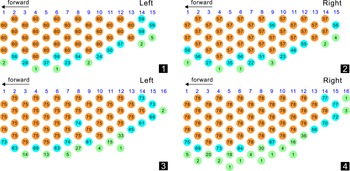
Of the 86 Omegops honggulelengensis specimens, 31 had both eyes, 29 had only an intact left eye, and 26 had only an intact right eye. The number of dorsoventral files on the visual surface of most specimens was 15, and a few specimens had only 14 or 13 files (Fig. 5.1, 5.2). The number of lenses in most files was up to four, but in a few specimens, that number reached five in the fourth and sixth files (Fig. 5.1, 5.2). For the total number of eye lenses, there were at least 40 and a maximum of 53; most specimens had lens numbers within the range of 45–49 (Fig. 6.1). Specimens with the same number of eye lenses often had diverse arrangement types (Supplemental file S3), but 49 eye lenses had only one pattern (i.e., 3/4/4/4/4/4/4/4/3/4/3/3/2/2/1), potentially representing the optimal arrangement of eye lenses in O. honggulelengensis. Another common arrangement type for eye lenses was 3/4/3/4/3/4/3/4/3/3/3/3/2/2/1 (45 lenses) (Supplemental file S3).
Schematic representations of eye lenses: (1, 2) Omegops honggulelengensis n. sp.; (3, 4) Omegops xiangi n. sp. From the Upper Devonian Hongguleleng Formation in western Junggar, NW China. The number in each circle represents the number of individuals carrying a lens in that position; brown indicates that all individuals had a lens in this position, light blue indicates that some individuals lacked a lens in this position, and light green indicates that some individuals had a new lens in this position.

The frequency distribution of eye lens numbers: (1) Omegops honggulelengensis n. sp.; (2) Omegops xiangi n. sp. From the Upper Devonian Hongguleleng Formation in western Junggar, NW China.

Among the 110 specimens of Omegops xiangi, 43 had both eyes well preserved, 32 had only an intact left eye, and 35 had only an intact right eye. Similar to O. honggulelengensis, the number of dorsoventral files on the visual surface of most O. xiangi specimens was 15, and a few specimens had 14 or 16 files (Fig. 5.3, 5.4). Most specimens had no more than five lenses in one file; however, a few specimens had up to six lenses in the second, fourth to sixth, and eighth files. The number of eye lenses varied greatly, ranging from 49 to 72, but most were concentrated between 54 and 61, and the numbers showed a normal distribution pattern (Fig. 6.2; Supplemental file S4). The most common eye-lens arrangement was 4/5/5/5/4/5/4/5/4/5/4/4/3/3/2 (57 lenses). Two additional arrangement patterns, 4/5/4/5/4/5/4/4/4/4/3/3/2/3/2 (56 lenses) and 4/5/4/5/4/5/4/4/4/4/3/4/3/3/2 (58 lenses), were also common (Supplemental file S4).
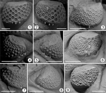
The diversity of eye lenses in Omegops is first reflected in the differences in the number of dorsoventral files. Although most of the Omegops specimens had 15 files, a few specimens (6/196) had one file more or one file fewer, and a very small number of specimens (1/196) had two files fewer (Fig. 7.8; Supplemental files S1, S2). Some specimens showed an increase or decrease in the number of eye lenses in each file (Figs. 5, 7.3, 7.7). The numbers of lenses on the ventral and posterior sides of the visual surface were most likely to change, but a few changes occurred on the dorsal side or in the middle part (Figs. 5, 7.2, 7.6). A small number of specimens also had irregular arrangements of lenses because of the increased number of lenses (Fig. 7.9). In some specimens with both eyes preserved, the numbers or arrangements of eye lenses in the left and right eyes were different (Fig. 7.1, 7.2, 7.4, 7.5; Supplemental files S1, S2).
Abnormal arrangement of eye lenses in the Late Devonian Omegops from western Junggar, Xinjiang. (1, 2) Omegops honggulelengensis (BGEG–HB–02): (1) left eye; (2) right eye. Lens number is asymmetrical in the seventh and ninth files. (3) Left eye of Omegops honggulelengensis (BGEG–HB–123). Lens number is up to five in the sixth file. (4, 5) Omegops xiangi (BGEG–HB–157): (4) left eye; (5) right eye. There are two lenses in the fourteenth and fifteenth files in the left eye, whereas there are three lenses and one lens in the same positions of the right eye. (6) Left eye of Omegops xiangi (BGEG–HB–34), missing one lens each in the seventh and eighth files. (7) Right eye of Omegops xiangi (BGEG–HB–34). The maximum number of lenses reached six in the eighth file. (8) Right eye of Omegops xiangi (BGEG–HB–140) with 16 vertical files. (9) Left eye of Omegops xiangi (BGEG–HB–04). The lenses of the tenth to twelfth files are irregularly arranged. The numbers in the figure represent the numbers of dorsoventral files. Scale bars = 2 mm.

Discussion
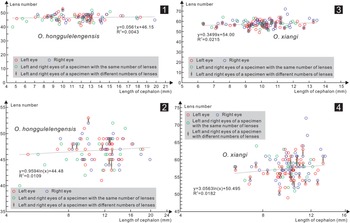
The diversity of eye lenses in the suborder Phacopina has been identified in several genera, and individual growth and development have been interpreted as an important reason for this diversity (Thomas, Reference Thomas1998; Crônier and Clarkson, Reference Crônier and Clarkson2001). For these specimens from western Junggar, most with only cephala preserved, it is difficult to distinguish between the meraspid and holaspid stages, but the cephalic size reflects the specimen's growth process. The eye-lens numbers of two new Omegops did not exhibit a distinctly linear change with the increase of cephalic length (Fig. 8). Therefore, the diversity of eye lenses of Omegops was likely not caused by individual growth. My material also reflects that the number of lenses in Omegops was likely fixed during certain ontogenetic stages and did not increase with the increase of body size.
Plot of eye lens number versus cephalic length. (1, 2) Omegops honggulelengensis n. sp. (3, 4) Omegops xiangi n. sp. From the Upper Devonian Hongguleleng Formation in western Junggar, NW China.

Selwood and Burton (Reference Selwood, Burton and Westermann1969) cited differences in the number of eye lenses as a distinguishing feature of sexual dimorphism in the Devonian trilobite Phacops schlotheimi schlotheimi (Bronn, Reference Bronn1825) but also listed other differences in the exoskeleton. However, the sexual dimorphism of trilobites is often considered as difference in a single feature (Whittington, Reference Whittington and Kaesler1997), and those differences enumerated by Selwood and Burton (Reference Selwood, Burton and Westermann1969) are more likely to represent different species or subspecies (Ramskold and Werdelin, Reference Ramsköld and Werdelin1991). Aside from the differences in the number of eye lenses, other characteristics of Omegops from Xinjiang are consistent. If the number of eye lenses were a secondary sexual characteristic, the eye lens number would show two similar arrangements or a bimodal pattern (considering the numbers of females and males in the same population are likely to be similar), with no or almost no intermediate transitional form. However, the numbers of eye lenses of Omegops are generally close to a normal distribution. In addition, the lens number and/or arrangement differed between eyes in the same individual in some specimens; therefore, sexual dimorphism cannot explain the eye-lens diversity.
I propose that the lens diversity of Omegops in western Junggar is likely comparable to other Phacopina, reflecting intraspecies variation (Crônier et al., Reference Crônier, Feist and Auffray2004, Reference Crônier, Budil, Fatka and Laibl2015), especially with an approximate normal distribution of the eye-lens number (Fig. 6). The causes of intraspecific variation are generally considered to be related to age or the living environment; that is, the number of lenses of specimens of the same species would change with evolution in different horizons or with different living environment (Crônier et al., Reference Crônier, Feist and Auffray2004): the lens numbers of phacopids that lived in deep-water environments were found to be lower than those of phacopids that lived in shallow-water environments (Feist et al., Reference Feist, McNamara, Crônier and Lerosey-Aubril2009). In the Hongguleleng Formation, the water gradually deepened from the lower to the middle member. However, trilobites in the middle member are all flattened molds without well-preserved eyes. It is therefore not clear whether evolution and water depth affected the lens numbers of Omegops. The material presented here was collected from the same horizon in two proximal sites with a consistent sedimentary environment, and the intraspecific diversity of eye lenses cannot be explained by evolution or differences in the water environment.
Considering the different numbers and arrangements of eye lenses in the left and right eyes, the increase or decrease in the number of dorsoventral files, abnormal arrangements of lenses, and the missing lenses in the middle part of the visual surface in some specimens from Xinjiang (Figs. 5, 7), I propose that eye-lens diversity likely represents malformed specimens. Malformations are not rare in trilobites but consist primarily of body segments: shortened, missing, abnormally fused, or bifurcated. Such malformation may have been caused by predatory attacks, unsuccessful molting, genetic or embryological malfunctions, or pathological conditions (Owen, Reference Owen1985; Babcock, Reference Babcock1993). More recently, abnormal ornamentations on exoskeletons have been identified as minor malformations that are believed to have been caused by developmental disorders (Bicknell and Smith, Reference Bicknell and Smith2021). There are few reported cases of ocular malformation (Fatka et al., Reference Fatka, Budil and Zicha2021 and their references), and the numbers and arrangements of eye lenses are affected in some malformed specimens (Schoenemann et al., Reference Schoenemann, Clarkson and Høyberget2017; Fatka et al., Reference Fatka, Budil and Zicha2021). However, in contrast to the Xinjiang specimens reported here, in addition to disturbance of the numbers or arrangements of lenses, the previously reported specimens had healed wounds on or near the visual surface, or the eyes became significantly smaller, which were interpreted as injuries caused by molting or predatory attacks (Schoenemann et al., Reference Schoenemann, Clarkson and Høyberget2017; Fatka et al., Reference Fatka, Budil and Zicha2021). In these Omegops specimens, no injuries were identified on the visual surfaces except for minor abnormalities such as missing or newly added eye lenses. Therefore, injuries can be excluded as an explanation, but the abnormalities may have been related to genetic or embryological malfunctions and/or pathological conditions (Owen, Reference Owen1985; Babcock, Reference Babcock1993). An interesting phenomenon is that there were no visible deformities on the exoskeletons of these trilobites; this may have been related to the higher sensitivity of eyes to environmental factors compared with other organs as the eyes are an important visual system of arthropods.
Eye-lens diversity also occurred in a variety of other members of the Phacopina besides Omegops from western Junggar (e.g., Lorenz, Reference Lorenz1991; Thomas, Reference Thomas1998; Crônier and Clarkson, Reference Crônier and Clarkson2001; Crônier et al., Reference Crônier, Feist and Auffray2004, Reference Crônier, Budil, Fatka and Laibl2015; Rustán and Balseiro, Reference Rustán and Balseiro2016). Even in the systematic classification of Phacopina, it was possible to identify the diversity of eye lenses in a few specimens (Zhou and Campbell, Reference Zhou and Campbell1990; McKellar and Chatterton, Reference McKellar and Chatterton2009). Thus, in taxonomic study, a large number of specimens is needed to obtain quantitative study on the numbers and arrangements of eye lenses in the Phacopina. Using only a single specimen or a small number of specimens may lead to great errors.
Conclusions
On the basis of 196 well-preserved new specimens, Famennian (Late Devonian) Omegops accipitrinus mobilis, Phacops circumspectans tuberculosus, and Omegops cornelius from western Junggar, Xinjiang, were reclassified as two new species of Omegops: O. honggulelengensis and O. xiangi. The former is distinguished from other species of Omegops by its wider axial glabellar furrows divergent angle, smooth postocular pads, 45–49 eye lenses, and five pleural ribs. The main distinguishing features of the latter are the maximum width of the glabella less than the cephalic length, 54–61 eye lenses, distinctive ornamentation of the intercalating ring and postocular pads, and six pleural ribs. Their eye lenses had considerable diversity; the total numbers presented approximate normal distribution patterns, with variations characterized mainly by increases or decreases in the number of files, asymmetrical distribution of the lenses in the left and right eyes, abnormal arrangements, or missing lenses in the middle part of the visual surface. These variations could have been caused by genetic or embryological malfunctions and/or pathological conditions rather than ontogeny, sexual dimorphism, evolution, or the water environment. My data also indicate that the eye-lens number did not increase with the increase of body size during certain growth stages of Omegops.
Acknowledgments
I appreciate much the constructive and critical comments from two anonymous reviewers, which aided in the further improvement of the manuscript. I thank Y.M. Gong, Z.H. Wang, R.Y. Fan, J.J. Song, Z. Shen, and X.S. Zhang for their help in the fieldwork. Thanks to D.J. Yuan and J.J. Song (NIGPAS) for their assistance when I observed the phacopids from the Hongguleleng Formation, which are housed in the Nanjing Institute of Geology and Palaeontology, Chinese Academy of Sciences. This work was supported by the National Natural Science Foundation of China (no. 42072041, 41702006).
Declaration of competing interests
The author declares none.
Data availability statement
Data available from the Dryad Digital Repository: https://doi.org/10.5061/dryad.sxksn037q.