Pompe disease is an autosomal recessive disorder caused by mutations in the acid alpha-glucosidase gene which leads to deficiency of the lysosomal acid alpha-glucosidase. This enzyme deficiency causes impaired glycogen degradation and lysosomal glycogen accumulation in the skeletal and cardiac muscle cells. Infantile-onset Pompe disease is characterised by respiratory insufficiency, hypertrophic cardiomyopathy, and generalised myopathy and hypotonia. Depending on residual enzyme activity, the disease manifests soon after birth or during the first months of life.Reference Owens, Wong, Bhattacharya and Ellaway1, Reference Löscher, Huemer and Stulnig2
Dilated cardiomyopathy is an extremely rare and curable complication of nutritional hypocalcaemic rickets. This form of the disease has better prognosis compared to idiopathic dilated cardiomyopathy.Reference Brown, Nunez, Russell and Spurney3, Reference Verma, Khadwal, Chopra, Rohit and Singhi4
In this article, we present the occurrence of dilated cardiomyopathy secondary to nutritional hypocalcaemic rickets and hypertrophic cardiomyopathy in an infant with infantile-onset Pompe disease.
Case report
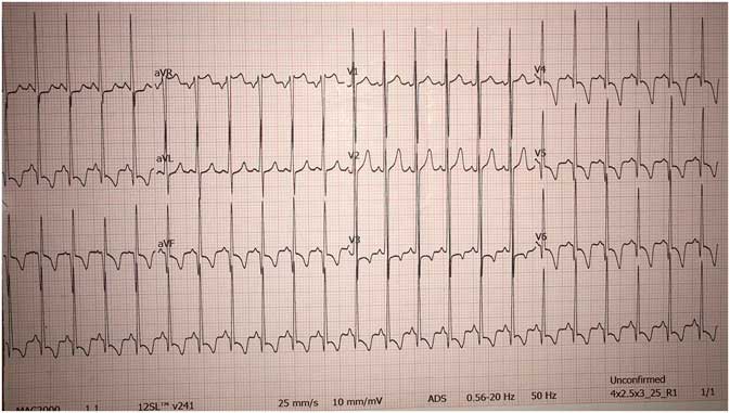
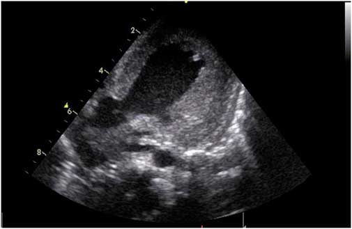
A 3-month-old female infant of a first cousin marriage was referred to our paediatric ICU with a 4-day history of fatigue, tachypnoea, tachycardia, hypoxia, and respiratory failure. She was born at 38 weeks of gestation, weighing 3580 g. Her mother had not taken any vitamin supplementation during the pregnancy. The patient did not have any vitamin D supplementation and complained about becoming tired when she was sucking during the past 15 days. On admission, she was lethargic (Glasgow Coma Scale: 12) and mildly hypotonic. She had subcostal and intercostal retractions and tachypnoea (68 breaths/minute). Oxygen saturation was 86% on room air and 95% under respiratory support given with a non-rebreathing reservoir mask. She had rales over the lung bases and the liver was palpable 5 cm below the costal margin. Non-invasive ventilation support with high-flow nasal cannula oxygen therapy was started soon after admission. She was tachycardic (182 beats/minute), hypotensive (68/34 mmHg), and her capillary refill time was 4 seconds. Electrocardiography revealed sinus tachycardia with a heart rate of 140/dk, left ventricle hypertrophy, and a P-R interval duration of 0.8 seconds (Fig 1). Chest radiography revealed an enlarged cardiac silhouette, with a cardiothoracic index of 0.64. Laboratory findings showed metabolic acidosis with high levels of lactate (5.2 mmol/L). Complete blood count, procalcitonin, and C-reactive protein levels were within normal ranges. Serum calcium was 7.9 mg/dL, albumin was 4.1 mg/dL, ionised calcium was 0.6 mmol/L (normal: 1.2–1.38 mmol/L), phosphorus was 2.9 mg/dL (normal: 4.5–6.7 mg/dL), alkaline phosphatase was 597 U/L (normal: 150–420 U/L), parathyroid hormone was 214 pg/mL (normal: 15–65 pg/mL), 25-hydroxy vitamin D was 3 ng/mL (normal: 17–54 ng/mL), troponin I was 0.375 ng/mL (normal: 0–0.16 ng/mL), and creatine kinase-MB was 39.8 U/L (0–25 U/L). Echocardiography revealed left ventricular enlargement and systolic dysfunction with left ventricular ejection fraction of 25%. In addition, the wall of both ventricles was thickened. The patient was diagnosed with nutritional hypocalcaemic rickets-related dilated cardiomyopathy and further investigations were planned for probable concomitant metabolic disorders. Medications for heart failure like adrenaline, milrinone, levosimendan, and furosemide were given at different times when required. In addition to these therapies, aspirin (4 mg/kg), vitamin D (intramuscular 75,000 IU), and intravenous calcium (50 mg/kg elemental calcium) treatments were started. On the fourth day, respiratory distress worsened, she was intubated, and invasive mechanical ventilation support was initiated. On the seventh day, a second dose of vitamin D (intramuscular 75,000 IU) was administered and intravenous calcium therapy was changed to enteral calcium lactate (75 mg/kg). Three days after administration of second vitamin D dose, echocardiography revealed elevated left ventricle systolic functions compared with initial echocardiography before Vitamin D supplementation and severe hypertrophy of both ventricles with diastolic dysfunction (Fig 2). On the 15th day of admission, lysosomal enzyme activity of the acid alpha-glucosidase was very low at 0.2 µmol/L/hour – normal >3.3 µmol/L/hour – confirming infantile-onset Pompe disease. With low enzyme activity, hypotonia, hepatomegaly, and hypertrophic cardiomyopathy, the patient was diagnosed with infantile-onset Pompe disease. Afterwards, homozygous c.896T >C (p.Leu299Pro) missense mutation was detected. The formal report procedure was initiated for obtaining the recombinant human acid alpha-glucosidase enzyme from abroad but the patient died because of sepsis and cardiorespiratory failure on the 25th day of admission.
Electrocardiography view of the patient.

Two-dimensional echocardiographic view of the patient revealing severe left ventricular hypertrophy.

Discussion
Calcium ions play an essential role in excitation and contraction of the cardiac muscles. Low serum levels of calcium secondary to nutritional hypocalcaemic rickets may affect ventricular contraction and cause dilated cardiomyopathy. Many case reports of nutritional hypocalcaemic rickets-related reversible dilated cardiomyopathy in childhood have been published in the current literature. Nutritional hypocalcaemic rickets-related dilated cardiomyopathy responds well to treatment and heart failure is curable with early diagnosis.Reference Brown, Nunez, Russell and Spurney3–Reference Olgun, Ceviz and Ozkan6 Brown et alReference Brown, Nunez, Russell and Spurney3 and Yilmaz et alReference Yilmaz, Olgun and Ciftel5 have reported patients with dilated cardiomyopathy and congestive heart failure associated with nutritional hypocalcaemic rickets. In both studies, the authors highlighted that all patients responded well to anti-congestive and inotropic agents as well as vitamin D supplementation with elementary calcium.Reference Brown, Nunez, Russell and Spurney3, Reference Yilmaz, Olgun and Ciftel5 In our patient, after administering elementary calcium and high dose intramuscular vitamin D, echocardiography revealed impaired left ventricle systolic functions and severe hypertrophy of the ventricles with diastolic dysfunction.
Infantile-onset Pompe disease is associated with a wide spectrum of non-specific clinical findings such as hypertrophic cardiomyopathy, muscle weakness, respiratory insufficiency secondary to hypotonia, aspiration, and pneumonia. The diagnosis is frequently made by 6 months of age. Without treatment, death generally occurs within the first 2 years of life, particularly as a result of cardiorespiratory failure.Reference Owens, Wong, Bhattacharya and Ellaway1, Reference Löscher, Huemer and Stulnig2, Reference Al-Hassnan, Khalifa and Bubshait7, Reference Felice8 Severe concentric hypertrophy, as in our patient, can also be seen affecting the septum, the right and the left ventricle.Reference Owens, Wong, Bhattacharya and Ellaway1, Reference Löscher, Huemer and Stulnig2, Reference Felice8 In studies and case reports, it has been reported that enzyme replacement therapy with recombinant human acid alpha-glucosidase prevents lysosomal glycogen accumulation and markedly improve clinical outcomes including motor development, ventilator-free survival, and hypertrophic cardiomyopathy in patients with infantile-onset Pompe disease.Reference Owens, Wong, Bhattacharya and Ellaway1, Reference Nicolino, Byrne and Wraith9–Reference Kishnani, Corzo and Leslie11 In our patient, the diagnosis was made when she was 3 months old. We believe that nutritional hypocalcaemic rickets masked the underlying infantile-onset Pompe disease by causing left ventricular enlargement and systolic dysfunction with mimicking dilated cardiomyopathy, and that administering high dose vitamin D and elementary calcium unmasked the underlying infantile-onset Pompe disease. Unfortunately, we were not able to administer enzyme replacement therapy to our patient and she died secondary to sepsis and cardiorespiratory failure.
In conclusion, nutritional hypocalcaemic rickets and infantile-onset Pompe disease must always be kept in mind among the causes of concomitant dilated cardiomyopathy and hypertrophic cardiomyopathy. If they are diagnosed and treated in time, dilated cardiomyopathy secondary to nutritional hypocalcaemic rickets and hypertrophic cardiomyopathy secondary to infantile-onset Pompe disease respond well to treatment.
Acknowledgements
None.
Financial Support
This research received no specific grant from any funding agency, or from commercial or not-for-profit sectors.
Conflicts of Interest
None.