Introduction
Percutaneous device closure is frequently undertaken for congenital and surgically placed interatrial communications. Reference Farb, Ibrahim and Zuckerman1–Reference Sarioglu, Sisli, Yalcinbas and Saygili3 Congenital tunnel-type interatrial communications are rare. Reference Torigoe, Sato and Sanada4–Reference Mahmoud, Hosny, Philip and Wagdy8 This report describes percutaneous device closure of a post-operative, iatrogenic tunnel-type interatrial communications that occurred during attempts to surgically close a residual atrial septal defect.
Case report
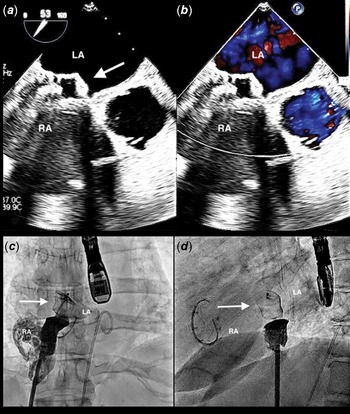
A 50-year-old woman, with a history of an atrial septal defect surgery at 11 months old, was diagnosed with an ascending aortic aneurysm. A presurgical transesophageal echocardiogram at another institution showed a residual atrial septal defect. Surgical resection of the aortic aneurysm and closure of the residual atrial septal defect were performed. The technique included placing cannulae in the superior and inferior vena cavae. Caval tapes were placed around the superior and inferior vena cavae. The old patch in the atrial septum was identified. The patch was imbricated with a double layer of running prolene suture. Immediately after separation from bypass, the patient had severe cyanosis and moderate-to-severe tricuspid regurgitation. The next day, she underwent a reoperation for atrial septal defect closure with a bovine pericardial patch and a tricuspid valve annuloplasty. The pericardial patch was large and was secured with running prolene suture from the rim of the coronary sinus to the entrance of the inferior vena cava, up the right atrial wall and then around the entrance of the superior vena cava. She continued to have moderate cyanosis and was transferred to our institution. After general anaesthesia, a transesophageal echocardiogram showed an interatrial tunnel-type communication with a diameter of 9–11 mm, coupled with a right-to-left shunt (Fig 1a and b). Cardiac catheterisation revealed a right atrial mean pressure of 12 mmHg, a left atrial mean pressure of 9 mmHg, a left upper pulmonary vein saturation of 97%, and an aortic saturation of 85%. There was a tunnel originating low in the right atrium and coursing cranially to the left atrium (Fig 1c and d). Balloon sizing of the tunnel demonstrated a diameter of 11–13 mm (Fig 1e and f) and a length of 2.5 cm. Echocardiographic images showed that the tunnel was not connected to the coronary sinus. Temporary balloon occlusion of the tunnel for diameter and distensibility assessment resulted in an increase in aortic saturation to 92–93%. The data were reviewed at a multidisciplinary conference, and the group recommended device closure. At repeat cardiac catheterisation, an 8-French delivery sheath was placed in the femoral vein and across the tunnel into the left atrium. With the aid of simultaneous transesophageal echocardiographic imaging, a 16-mm Amplatzer Vascular Plug II was delivered inside the tunnel. Following a gentle push–pull manoeuvre to show stability, the device was released. At the end of the catheterisation procedure, there was no residual right-to-left shunt by colour flow Doppler and right atrial angiography (Fig 2), and the aortic saturation was 92%. The patient received 3 doses of Ancef over 24 hours. She was discharged on daily aspirin (325 mg). In a 3-month follow-up clinic visit, she was doing well with an oxygen saturation of 92%.
(a and b) Transesophageal images of the right atrial to left atrial tunnel: (a) Relatively circular tunnel on the left side of the atrial septum (arrow). (b) Longitudinal view of the tunnel with right to left flow (asterisks). (c and d) Angiographic views of the tunnel: (c) AP view. (d) Lateral view. LA = left atrium, RA = right atrium, T = tunnel, arrow. (e and f) Balloon sizing of the tunnel: (e) AP view. (f) Lateral view.

(a and b) Transesophageal echocardiographic images showing the device inside the tunnel: (a) 2D imaging. (b) Colour flow. The device (arrow) is well seated inside the tunnel. There is no residual flow. (c and d) Proximal tunnel angiogram after device deployment: (c) AP view. (d) Lateral view. The device (arrow) is in good position. There is no residual right to left flow.

Discussion
Congenital interatrial tunnel-type communications are rare. Torigoe and colleagues described an extracardiac interatrial tunnel in conjunction with an atrial septal defect. Reference Torigoe, Sato and Sanada4 The tunnel drained from the left atrium anteriorly to the right atrium. A septal occluder was used to close both the atrial septal defect and the extracardiac tunnel. Reference Torigoe, Sato and Sanada4 Waldman and colleagues described a tunnel structure connecting the right atrium to the left atrial appendage discovered at autopsy in a 1.8-kg dysmorphic premature infant with a 10q26 chromosomal abnormality. Reference Waldman, McFeeley and Bornikova5 Kanadasia and colleagues described an interatrial venous channel in a patient with a secundum atrial septal defect. Reference Kanadaşı, Özbarlas, Demirtaş, Usal and Akpınar6 Further, Lyon and Hoyer described a left atrial appendage-to-right atrial appendage fistula (with tunnel-type communication characteristics) occurring with juxtaposed atrial appendages. Reference Lyon and Hoyer7 In this case, the unusual communication was closed with a 20-mm Amplatzer Vascular Plug II (St. Jude Medical, St. Paul, MN). Reference Lyon and Hoyer7 Also, Mahmoud and colleagues reported a tunnel-type connection in a patient with cortriatriatum. Reference Mahmoud, Hosny, Philip and Wagdy8 The tunnel connected the right atrium with the antero-inferior chamber in the left atrium below the cortriatriatum membrane.
A patent foramen ovale may have tunnel characteristics when associated with significant length. The patent foramen ovale tunnel originates in the fossa ovalis. In our case, the right atrial origin of the tunnel was inferior suggesting a low secundum atrial septal defect or an inferior vena caval-type sinus venous atrial septal defect that had undergone several attempts at surgical closure. The mechanism for the tunnel’s creation was unclear. We speculate that the surgical patch missed the lower end of the defect or possibly the tie around the inferior vena cava cannula may have covered the lower end of the defect, resulting in a defect from low right atrium communicating more cranially into the left atrium.
As the patient had undergone two previous surgical procedures and had a persistent significant right-to-left shunt, we opted for transcatheter closure. We considered using an atrial septal occluder and opening the left atrial disc in the left atrium, pulling to reduce the length of the tunnel, and placing the right-sided disc entirely on the right atrial side of the tunnel. However, we concluded that the tunnel length of at least 2.5 cm precluded this approach. Rather, as the tunnel lacked significant distensibility noted during balloon sizing, we selected an Amplatzer Vascular Plug II for closure. Prior to and following device placement, we carefully confirmed by echocardiography the patency of the coronary sinus.
In conclusion, we describe successful closure of an iatrogenic interatrial tunnel with an Amplatzer Vascular Plug II.
Acknowledgements
The authors acknowledge the editorial work of Dr. Humberto Restrepo.
Financial support
This research received no specific grant from any funding agency, commercial, or not-for-profit sectors.
Competing interests
None.
Ethical standard
The authors assert that this work is exempt of Institutional Review Board for informed consent and approval.