Introduction
Laryngeal squamous cell carcinoma is a common malignant tumour of the head and neck region.Reference Chu and Kim 1 In addition to invasion and metastasis, deregulation of cell proliferation and apoptosis underlies the development of squamous cell cancer in the head and neck.Reference Tímár, Csuka, Remenár, Répássy and Kásler 2 However, the pathogenetic mechanisms that regulate these biological characteristics of laryngeal squamous cell carcinoma cells are still not well known. The identification of potential biomarkers which could reflect the prognosis of laryngeal cancer is currently a popular research topic.
Focal adhesion kinase is a non-receptor tyrosine kinase which is located at focal contact sites where the cell interacts with the extracellular matrix. The 125 kDa focal adhesion kinase is encoded by the focal adhesion kinase gene located on human chromosome 8q24, and its activity is stimulated in response to extracellular ligands. Focal adhesion kinase is now known to be a key molecular integrator of multiple cues within the extracellular milieu, which modulates cell adhesion, angiogenesis and migration.Reference Golubovskaya 3 Previous studies have demonstrated that focal adhesion kinase is involved in regulating cellular proliferation in several different types of tumours.Reference Wang, Ma, Yang, Zhang, Wang and Liu 4 – Reference Huang, Wan, Liao, Hu, Wei and Wang 6 Other authors have found that focal adhesion kinase influences cell apoptosis in a variety of tumours.Reference Wang, Ma, Yang, Zhang, Wang and Liu 4 , Reference Lei, Ye, Yang, Wang, Jiang and Jiang 5 , Reference Xia, Lv, Hong, Li, Tao and Chen 7 , Reference Beierle, Ma, Trujillo, Kurenova, Cance and Golubovskaya 8 To the best of our knowledge, no previous studies have assessed the in vivo relationship between focal adhesion kinase expression and cellular proliferation and apoptosis within laryngeal squamous cell carcinoma.
Considering its biological features, focal adhesion kinase could be regarded as a potential target for cancer therapy.Reference Golubovskaya 3 Accordingly, several clinical trials have explored its influence on the prognosis of various human malignancies, including gastric carcinoma, hepatocellular carcinoma and extrahepatic bile duct carcinoma.Reference Park, Lee, Yoon, Kim, Kim and Yang 9 – Reference Hayashi, Aishima, Inoue, Nakata, Morimatsu and Nagai 11 Nevertheless, according to the recent literature, only a small number of in vivo studies have focused on the prognostic value of focal adhesion kinase in human samples of laryngeal cancer.Reference Canel, Secades, Rodrigo, Cabanillas, Herrero and Suarez 12 – Reference Rodrigo, Dominguez, Suárez, Canel, Secades and Chiara 14 Moreover, the correlation between focal adhesion kinase expression and local lymph node metastasis in patients with laryngeal squamous cell carcinoma is still a controversial issue.Reference Canel, Secades, Rodrigo, Cabanillas, Herrero and Suarez 12 , Reference Rodrigo, Cabanillas, Secades, Canel, Chiara and Suárez 13
In the current study, we examined the expression of focal adhesion kinase in 86 cases of laryngeal squamous cell carcinoma, by immunohistochemistry, and evaluated its association with clinicopathological parameters, apoptotic and proliferative indices, and overall survival.
Materials and methods
Patients and tissue samples
This study included 86 patients who underwent surgery for laryngeal squamous cell carcinoma at the department of otolaryngology, head and neck surgery at Shanghai Jiaotong University Affiliated First People's Hospital between January 1997 and December 2008. Informed consent was obtained from either the patients or their family.
The patients consisted of 81 men and 5 women, with ages ranging from 37 to 84 years (median age, 51 years). None of the patients had undergone radiation or chemotherapy prior to surgery.
All the histopathological samples used in this study were diagnosed as laryngeal squamous cell carcinoma by experienced pathologists.
Samples were classified based on the anatomical site of the primary tumour; there were 18 cases of supraglottic laryngeal carcinoma, 66 cases of glottic carcinoma and 2 cases of subglottic carcinoma.
Cancers were staged after surgical resection of the primary tumour, according to the 2002 tumour-node-metastasis (TNM) classification of the International Union Against Cancer. There were 26 (30 per cent) cases in stage I, 37 (43 per cent) in stage II, 14 (16 per cent) in stage III and 9 (11 per cent) in stage IV. Cases were divided into two groups: those in stages I to II and those in stages III to IV.
Cases were also graded histologically according to the degree of differentiation of the tumour. There were 29 well differentiated cases (grade I), 44 moderately differentiated cases (grade II), and 13 poorly differentiated cases (grade III).
In addition, normal laryngeal mucosa was collected from 32 patients without cancer, after obtaining informed consent.
The study protocol was approved by the hospital ethics committee.
Immunohistochemistry
Paraffin-embedded tumour tissue specimens were obtained from the pathology department archives. Tissue sections (5 µm) were deparaffinised in xylene and rehydrated through a graded series of ethanol solutions. After rinsing in phosphate-buffered saline, tissue sections were heated in 0.01 M sodium citrate buffer (10 mmol/l, pH 6.0) for 18 minutes at 500 W in a microwave oven, and then cooled to room temperature for antigen retrieval. Next, slides were rinsed in phosphate-buffered saline and endogenous peroxidase activity was blocked using 3 per cent hydrogen peroxide in methanol for 10 minutes. To reduce non-specific staining, the sections were incubated in 10 per cent normal goat serum for 30 minutes.
Tissue sections were then incubated with one of the following primary antibodies: either rabbit anti focal adhesion kinase polyclonal antibody (Epitomics, Burlingame, California, USA) at 1:400 dilution overnight at 4°C; or rabbit anti-Ki-67 polyclonal antibody (Boster, Wuhan, China) at 1:800 dilution overnight at 4°C.
Immunohistochemical staining was carried out by a two-step immunohistochemical procedure using the Dako EnVision+ System (Dako, Carpinteria, California, USA), following the manufacturer's instructions. After washing with water, nuclei within the sections were counterstained with haematoxylin.
Negative controls were processed in the same manner, using phosphate-buffered saline in place of the primary antibody.
Apoptosis detection
The apoptotic index was determined by analysing DNA fragmentation in the cell nuclei using the terminal deoxynucleotidyl transferase mediated deoxyuridine triphosphate nick end labelling (‘TUNEL’) method, and using an ApopTag Peroxidase In Situ Apoptosis Detection kit (S7100; Chemicon International, Temecula, California, USA), according to the manufacturer's instructions. After dewaxing, tissue sections were incubated with proteinase K (20 g/ml) for 15 minutes at room temperature. Sections were then quenched in 3.0 per cent hydrogen peroxide in phosphate-buffered saline for 5 minutes at room temperature, treated with equilibration buffer for 1 minute at room temperature, and mixed with terminal deoxynucleotidyl transferase enzyme for 1 hour at 37°C. After that, the sections were incubated with anti digoxigenin conjugate for 30 minutes, followed by diaminobenzidine solution for 10 minutes at room temperature, and counterstaining with haematoxylin.
Evaluation of staining
The extent of immunostaining was assessed independently by two investigators, without prior knowledge of the patient's history. A semiquantitative scoring system was used to evaluate immunohistochemical staining for focal adhesion kinase, as described in a previous study.Reference Hayashi, Aishima, Inoue, Nakata, Morimatsu and Nagai 11 For statistical purposes, tissue specimens were classified as positive or negative. In order to quantify cell nuclei staining (as detected by Ki-67 immunohistochemistry) and apoptosis (as detected by terminal deoxynucleotidyl transferase mediated deoxyuridine triphosphate nick end labelling), a minimum of 1000 total neoplastic cells in the representative areas of each section were observed and counted on a ×400 objective. Subsequently, proliferative and apoptotic indices were expressed as the number of immunostained nuclei or apoptotic cells, respectively, per 100 total cells.
Statistical analysis
Categorical variables were analysed using the chi-square or Fisher's exact test. Quantitative measurements from different groups were compared using Student's t-test or one-way analysis of variance. Univariate analysis of overall survival and survival curves was conducted using the Kaplan–Meier test. Multivariate analysis was determined by the Cox proportional hazards regression model. Data were analysed using the Statistical Package for the Social Sciences version 13.0 statistical software program (SPSS Inc, Chicago, Illinois, USA). A probability value of less than 0.05 was regarded as statistically significant.
Results and analysis
Proliferative and apoptotic indices
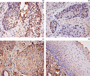
The proliferative and apoptotic indices were determined by measuring the proportion of positively stained cell nuclei and apoptotic cells, respectively (Figure 1a and 1b), as outlined above. In our study, the mean ± standard deviation for the proliferative and apoptotic indices of the laryngeal cancer samples analysed were 25.24 ± 6.65 and 1.44 ± 0.32, respectively.
Photomicrographs showing immunohistochemical analysis of laryngeal squamous cell carcinoma: (a) expression of proliferating cell nuclear antigen (Ki-67) in cell nuclei (×400); (b) detection of apoptotic cells by the terminal deoxynucleotidyl transferase mediated deoxyuridine triphosphate nick end labelling method (×400); and (c) expression of focal adhesion kinase at the cell membrane and in the cell cytoplasm (×400). (d) Photomicrograph showing negative immunostaining for focal adhesion kinase expression in normal laryngeal epithelium (×400).

Relation of focal adhesion kinase to clinicopathological parameters
Focal adhesion kinase immunostaining was observed in 63 (73.26 per cent) of the 86 laryngeal cancer samples, being localised at the cell membrane and also present in the cytoplasm of laryngeal cancer cells (Figure 1c). Although focal adhesion kinase staining was also detected in 5 (15.63 per cent) of the 32 normal laryngeal mucosa specimens, the proportion was significantly lower compared with the laryngeal cancer cells (p < 0.05). In the normal laryngeal mucosa, focal adhesion kinase was located mainly in the basal layers of the epidermis, and was distributed on cell membranes and in the cell cytoplasm.
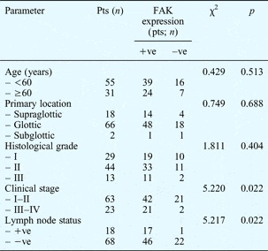
In laryngeal cancer tissues, focal adhesion kinase expression correlated closely with the patient's clinical stage and the presence of lymph node metastasis (p < 0.05); however, there was no significant association with age, primary location or tumour histological grade (p > 0.05) (Table I).
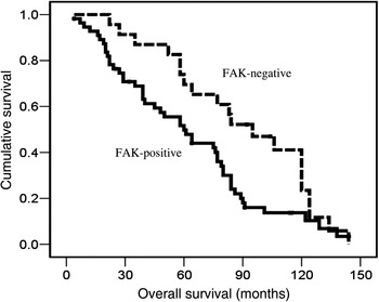
Kaplan–Meier disease-specific survival curves for patients with laryngeal squamous cell carcinoma negative and positive for focal adhesion kinase (FAK) immunostaining (p = 0.031).

Correlation between focal adhesion kinase expression and clinicopathological parameters*

* In 86 cases of laryngeal squamous cell carcinoma. Pts = patients; FAK = focal adhesion kinase; +ve = positive; –ve = negative
Relation of focal adhesion kinase to proliferative and apoptotic indices
In the laryngeal carcinoma specimens, focal adhesion kinase immunoreactivity was positively correlated with proliferative index (p < 0.05). However, lower apoptotic index values were significantly associated with higher levels of focal adhesion kinase expression (p < 0.05) (Table II).
Association between focal adhesion kinase expression and apoptotic and proliferative indices

FAK = focal adhesion kinase; +ve = positive; –ve = negative; Apo = apoptotic; SD = standard deviation; Prol = proliferative
Survival analysis
Clinical follow-up data were available for 79 of the patients with laryngeal squamous cell carcinoma who had undergone surgical resection. Seven patients had been lost to follow up and were thus excluded from the survival analysis.
The median patient survival time was 64.0 months (range, 4.0–144.0 months). The three- and five-year survival rates were 72.2 and 53.2 per cent, respectively.
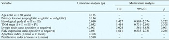
Univariate survival analysis indicated that overall survival was significantly better in the focal adhesion kinase negative group compared with the focal adhesion kinase positive group (p = 0.031) (Figure 2, Table III). However, multivariate analysis by Cox regression indicated that the level of focal adhesion kinase expression was not related to the prognosis of patients with laryngeal squamous cell carcinoma (p = 0.265) (Table III).
Univariate and multivariate analysis of prognostic factors*

* In patients with laryngeal squamous cell carcinoma. HR = hazard ratio; CI = confidence interval; TNM = tumour-node-metastasis; FAC = focal adhesion kinase
Discussion
It is now clear that conventional clinicopathological prognostic variables, such as TNM staging and histological grade, are not sufficient to accurately estimate the prognosis of patients with laryngeal squamous cell carcinoma.Reference Hussein and Cullen 15 Numerous studies have indicated that certain biological compounds not only reflect the biological characteristics of tumours but may also help clinicians to formulate individual treatment schemes and to evaluate the prognosis of patients with laryngeal squamous cell carcinoma.Reference Li, Yang, Wang and Tang 16 – Reference Chen, Zhou, Wan, Dou and Tian 18 Ongoing research in pursuit of these goals is thus an important priority.
Focal adhesion kinase is a cytoplasmic protein tyrosine kinase which has been confirmed as a key integrator mediating a series of cellular biological processes known to occur in cancer, such as migration, adhesion and angiogenesis.Reference Golubovskaya 3 There is now growing evidence that analysis of focal adhesion kinase could play a vital role in predicting the progression and prognosis of many malignant neoplasms.Reference Park, Lee, Yoon, Kim, Kim and Yang 9 – Reference Hayashi, Aishima, Inoue, Nakata, Morimatsu and Nagai 11
Interestingly, there is substantial controversy over the extent of focal adhesion kinase expression in tumour tissue versus normal controls. On the one hand, most studies had reported stronger focal adhesion kinase expression in tumour cells than in normal epithelial cells.Reference Cai, Han, Zhuo, Xiong, Dong and Li 10 , Reference Kamo, Naomoto, Shirakawa, Yamatsuji, Hirota and Fujiwara 19 On the other hand, Hayashi and colleagues found focal adhesion kinase expression to be down-regulated in tumour specimens compared with normal controls.Reference Hayashi, Aishima, Inoue, Nakata, Morimatsu and Nagai 11 In the present study, we found higher levels of focal adhesion kinase expression in laryngeal cancer specimens than in normal epithelial tissue, in agreement with previous laryngeal cancer studies.Reference Canel, Secades, Rodrigo, Cabanillas, Herrero and Suarez 12 , Reference Rodrigo, Dominguez, Suárez, Canel, Secades and Chiara 14
Focal adhesion kinase expression may vary in different tumour types, and this may contribute to the phenotypic differences observed. In the current study, we observed a positive correlation between focal adhesion kinase expression and clinical stage, supporting the idea that focal adhesion kinase may be involved in malignant progression of laryngeal squamous cell carcinoma.
To our best knowledge, the relationship between focal adhesion kinase expression and cervical lymph node metastasis in laryngeal carcinoma has not yet been fully elucidated. Canel et al. have clearly demonstrated that focal adhesion kinase expression is an effective index with which to estimate the presence of cervical lymph node metastases in laryngeal squamous cell carcinoma patients.Reference Canel, Secades, Rodrigo, Cabanillas, Herrero and Suarez 12 In contrast, Rodrigo et al. reported that focal adhesion kinase expression had little value as a predictor of lymph node metastases in laryngeal cancer.Reference Rodrigo, Cabanillas, Secades, Canel, Chiara and Suárez 13 Our results demonstrated that focal adhesion kinase expression was positively associated with lymph node metastasis in laryngeal squamous cell carcinoma, consistent with the work of Canel et al. Reference Canel, Secades, Rodrigo, Cabanillas, Herrero and Suarez 12 In other words, focal adhesion kinase may be an effective biomarker with which to select those laryngeal squamous cell carcinoma patients who are at higher risk of lymph node metastasis.
-
• Focal adhesion kinase modulates tumour cell adhesion, angiogenesis, migration, proliferation and apoptosis
-
• Its role in gastric, hepatocellular and extrahepatic bile duct carcinoma has been assessed
-
• This study found an influence on laryngeal squamous cell carcinoma (SCC) progression
-
• Effects included suppression of apoptosis and promotion of cell proliferation
-
• Focal adhesion kinase may be a potential biomarker for laryngeal SCC prognosis
There have only been a few studies of the relationship between focal adhesion kinase expression and the survival of patients with laryngeal carcinoma. Canel et al. and Rodrigo et al. found no significant correlation between focal adhesion kinase expression and the survival of patients with squamous cell carcinoma of the supraglottic larynx.Reference Canel, Secades, Rodrigo, Cabanillas, Herrero and Suarez 12 , Reference Rodrigo, Cabanillas, Secades, Canel, Chiara and Suárez 13 In the current study, we observed that up-regulation of focal adhesion kinase expression was closely associated with poor overall survival in patients with laryngeal squamous cell carcinoma; however, multivariate analysis indicated that focal adhesion kinase expression cannot be used as an independent prognostic marker in laryngeal carcinoma patients. Consequently, it is possible to state that detection of a high level of focal adhesion kinase expression can assist the identification of laryngeal cancer patients with worse overall survival rates. The inconsistencies between different authors' conclusions, as mentioned above, may relate to immunohistochemical differences between various types of primary laryngeal cancer. As stated above, data from the current study provide evidence that assessment of focal adhesion kinase expression may assist the prediction of disease progression and prognosis of laryngeal carcinoma patients.
The current understanding is that a balance between cell proliferation and apoptosis is essential for normal tissue development and homeostasis.Reference Alenzi 20 Disturbance of this balance usually underlies tumourigenesis and neoplastic development, and involves alteration of the expression of multiple genes.Reference Hipfner and Cohen 21 Previous investigators have focused on the relationship between focal adhesion kinase expression and the apoptotic and proliferative activities within a variety of human tumours. Xia et al. conducted experimental research which demonstrated that elevated expression of focal adhesion kinase during rat tongue carcinogenesis correlated strongly with proliferation inhibition and apoptosis promotion within cancer cells.Reference Xia, Lv, Hong, Li, Tao and Chen 7 Various studies have indicated that down-regulation of focal adhesion kinase expression may represent a potential therapeutic approach, retarding the unlimited proliferation and apoptosis suppression seen in various neoplasms such as malignant glioma, colon carcinoma and breast cancer.Reference Wang, Ma, Yang, Zhang, Wang and Liu 4 – Reference Huang, Wan, Liao, Hu, Wei and Wang 6 However, the effect of focal adhesion kinase on cellular apoptosis and proliferation within laryngeal squamous cell carcinoma still remains unclear. In the current study, we investigated whether focal adhesion kinase contributes to apoptosis inhibition and proliferation promotion in laryngeal cancer, by assessing focal adhesion kinase expression in laryngeal cancer cells. Our data indicate that unregulated focal adhesion kinase expression correlates closely with decreased cell apoptosis and increased proliferation, suggesting that aberrant focal adhesion kinase expression in laryngeal squamous cell carcinoma may lead to up-regulation of proliferation and down-regulation of apoptosis.
Further in vitro research is necessary to elucidate this relationship and to define its molecular mechanisms. Our study data indicate that focal adhesion kinase assists malignant development of laryngeal squamous cell carcinoma through inhibiting cellular apoptosis and promoting proliferation.
Conclusion
Our study findings indicate that focal adhesion kinase may play a potential role in the progression of laryngeal squamous cell carcinoma, due to multiple biological activities including suppression of cellular apoptosis and promotion of proliferation.
Our data provide evidence that focal adhesion kinase may be an effective biomarker with which to predict the prognosis of laryngeal squamous cell carcinoma patients.
Acknowledgements
We thank Dr Jia-Wei Chen, Department of Pathology, Shanghai Jiaotong University Affiliated First People's Hospital, for the database of clinical indices, and Miss Yu-Ying Chen, Morphology and Cell Chemistry Laboratory of Shanghai Jiao Tong University School of Medicine, for technical support in immunohistochemical staining. This work was supported by a grant from the Shanghai Science and Technology Development Fund, China (grant number 09411951000).