Introduction
An apparently undescribed, helotialean lichenicolous fungus has been collected several times on thalli and apothecia of Phaeophyscia species. Because of the subcylindrical asci with uniseriate, shortly ellipsoid ascospores, it strongly resembles the monotypic genus Geltingia Alstrup & D. Hawksw. (Alstrup & Hawksworth Reference Alstrup and Hawksworth1990). However, the general macroscopical appearance is not that of Geltingia associata (Th. Fr.) Alstrup & D. Hawksw., and the asci and the excipular structure are different. Several further species from other genera are considered to be congeneric with the new fungus on Phaeophyscia. All these species were compared with the similar helotialean genus Gelatinopsis Rambold & Triebel, the type species of which differs by strongly convex, immarginate, poorly delimited ascomata, a different excipular structure and pigmentation, and slightly different asci. They are most reminiscent of the genus Llimoniella Hafellner & Nav.-Ros., but differ by the absence of typical excipular and epihymenial pigments characterizing that genus. However, other morphological and anatomical characters suggest that these species should be included in an enlarged concept of Llimoniella. The aim of this paper is therefore to describe or combine these species in Llimoniella. Several additional species of Llimoniella have been studied, and a revised key to all known Llimoniella species is provided. As a detailed study of Geltingia associata was necessary to support the distinctiveness of the two genera, a redescription and illustrations of G. associata are also presented.
Material and Methods
The specimens examined are located in the institutional herbaria ASU, BCN, BG, BR, C, E, HBG, LE, M, MSC, NY, OTA and UPS, and in the personal collections of A. Aptroot, P. Diederich, J. Etayo, M. G. Halici, K. Kalb and P. van den Boom. The morphological characters of dry herbarium specimens have been studied and illustrated using a dissecting microscope. The microscopical examination was carried out using hand-cut sections mounted in water, 5% KOH (K), concentrated nitric acid (N), Congo Red, lactophenol Cotton Blue (LCB), or Lugol's reagent, without (I) or with (K/I) pre-treatment with KOH. For macroscopical and some microscopical photographs (e.g., asci, paraphyses), several images with different parts in focus were combined into one picture using the free software CombineZP (developed by Alan Hadley). Photographs of ascomatal sections were obtained by stitching together many photos representing parts of the section examined at a high magnification.
The following specimens have been examined and compared with the species studied: Gelatinopsis geoglossi: USA: Connecticut: Sharon Audubon Center, 2 miles SE of Sharon, on Trichoglossum farlowii, 1969, C. Rogerson & G. Smith (NY). North Carolina: Blue Ridge Mountains, Highlands Biological Station, Highlands, Macon Co., 1961, R. H. Petersen & C. Rogersen (NY).
Llimoniella fuscatae: Great Britain: Wales: VC 43, Radnor, N of Rhayader, Gilfach Farm, on Acarospora fuscata, 1997, S. P. Chambers (E, hb. Diederich).
Results
The new Llimoniella phaeophysciae group
The informal Llimoniella phaeophysciae group, including L. phaeophysciae and four related species are introduced here, and the relationships between these species and Llimoniella s.str. discussed.
Llimoniella phaeophysciae Diederich, Ertz & Etayo sp. nov
Llimoniella species in thallis Phaeophysciae vigens, insignis excipulo et epihymenio brunneo K−, hymenio non insperso, ascis subcylindricis apice applanato pariete apicaliter tenui 8-spori, et ascosporis uniseriatis subglobosis ad breviter ellipsoideis 7–11 × 6–9 μm.
Typus: USA, Arizona, near San Francisco Peaks, on side of hwy 89 just N of Sunset Crater, 35.4029° N, 111.5762° W, alt. c. 6500 feet, on sheltered Juniperus bark, on Phaeophyscia hirsuta s. lat., 9 June 2008, J. Hollinger 20080609.12 (ASU—holotypus; hb. Diederich—isotypus).
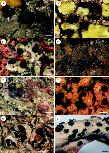
A–E, Llimoniella phaeophysciae group, ascomata of the five species of the group; A, L. phaeophysciae on Phaeophyscia hirsuta (holotype); B, L. acarosporicola on Acarospora socialis (isotype); C, L. groenlandiae on thallus squamules of Caloplaca group holocarpa (F. Berger 14961); D, L. heppiae on Heppia despreauxii (holotype); E, L. terricola on Epiphloea byssina (isotype of Gelatinopsis leptogii). F, L. placopsidis on Placopsis elixii, ascomata (holotype). G & H, Geltingia associata ascomata; G, on Ochrolechia sp. (T. Tønsberg 24700); H, on Thamnolia vermicularis (M. Zhurbenko 95307). Scales: A – H = 200 μm.

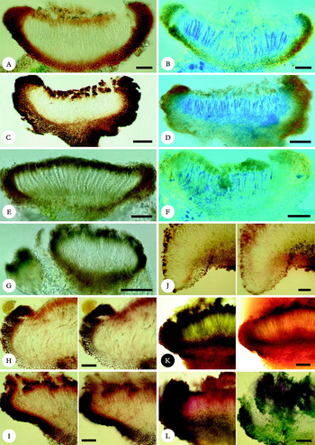
A & B, Llimoniella phaeophysciae, sections through ascoma (holotype); A, observed in water; B, examined in LCB (staining asci and lumina of excipular cells), pre-treated with N (holotype); note the hyaline outer layer and the radiating cells of the lateral exciple. C & D, L. placopsidis (holotype), same methods of examination; note the strong pigmentation of the paraphyses apices in water. E & F, Geltingia associata (T. Tønsberg 24700); E, examined in K; F, in LCB, pre-treated with N; note the thick, dark epihymenium, the lateral exciple made of more or less parallel or slightly interwoven hyphae with poorly stained lumina in LCB, and the pale, sometimes almost indistinct lower exciple. G, Rhymbocarpus roccellae observed in water (T. H. Nash III 26361, hb. Diederich), note the exciple made of almost parallel or slightly interwoven hyphae, especially visible in the left, young ascoma, and the greenish epihymenial pigment. H & I, sections through ascomata of Llimoniella s.str. in water (left), and K-reaction (right); H, L. adnata (M. Zhurbenko 92198, hb. Diederich); I, L. scabridula, the generic type (Santesson Fungi Lichenicoli Exsiccati 211, hb. Diederich); note the pale purplish violet reaction of the epihymenium and the upper exciple, and the dark brown lower exciple in K. J–L, Llimoniella ramalinae, sections through ascomata showing variability of infraspecific pigmentation; J, poorly pigmented ascoma, pale K+ purplish reaction (New Zealand, South Island, Allisson Reserve, on R. unilateralis, J. Bannister s. n., OTA); K, greenish hymenium and reddish brown exciple, strong orange red to purplish reaction (dissolving in K) (New Zealand, South Island, on R. unilateralis, J. Bannister 1793, OTA); L, deep purplish epihymenium and upper exciple, greenish lower exciple, giving a strong green and partly an additional violet K-reaction (pigments not dissolving in K, shown here in a squash preparation several minutes after adding K) (New Zealand, South Island, on R. celastri, J. Bannister 1278, OTA). Scales: A–L = 50 μm.

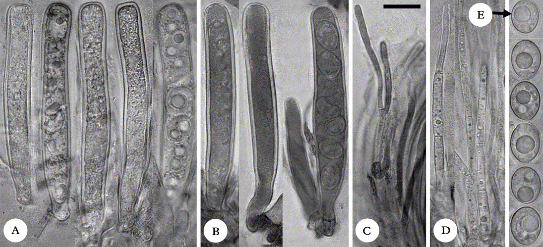
Llimoniella phaeophysciae, asci, paraphyses and ascospores. A, D & E, in water (holotype). B & C, in Congo Red after pre-treatment with 5% KOH (P. Conkin). Note the subcylindrical asci with a more or less applanate apex and an apically thinner wall. Scale = 10 μm.

Ascomata erumpent, sessile, roundish, dispersed or more frequently in groups of up to 15, 120–350 μm diam., with a distinct, prominent, blackish, matt or slightly shiny margin of 40–60 μm and a dark reddish brown to blackish, matt, applanate disc. Exciple reddish to orange-brown, K− or slightly darker, N+ brighter orange, laterally of thick-walled, elongate, rarely branched, radiating hyphae with swollen end cells, 25–65 μm thick, basally of conglutinated, thick-walled and more or less isodiametric cells 7–11·5 μm diam., 25–60 μm thick, occasionally prolonged into a brown, immersed ‘stipe’; without hairs; externally sometimes bordered by a thin, hyaline necrotic layer. Subhymenium colourless. Epihymenium pale to orange-brown, K−, N+ brighter orange. Hymenium colourless to yellowish, not inspersed, 100–130 μm tall, I−, K/I−. Paraphyses septate, simple or more rarely branched in the upper part, apically not or slightly swollen, 1·5–3 μm thick, cells with many small oil guttules. Asci subcylindrical, apically more or less applanate, (3–)8-spored, wall laterally c. 0·4–0·6 μm thick, apically distinctly thinner, c. 0·3–0·4 μm, without ocular chamber, without croziers, 55–75 × 7·5–9 μm, I−, K/I−. Ascospores uniseriate in the ascus, non-septate, colourless, subglobose to shortly ellipsoid, with 1(–3) large lipid guttule(s), 7–11 × 6–9 μm, wall smooth, c. 0·3 μm thick, without perispore.
Conidiomata unknown.
The new species is distinguished from most Llimoniella species discussed below by the broad ascospores, more than 6 μm in width, and from L. acarosporicola by the constantly rounded ascomata. Macroscopically, L. phaeophysciae could be confused with Buelliella physciicola Poelt & Hafellner, a lichenicolous fungus confined to Phaeophyscia species, which is readily distinguished by the eventually brownish, 1-septate ascospores (Hafellner Reference Hafellner, Nash, Ryan, Diederich, Gries and Bungartz2004). The new species should also be compared with Phaeopyxis muscigenae Alstrup & E. S. Hansen, described from Physconia muscigena in Greenland (Alstrup & Hansen Reference Alstrup and Hansen2001). That species has similar more or less globose, but slightly smaller ascospores, c. 5–8 μm diam., but they are biseriate in much broader, clavate asci, 48–54 × 14–20 μm, ascomata are larger, up to 0·7 mm diam., and the hymenium is lower, 55–60 μm tall.
Distribution and hosts. The new species is known from Europe (Italy and Spain) and the USA (Arizona) and appears to be confined to Phaeophyscia species (thallus and apothecia), on which it does not cause any visible damage. The known hosts are P. ciliata, P. hirsuta s. lat. (incl. P. cernohorskyi) and P. nigricans. Marcos Laso (Reference Marcos Laso2001) reported Geltingia associata (as Lecidea associata) from Salamanca (Spain) on P. nigricans (material in SALA). Although we did not examine any corresponding material, it is very likely to belong to Llimoniella phaeophysciae as well.
Additional specimens examined. Italy: Calabria: N of Catanzaro, W of Pagliarelle, along road Pagliarelle to Petto di Mandra, on Phaeophyscia ciliata, 1990, P. van den Boom 10306 (hb. van den Boom).—Spain: Aragón: Zaragoza, Biel, alt. 690 m, encinar de Callau, on Tilia, on P. nigricans, 2004, J. Etayo 22087 (hb. Etayo). Castilla y León: Soria, Soria capital, paseo por el Duero camino a S. Saturio, olmeda, alt. 900 m, on Aesculus hippocastanum, on P. nigricans, 2001, J. Etayo 18371 & E. Ros (hb. Etayo).—USA.: Arizona: c. 60 miles SE of Flagstaff, Navajo, 34.6155° N, 110.5469° W, on P. cernohorskyi, 2002, P. Conklin (ASU).
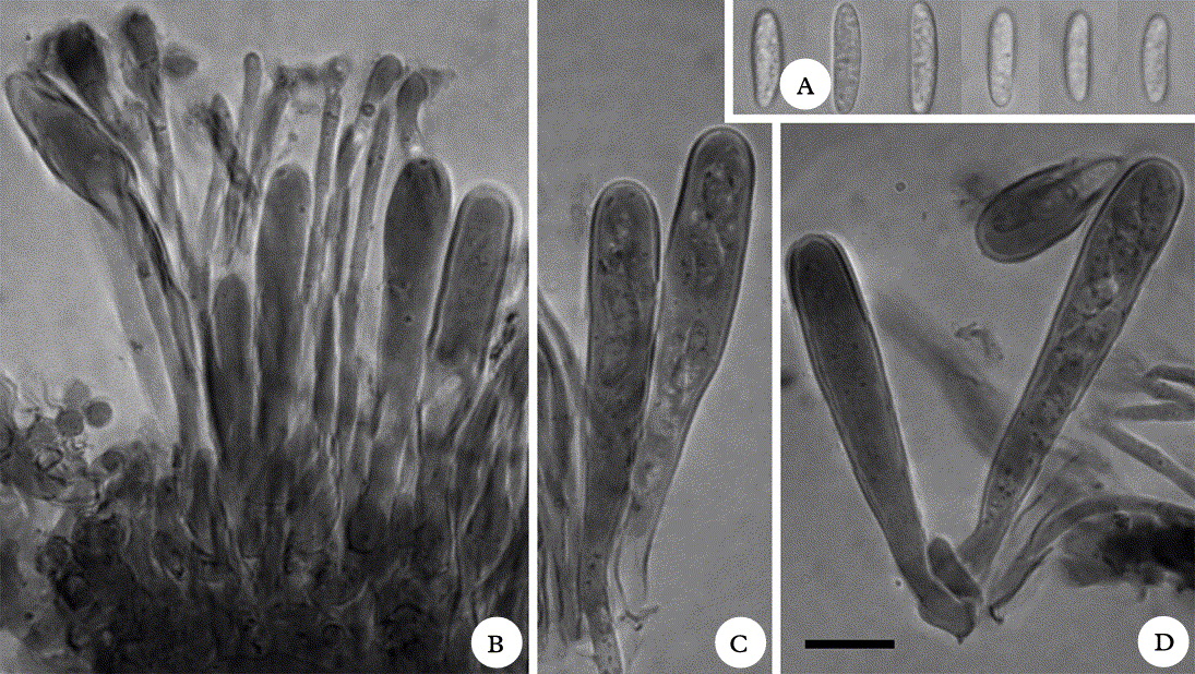
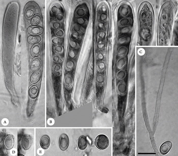
Discussion. Llimoniella phaeophysciae resembles the monotypic lichenicolous genus Geltingia [type species: G. associata] by the subcylindrical asci, the uniseriate, shortly ellipsoid ascospores and the same excipular and epihymenial pigment. Both taxa differ nevertheless by several important characters. Asci of L. phaeophysciae are apically applanate, the wall is uniformly thin and apically even thinner, and an ocular chamber is always missing (Fig. 3A & B); those of G. associata are apically rounded, the wall is thickened near the apex, and a distinct ocular chamber is usually visible (Fig. 5A & B). Ascospores are smooth and very thin-walled in L. phaeophysciae (Fig. 3E), but verruculose in G. associata with a distinctly thicker wall (Fig. 5D & E). Although the excipular pigment appears to be the same in both genera, the excipular anatomy differs: the lateral exciple of L. phaeophysciae consists of strongly conglutinated radiating hyphae with more or less isodiametric cells and with enlarged or swollen end cells [= outer cells of exciple], comparable to the exciple of typical Llimoniella species (Diederich & Etayo Reference Diederich and Etayo2000) (Fig. 2A & B); the lateral exciple of G. associata consists of elongate, filiform, more or less parallel or irregularly arranged, but never radiating hyphae, without swollen end cells (Fig. 2E & F). The basal exciple of L. phaeophysciae is strongly developed and dark (pigmentation comparable to the lateral exciple), sometimes prolonged into an immersed, dark stipe; that of G. associata is pale, often poorly distinct. Finally, ascomata in L. phaeophysciae are always rounded, regular in shape and size, more or less superficial, with a more or less matt disc and a prominent margin (Fig. 1A), whilst those of G. associata are frequently elongate, especially when growing on the margin of the host apothecia, often irregular in size and form, immersed to erumpent, with a typically shiny disc and a less prominent, sometimes almost undulate margin (Fig. 1G & H). Llimoniella phaeophysciae is distinguished from almost all other known lichenicolous helotialean fungi with dark ascomata by the subcylindrical asci with an applanate apex and no visible internal apical structures, and uniseriate, shortly ellipsoid ascospores (Fig. 3).
After carefully comparing the new species with other lichenicolous members of Helotiales, we realized that Llimoniella phaeophysciae is not only superficially, but also microscopically very similar to Gelatinopsis leptogii Ertz & Diederich (Ertz & Diederich Reference Ertz and Diederich2006). The ascomata of G. leptogii are reminiscent of those of L. phaeophysciae, although sometimes slightly immersed in the host thallus (Fig. 1E), the excipular anatomy and pigmentation are the same, the asci are subcylindrical, thin-walled and frequently apically applanate, and the ascospores are shortly ellipsoid. There is little doubt that both species are congeneric. Similarly, the very recently described G. acarosporicola Kocourk. & K. Knudsen has comparable ascomata (they differ in being frequently elongate or angular) (Fig. 1B), a similar exciple, subcylindrical to clavate asci with a thin wall, and 1(–2)-seriate, shortly ellipsoid ascospores (Kocourková & Knudsen Reference Kocourková and Knudsen2009). Although asci of G. acarosporicola are apically not applanate, the species is so close to G. leptogii that it must be considered as congeneric. Gelatinopsis heppiae Nav.-Ros., Hladun & Llimona, recently described from Spain (Navarro-Rosinés et al. Reference Navarro-Rosinés, Hladun and Llimona2008) (Fig. 1D), is very similar to the Llimoniella species discussed here, but differs in the more deeply pigmented orange to reddish brown exciple and epihymenium, and the distinctly swollen (up to 6 μm) and pigmented paraphyses apices. These differences hardly justify a distinct genus. In conclusion, these three lichenicolous Gelatinopsis species are congeneric with the new Llimoniella phaeophysciae forming an obviously natural group of fungi (hereafter called the Llimoniella phaeophysciae group) that also includes species in which asci are apically larger and rounded.
There are two other known lichenicolous Gelatinopsis species, G. ericetorum (Körb.) Rambold & Triebel and G. roccellae Etayo, Paz-Bermúdez & Diederich. The type of the latter species differs in the immersed, immarginate ascomata growing on the thallus of Roccella, clavate to subcylindrical asci that are apically not applanate, and ellipsoid to ovoid, 1–2-seriate ascospores. The exciple and epihymenium in the type specimen are brownish, K− (Etayo et al. Reference Etayo, Paz-Bermúdez and Diederich2001). In a Mexican specimen of the same species (T. H. Nash III 26361), an additional olivaceous pigment is present in the epihymenium and the upper exciple. The same olivaceous pigment is also present in the epihymenium of G. ericetorum, a lichenicolous species developing blackish ascomata over the thallus of Dibaeis baeomyces. Both species differ from the Llimoniella phaeophysciae group by the presence of the olivaceous epihymenial pigment (absent in some specimens of G. roccellae), more or less immersed and immarginate ascomata with a flat disc, and an exciple entirely made up of more or less parallel to interwoven hyphae. Asci in both species are clavate to subcylindrical, never with a flattened apex, and the wall in G. ericetorum is apically slightly thickened (Baral & Marson Reference Baral and Marson2001). The ascomata of both are never concave, like those of all non-lichenicolous Gelatinopsis species, including the type G. geoglossi (Ellis & Everh.) Rambold & Triebel (Baral & Marson Reference Baral and Marson2001). This is not the case with Llimoniella, in which all species have a slightly to strongly prominent margin at least when young. The ascomata of G. roccellae might appear concave, as they are sometimes deeply immersed and surrounded by the host thallus, but the hymenial disc is always flat and immarginate. Gelatinopsis ericetorum was included in Gelatinopsis by Rambold & Triebel (Reference Rambold and Triebel1990) with some hesitation, a decision that was eventually accepted by Baral & Marson (Reference Baral and Marson2001). All other, non-lichenicolous Gelatinopsis species studied by Baral & Marson (Reference Baral and Marson2001), including the generic type G. geoglossi (Ellis & Everh.) Rambold & Triebel, have an apically distinctly thickened ascus wall. The type species has black (when dry), superficial ascomata that are strongly convex, irregularly delimited and often conglutinated, and always immarginate (several specimens studied by us, see under Material and Methods), and are macroscopically very distinct from those of G. ericetorum and G. roccellae. Instead, these two lichenicolous taxa are very similar to several species of Rhymbocarpus Zopf with a reduced exciple, including the generic type R. geographici (J. Steiner) Vouaux, and they will consequently be included in Rhymbocarpus below.
A further lichenicolous helotialean fungus, Geltingia groenlandiae Alstrup & D. Hawksw., was initially described in an enlarged concept of Geltingia Alstrup & D. Hawksw. (Alstrup & Hawksworth Reference Alstrup and Hawksworth1990), and then transferred to Llimoniella (Kümmerling et al. Reference Kümmerling, Triebel and Rambold1993). When Diederich & Etayo (Reference Diederich and Etayo2000) revised the genus Llimoniella, they considered that the species did not belong to that genus because of the lack of excipular and epihymenial pigments characteristic of Llimoniella. They proposed that the available material might represent poorly developed ascomata of a species of Unguiculariopsis, in which excipular hairs were missing. The species was consequently transferred to that genus. With the discovery of more material, it became evident that ascomata of this species are usually blackish and never have excipular hairs (Fig. 2C) and that it cannot remain in Unguiculariopsis. Macroscopically, this species is a typical member of the Llimoniella phaeophysciae group. It also has the same excipular anatomy and pigmentation, and entirely thin-walled asci. The main differences are the slightly broader, subcylindrical to elongate clavate asci with a rounded apex, the narrowly ellipsoid 2-seriate ascospores and a hymenium that is inspersed in the upper half. We conclude that this species also belongs to the L. phaeophysciae group and that the narrowly ellipsoid form of the ascospores does not allow them to be uniseriate in the asci, which as a consequence are not subcylindrical but elongate claviform.
The Llimoniella phaeophysciae group is therefore characterized as follows: ascomata dark brown to blackish, more or less matt, with an often prominent margin, at least when young; excipulum of shortly elongate to isodiametric cells, sometimes laterally radiating with enlarged end cells; an orange- brown excipular and epihymenial pigment not reacting in K and N (but becoming brighter in N); entirely thin-walled, elongate clavate to subcylindrical asci, in some species with a flattened apex, that are I− and K/I−, and smooth- and thin-walled and, simple, colourless ascospores. The differences from Geltingia and Gelatinopsis are discussed above. The genus Rhymbocarpus is distinguished by a greenish, K+ olivaceous epihymenial and occasionally excipular pigment, a lateral exciple made of elongate, narrow, almost filiform hyphae, the presence of excipular hairs in some species and ascomata in several species (including the generic type) more or less immersed with a punctiform disc, in other species becoming superficial, the disc often partly or mostly covered by the exciple (Diederich & Etayo Reference Diederich and Etayo2000).
Llimoniella s. str. is distinguished by a K+ violet epihymenial and excipular pigment. In several species of Llimoniella s. lat., a more reddish or purplish pigment, and sometimes an additional, pale olivaceous, K+ bright green pigment occurs. In a few species, a slight, but distinct thickening of the apical ascal wall or even an ocular chamber have been observed, and several species (possibly not closely related to the generic type) have very long and narrow, multiguttulate and/or septate ascospores (Diederich & Etayo Reference Diederich and Etayo2000). The different pigmentation of the species of the Llimoniella phaeophysciae group and the absence of a positive K-reaction might suggest a distinct genus. However, within many genera, the observed pigmentation of the different species is variable (e.g., Skyttea, see Diederich & Etayo Reference Diederich and Etayo2000), and this character alone does not justify the distinction of two genera. As all other characters are in agreement with the type of Llimoniella, we propose to include the species of the L. phaeophysciae group within Llimoniella s. lat.
Llimoniella acarosporicola (Kocourk. & K. Knudsen) Diederich & Ertz comb. nov
Basionym: Gelatinopsis acarosporicola Kocourk. & K. Knudsen, Bryologist 112: 364 (2009); type: USA, California, Orange County, Santa Ana Mountains, Fremont Canyon, 33°47′14″ N, 117°43′03″ W, alt. 294 m, on Acarospora socialis on sandstone slab, 15 x 2007, K. Knudsen 9236 (UCR—holotype; MSC, PRM, hb. Diederich—isotype!).
(Fig. 1B)
For a description and illustrations, see Kocourková & Knudsen (Reference Kocourková and Knudsen2009).
Notes: This species differs mainly from L. phaeophysciae by the more irregular, often angular or laterally compressed ascomata. It is very similar to Llimoniella fuscatae Hafellner & Obermayer, a species confined to the same host genus Acarospora, but differs by the entirely K− epihymenium and exciple (in L. fuscatae, a pale, but distinct violaceous reaction is visible, at least in thick sections), the more irregular and often angular ascomata, the less prominent ascomatal margin and a different host selection (Acarospora socialis, versus A. fuscata).
Distribution and host. Known from two localities in California, always on the thallus of Acarospora socialis, on which the fungus does not cause any visible damage.
Llimoniella groenlandiae (Alstrup & D. Hawksw.) Triebel & Hafellner
In Kümmerling et al., Bibl. Lichenol. 53: 156 (1993).—Geltingia groenlandiae Alstrup & D. Hawksw., Meddr Grønland, Biosci. 31: 33 (1990).—Unguiculariopsis groenlandiae (Alstrup & D. Hawksw.) Etayo & Diederich, Lichenologist 32: 472 (2000); type: Greenland, Narsaq distr., 1 km S of Qagssiarssuk, 61°08′ N, 45°32′ W, alt. 140 m, on Caloplaca citrina s.l., viii 1980, V. Alstrup 801120 (C—holotypus!).
=? Llimoniella caloplacae S.Y. Kondr. & Khodos., Mycologia Balcanica 3: 96 (2006); type: Ukraine, Khersons oblast, Belozersky district, near Shirokaya Balka village, on loess slopes, on Caloplaca borysthenica areoles and apothecia, 7 May 2002, A. Khodosovtsev (KW—holotypus; LD, KHER—isotypi, n.v.).
(Fig. 1C)
For a description and additional illustrations, see Alstrup & Hawksworth (Reference Alstrup and Hawksworth1990: 33–35).
Notes. The two recent collections both have a hymenium that is strongly inspersed with small oil droplets in the upper half. However, we did not re-examine the very small type specimen (studied by Diederich & Etayo Reference Diederich and Etayo2000), and thus cannot confirm if the hymenium in the type is also inspersed. This character, also present in L. pyrenulae, seems to distinguish L. groenlandiae from all other known species of the Llimoniella phaeophysciae group and should be carefully checked in additional specimens. A Dutch specimen of L. groenlandiae (A. Aptroot 50330) is unusual within the L. phaeophysciae group in possessing paler ascomata, but all other characters, including the inspersed hymenium and the host selection, confirm that it does belong to L. groenlandiae. Llimoniella caloplacae, described from the same host genus, is probably a synonym of L. groenlandiae, but unfortunately our request for the loan of any original material was unsuccessful (no answer from KW and LD).
Distribution and hosts. Known from Austria, Greenland, The Netherlands and Sweden, possibly also Ukraine (type of L. caloplacae). Commensalistic on Caloplaca citrina s. lat., C. flavocitrina and an unidentified specimen of the C. holocarpa group with pale yellowish, convex thallus squamules; possibly also on C. borysthenica (see above). An additional specimen of L. groenlandiae, mentioned by Alstrup & Hawksworth (Reference Alstrup and Hawksworth1990) on Lepraria neglecta, proved to be typical Rhymbocarpus neglectus (Vain.) Diederich & Etayo on Lepraria alpina (Kümmerling et al. Reference Kümmerling, Triebel and Rambold1993). Diederich & Etayo (Reference Diederich and Etayo2000) also mentioned two specimens on Fulgensia bracteata and F. fulgens (thalli). We were able to re-examine one of these [Greenland: CW, Søndre Strømfjord Airport, S-slope of Mt. Hassel, NE of airport, alt. 150 m, on a sterile thallus of F. bracteata, 1991, V. Alstrup 91072 (C 1212).]. The specimen is in an extremely poor condition, and we did not observe any healthy asci or ascospores. The hymenium is not inspersed, and thus it is not certain if the specimen really belongs to L. groenlandiae. The second specimen from Sweden could not be located. We would therefore prefer to examine additional material on Fulgensia before definitely including such specimens in L. groenlandiae.
Additional specimens examined. Austria: Oberösterreich: Aschbach an der Donau, Bahnhofgelände, Gleis vor Lagerhaus, on Caloplaca gr. holocarpa, 2000, F. Berger 14961 (hb. Berger).—The Netherlands: Gelderland: Betuwe, Ingen, on brick wall of church, on C. flavocitrina, 2001, A. Aptroot 50330 (hb. Aptroot).
Llimoniella heppiae (Nav.-Ros., Hladun & Llimona) Diederich & Ertz comb. nov
Basionym: Gelatinopsis heppiae Nav.-Ros., Hladun & Llimona, Revista Catalana de Micologia 30: 60 (2008); type: Spain, Aragón, prov. Zaragoza, Osera, carretera a Monegrillo, lomas a la derecha de la carretera, 41°33′35″ N, 0°33′20″ E, alt. 200–210 m, en el talo de Heppia despreauxii, suelos arcilloso-yesosos, 3 June 2003, X. Llimona (BCN-lich. 15840—holotype!; 15841—isotype).
(Fig. 1D)
For a description and illustrations, see Navarro-Rosinés et al. (Reference Navarro-Rosinés, Hladun and Llimona2008).
Notes. This species is distinguished from the other members of the Llimoniella phaeophysciae group by its more strongly pigmented epihymenium, and especially by the strongly swollen and brown paraphyses apices.
Distribution and host. Known only from the type locality in Spain on the thallus of Heppia despreauxii, on which the fungus does not cause any visible damage.
Llimoniella terricola (Rehm ex Arnold) M. Schultz, Diederich & Ertz comb. nov
Basionym: Physma terricolum Rehm ex Arnold, Flora 68: 216 (1885).—Psorotichia terricola (Rehm ex Arnold) Lettau, Hedwigia 52: 179 (1912).— Biatora terricola Rehm ex Arnold, Flora 51: 521 (1868), nom illegit. [later homonym of Biatora terricola (Anzi) Th. Fr., Lichenes Spitsbergenses: 37 (1867)]; type: [Germany, Bavaria,] auf Erde eines schattigen Waldgrabens bei Sugenheim in Franken, [on Epiphloea byssina,] Sommer 1867, H. Rehm, in Arnold Lich. Exs. 387 (M—lichenicolous ascomata —lectotypus, hic designatus!).
Syn. nov.: Gelatinopsis leptogii Ertz & Diederich, Lichenologist 38: 515 (2006); type: Belgium, Oret (près de Mettet), hameau de Coroy, alt. 245 m, carrière de sable, sol argilo-sablonneux, on Epiphloea byssina, 9 February 2003, D. Ertz 3040 & J.-P. Duvivier (BR—holotype!; hb Diederich—isotype!).
(Fig. 1E)
For a description and additional illustrations, see Ertz & Diederich (Reference Ertz and Diederich2006).
Notes. Physma terricolum, described as a terricolous lichen, is identical to the recently described Gelatinopsis leptogii, and is lichenicolous on a reduced thallus of Epiphloea byssina. As the epithet terricolum was used for these two organisms, and as the original description clearly refers to the lichenicolous ascomata and not to the sterile host thallus, we herewith lectotypify the name on the lichenicolous fungus.
Distribution and host. Known from Belgium and Germany, always on the thallus ofEpiphloea byssina (= Leptogium byssinum), on which the fungus does not cause any visible damage.
A new species of Llimoniella and new observations on L. pyrenulae and L. ramalinae
Llimoniella placopsidis Diederich & Fryday sp. nov
Llimoniella species in thallis Placopsidis vigens, insignis excipulo et epihymenio brunneo K+ purpureo, hymenio non insperso, paraphysibus apicaliter incrassatis brunneis, ascis elongate clavatis apice rotundato pariete apicaliter tenui 8-spori, et ascosporis 1–2-seriatis anguste ellipsoideis 9–9·5 × 2·5–3 μm.
Typus: New Zealand, South Island, W of Makarora, Lake Lucidus, 44°15′ S, 168°53′ E, alt. 780 m, on Placopsis elixii, [no date indicated; c. 2000], A. Knight s. n. (OTA—holotypus; hb. Diederich—isotypus).
Llimoniella placopsidis, asci, paraphyses and ascospores (holotype). A, in water. B–D, in Congo Red after pre-treatment with 5% KOH. Note the multiguttulate ascospores and the reduced pigmentation of paraphysis apices after KOH. Scale = 10 μm.

Ascomata erumpent, sessile, roundish or slightly elongate, often irregular in form, dispersed or more rarely in groups, 100–300(–350) μm diam., with a distinct, prominent, blackish, matt or slightly shiny, often uneven or cracked margin of 45–65 μm, sometimes almost disappearing when old, and a blackish, matt, applanate disc with a rough surface. Exciple dark orange to reddish brown, K+ purplish, N+ orange-brown, laterally of thick-walled, elongate, rarely branched, radiating hyphae, or sometimes of almost isodiametric cells, 25–50 μm thick, basally of conglutinated, thick-walled cells that are internally elongate, centrally and externally more or less isodiametric, 3·5–6 μm diam., 35–70 μm thick, occasionally prolonged into a brown, immersed ‘stipe’ and up to 100 μm; without hairs. Subhymenium colourless. Epihymenium dark brown, K+ purplish, N+ orange-brown. Hymenium colourless to yellowish or brownish, not inspersed, 45–70 μm tall, I−, K/I−. Paraphyses septate, simple or more rarely branched in the upper part, 1–1·5 μm thick, apically distinctly swollen and brown, up to 4·5 μm thick. Asci elongate clavate to subcylindrical, apically rounded, 6–8-spored, wall laterally and apically c. 0·5 thick, without ocular chamber, without croziers, 35–58 × 6–8 μm, I−, K/I−. Ascospores 1–2-seriate in the ascus, non-septate, colourless, narrowly ellipsoid, multiguttulate, 9–12 × 2·5–3 μm, wall smooth, very thin, without perispore.
Conidiomata unknown.
The new species is distinguished from the Llimoniella species with narrow, aseptate ascospores and a positive K-reaction of the exciple and epihymenium (mainly L. pertusariae Diederich & Etayo and L. pyrenulae Diederich & Etayo), and by the shorter ascospores. Llimoniella pyrenulae is furthermore distinguished by the inspersed hymenium, and L. pertusariae by the larger apothecia, 300–600 μm diam. Galloway (Reference Galloway2001) reported a lichenicolous fungus with “small, scattered, coal-black apothecia (to 0·1 mm diam.), with a brown hypothallus (sic!) and epithecium, and cylindrical, colourless, simple ascospores 10–13·5 × 1·5–2·5 μm” on thalli of Placopsis elixii in New Zealand. Although we did not examine these specimens, they are likely to belong to L. placopsidis as well.
Distribution and hosts. Llimoniella placopsidis is known from two localities in New Zealand (South Island), where it grows on the thallus and the cephalodia (but not on the apothecia) of Placopsis elixii and P. fuscidula that are not visibly damaged.
Additional specimen examined. New Zealand: South Island: Otago, rain forest along Morrison Creek, Leith Valley Road, north side of Dunedin, Waikouaiti County, 500–800 ft., on Placopsis fuscidula, 1972, H. A. Imshaug 55944 (MSC).
Llimoniella pyrenulae Diederich & Etayo
Several atypical specimens have been examined. In R. C. Harris 41988, ascospores are particularly short, 8–10 × 2·5–3 μm, compared to 15–25 × 2·5–3·5 μm in typical L. pyrenulae (Diederich & Etayo Reference Diederich and Etayo2000). Asci in all specimens have rounded apices, except in R. Santesson 27716, in which they are apically slightly flattened, and even strongly flattened when damaged (similar to those of L. phaeophysciae).
The pigmentation is variable and more complex than indicated by Diederich & Etayo (Reference Diederich and Etayo2000). A greenish pigment (probably K−, but difficult to examine, as it is intermixed with other pigments giving a strong reaction) is present in two specimens (R. Santesson 27716 and R. C. Harris 25030A). Three specimens recently examined by us react K+ violet and K+ aeruginose green (both pigments not dissolving in K; the K+ violet reaction is stronger than that observed in L. adnata and L. scabridula and not disappearing and probably represents a distinct pigment) (isotype in hb. Diederich, R. C. Harris 15729A and 41988). Two other specimens react K+ orange to purplish red (pigment dissolving) (R. C. Harris 15729A and 25030A). One specimen appears K− (R. Santesson 27716), similar to species of the L. phaeophysciae group, possibly due to a very low concentration of pigments.
There is one taxonomically important character, an inspersed hymenium, that all these specimens have in common and this is a strong argument that they all belong to the same species, which is much more variable than initially believed.
Distribution and hosts. The species is known from Australia, the Dominican Republic, Kenya, Panama and the USA (Florida and Missouri), and is likely to be widespread in the Tropics. All known specimens are lichenicolous on Pyrenula species on which the fungus does not cause any visible damage. In specimen R. C. Harris 41988, the fungus grows exclusively on P. concatervans, and not on contiguous thalli of P. leucostoma.
Additional specimens examined. Dominican Republic: Prov. La Vega: Island in N branch of Yaque River, 1 km from Monabao on road to La Angostura, on Pyrenula quassiaecola, 1982, R. C. Harris 15729A (NY).—Panama: Veraguas, Bahìa Honda, Cascada beach, way from the beach to the waterfall, 0–20 m, on Pyrenula sp., 2001, J. Etayo 18589 (hb. Etayo).—USA: Florida: Lee Co., Sanibel Island, ‘Ding’ Darling National Wildlife Refuge, on Pyrenula sp., 1977, R. Santesson 27716 (UPS); De Soto Co., along CR 760 at Peace River, 1·2 miles W of US 17 at Nocatee, 27°09′ N, 81°54′ W, floodplain forest, on P. concatervans, 1998, R. C. Harris 41988 (NY); Liberty Co., swamp forest, Ochlockonee River at W side of Forest Service Hwy. 13 crossing, Apalachicola National Forest, on P. thelomorpha, 1990, R. C. Harris 25030A (NY).
Llimoniella ramalinae (Müll. Arg.) Etayo & Diederich
(Fig. 2J–L)
Examination of the rich material of this species (see below) revealed a great variability in pigmentation. The hymenium observed in water can be almost colourless, yellowish brown, purplish or greenish, and the brown exciple can have a reddish or greenish tinge. In K, the entire ascomatal section can turn strongly orange to purplish red (pigment dissolving), or pale to strongly violet (pigment not dissolving), and an additional K+ aeruginose green pigment (not dissolving) might be absent or present, and then frequently gives a strong reaction. As all other characters are the same in all the material, it rapidly became clear that this is a variable species in which chemical variability does not reflect the presence of several taxa. The variable pigmentation is similar to that observed in L. pyrenulae (see above), suggesting that both might be closely related.
Distribution and hosts. This species was reported by Diederich & Etayo (Reference Diederich and Etayo2000) from two Australian localities (Victoria and Western Australia), both on Ramalina celastri. Dr J. Bannister kindly sent us many Ramalina specimens from New Zealand (all kept in OTA) infected by lichenicolous fungi, and these provided more accurate information on frequency, distribution and host selection. A total of 38 specimens infected by L. ramalinae have been examined. Amongst them, 23 are on R. celastri, whilst 15 grow on R. unilateralis, suggesting that the fungus is confined to these two hosts. Eleven specimens have been collected in the North Island and 27 in the South Island. A further specimen on R. unilateralis is a new record for Tasmania. As R. celastri has a palaeotropical distribution and R. unilateralis is also known from S Africa and S America (Galloway Reference Galloway2007), L. ramalinae should be carefully searched for in other parts of the world where the known hosts occur.
Selected specimens examined. New Zealand: North Island: White Pine Bush Reserve, on R. celastri, 1999, J. Bannister 1571 (OTA 61312); Esk Forest, on R. unilateralis, 2000, J. Bannister 1611 (OTA 61315); Whakamaru, on R. celastri, 2000, J. Bannister 1646 (OTA 61313); Ueka Scenic Reserve, on R. celastri, 2000, J. Bannister 1681 (OTA 61314). South Island: Akatore, on R. unilateralis, 2000, J. Bannister 1588 (OTA 61316).
A revised key to the species of Llimoniella
1 Ascomatal sections with a brown excipular and epihymenial pigment (no other pigment visible), not distinctly reacting with K (Llimoniella phaeophysciae group) ... 2
Ascomatal sections frequently with an additional purplish, orange brown or more rarely greenish pigment, K+ violet, purplish, reddish and/or green (reactions sometimes weak) ... 6
2(1) Paraphyses apically strongly swollen, 5–6 μm thick, deep orange to reddish brown; ascospores 10·5–13(–13·5) × (4–)4·5–5·5 μm; on Heppia ... L. heppiae
Paraphyses apically not or slightly swollen, up to 3 μm thick, hyaline or rarely pale brown; ascospores mostly shorter ... 3
3(2) Ascospores narrowly ellipsoid, 8·5–10(–11) × 2·5–3(–3·5) μm; upper part of the hymenium inspersed with numerous oil droplets; on Caloplaca and possibly Fulgensia ... L. groenlandiae
[rare forms of L. pyrenulae with ascomatal sections not reacting in KOH may key out here; they also have an inspersed hymenium, but are distinguished by usually much longer ascospores, (8–)15–25 × 2·5–3·5 μm and a different host selection (Pyrenula spp.)]
Ascospores subspherical to ellipsoid, at least 4·5 μm broad; hymenium not inspersed ... 4
4(3) Ascospores (6–)7–8(–9) × (4–)4·5–5·5(–6); on Epiphloea ... L. terricola
Ascospores at least 6 μm broad, often longer ... 5
5(4) Ascomata always rounded; ascus apex applanate; on Phaeophyscia ... ... L. phaeophysciae
Ascomata rounded or more often elongate, typically angular; ascus apex rounded; on Acarospora ... L. acarosporicola
6(1) Epihymenium and upper exciple K+ shortly violet (reaction distinct, but sometimes pale) (Fig. 2H & I); basal exciple K+ dark brown (reddish tinge disappearing); never K+ green; ascospores 1–2(–3)-guttulate, broadly ellipsoid, aseptate, 9–13 × 4–7 μm; on terricolous hosts (Llimoniella s. str.) ... 7
Epihymenium and exciple K+ purplish (dissolving) (Fig. 2J), reddish (Fig. 2K) or violet (reaction frequent, but not always strong); basal exciple usually not dark brown in K (often with a red tinge); frequently with an additional K+ green pigment (Fig. 2L); ascospores multiguttulate, narrowly ellipsoid, elongate, fusiform or sigmoid, septate or not, (8–)12–32 × 2·5–4 μm (except in L. fuscatae, in which ascospores are broadly ellipsoid; distinguished from Llimoniella s. str. by an exciple remaining reddish brown in K and multiguttulate ascospores); on corticolous or saxicolous hosts (Llimoniella s. lat.) ... 9
7(6) Ascospores relatively narrow, 9–11 × 4–5 μm; ascomatal margin scabrous; on terricolous Acarospora nodulosa and A. placodiiformis ... L. scabridula
Ascospores at least 5·5 μm broad; ascomatal margin not scabrous ... 8
8(7) Hymenium 100–120 μm; asci 80–90 × 8–9 μm; ascospores 11–13 × 6–7 μm, 1–2- guttulate; ascomata regularly roundish; on Placidium (border of squamules) ... ... L. adnata
Hymenium 60–80 μm; asci 50–70 × 12–16 μm; ascospores slightly shorter, 9–12 × 5·5–7·5 μm, multiguttulate; ascomata more irregular in form, never roundish; on Acarospora fuscata ... see 9 [L. fuscatae]
9(6) Ascospores broadly ellipsoid, 9–12 × 5·5–7·5 μm; on Acarospora fuscata ... L. fuscatae
Ascospores narrowly ellipsoid, elongate, fusiform or sigmoid, septate or not, (8–)12–32 × 2·5–4 μm ... 10
10(9) Hymenium inspersed; ascospores aseptate, (8–)15–25 × 2.5–3.5 μm; on Pyrenula ... L. pyrenulae
Hymenium not inspersed ... 11
11(10) Ascospores 1–3-septate, sigmoid, 15–32 × 3–4 μm; on Ochrolechia and Pertusaria cf. borealis ... L. vinosa
Ascospores septate or not, straight or curved, but never sigmoid, less than 17 μm long ... 12
12(11) Ascospores 1-septate, straight or slightly curved, 12–16 × 3–4 μm; on Ramalina celastri and R. unilateralis ... L. ramalinae
Ascospores aseptate (exceptionally up to 3–septate when overmature) ... 13
13(12) Ascospores 12–17 × 2·5–4 μm; ascomatal margin smooth or concentrically striate; ascomata 300–600 μm diam.; on Pertusaria glaucomela and P. ophthalmiza ... L. pertusariae
Ascospores 9–12 × 2·5–3 μm; ascomatal margin often uneven or cracked; ascomata 150–300(–350) μm diam.; on Placopsis ... L. placopsidis
Excluded species
Llimoniella muralicola Halici
Mycotaxon 105: 204 (2008); type: Turkey, Denizli, Dikmen village, Babadağ, 38°43′ N, 28°52′ E, 2065 m, on the thallus of saxicolous Lecanora muralis, 14 June 2007, M. Kocakaya (hb. M.G. Halici 0.5268, Erciyes University, Science & Art Faculty, Biology Department, Kayseri, Turkey—holotype!).
The type specimen is relatively small (one large and more than ten smaller ascomata) and in a poor condition (the hymenium of the large ascoma is parasitized by a fungus with a brown mycelium, and a smaller ascoma examined by us was immature). Thus we did not see any healthy asci. As the ascomata are strongly urceolate, initially almost closed, later with a relatively thin undulating margin mostly covering the disk, the species certainly does not belong to Llimoniella s. lat. Macroscopically, the ascomata are reminiscent of those of some Unguiculariopsis species [e.g., U. thallophila (P. Karst.) W. Y Zhuang], but the excipular hairs characteristic of Unguiculariopsis are poorly developed. We prefer therefore to exclude the species from Llimoniella, and to await the discovery of richer material before transferring it toUnguiculariopsis.
New combinations in Rhymbocarpus
From the discussion above, and after examination of specimens of the generic type Gelatinopsis geoglossi, it became clear that the two remaining lichenicolous species of Gelatinopsis should best be included in the genus Rhymbocarpus. Both have brown to blackish ascomata that are entirely or half immersed and without any visible margin, a relatively thin and reduced, poorly pigmented exciple made of elongate, more or less parallel hyphae, a brown excipular pigment and a greenish pigment in the upper exciple and epihymenium (observed in all specimens, except the type of G. roccellae) (Fig. 2G). They are both very similar to the generic type, Rhymbocarpus geographici (J. Steiner) Vouaux.
Rhymbocarpus ericetorum (Körb.) Etayo, Diederich & Ertz comb. nov
Basionym: Nesolechia ericetorum Körb., Parerga Lich: 461 (1865).—Lecidea ericetorum (Körb.) H. Olivier, Princ. Par. Lich. Fr., Suppl.: 6 (1907).—Phacopsis ericetorum (Körb.) Vouaux, Bull. Soc. Mycol. France 30: 144 (1914).—Gelatinopsis ericetorum (Körb.) Rambold & Triebel, Notes RBG Edinb. 46: 376 (1990); type: Poland, Silesia, prope praedium Paulinum circa Hirschbergam, on Dibaeis baeomyces, 1862, G. W. Körber (Körber Lich. Sel. Germ. 300, sub Nesolechia ericetorum) (M—lectotype, selected by Rambold & Triebel Reference Rambold and Triebel1990: 376, n.v.; E—isolectotype!).
For a modern description and illustrations, see Rambold & Triebel (Reference Rambold and Triebel1990) and Baral & Marson (Reference Baral and Marson2001).
Rhymbocarpus roccellae (Etayo, Paz-Bermúdez & Diederich) Etayo, Diederich & Ertz comb. nov
Basionym: Gelatinopsis roccellae Etayo, Paz-Bermúdez & Diederich, Lichenologist 33: 473 (2001); type: Spain, A Coruña, Illa de Sálvora, Praia do Almacén, on granite, on Roccella phycopsis, June 1996, G. Paz-Bermúdez (SANT-Lich 9862—holotype!; hb. Diederich, hb. Etayo—isotypes!).
(Fig. 2G)
For a description and illustrations, see Etayo et al. (Reference Etayo, Paz-Bermúdez and Diederich2001).
Additional specimens examined. Canary Islands: Fuerteventura: Parc Natural de Jandia, on Roccella, 2001, P. & B. van den Boom 25917 (hb. van den Boom).—Mexico: Baja California Sur: Punta Abreojos, on Roccella babingtonii, 1985, T. Nash 26361 (ASU, hb. Diederich).
Redescription of Geltingia associata
The genus Geltingia was described for three species of lichenicolous fungi (Alstrup & Hawksworth Reference Alstrup and Hawksworth1990), two of which were later considered not to be congeneric with the type species (Diederich & Etayo Reference Diederich and Etayo2000). After the exclusion of these two species, the genus Geltingia became monotypic. Although a modern description of G. associata was given by Rambold & Triebel (Reference Rambold and Triebel1990), we present here a re-description, based on the study of recent material, and focusing on the characters that distinguish the genus from Llimoniella phaeophysciae. The type specimen of Geltingia associata has not been examined by us, but was studied by Rambold & Triebel (Reference Rambold and Triebel1990), who also gave a list of synonyms and specimens. The genus Geltingia belongs to the Helotiales, but currently cannot be assigned to any family.
Geltingia associata (Th. Fr.) Alstrup & D. Hawksw
Lecidea associata Th. Fr., Kongl. Svensk Vetenskapsakad. Handl. 7 (2): 42 (1867); type: Spitsbergen, Danskøn, 1861, A. J. Malmgren (UPS—lectotype, selected by Rambold & Triebel Reference Rambold and Triebel1990: 380, n.v.).
Geltingia associata, asci, paraphysis and ascospores (T. Tønsberg 24700). A & C, in Congo Red after pre-treatment with 5% KOH; B & E, in Lugol's reagent after pre-treatment with 5% KOH, note the thickened apical ascus wall with a distinct ocular chamber in B; D, ascospore in water (left in optical section, showing thick wall; right in surface view, showing verruculose ornamentation). Scale = 10 μm.

Ascomata immersed, occasionally erumpent and breaking through the host cortex, margins frequently covered by fragments of that cortex, roundish or more often elongate to irregular in form, sometimes with an undulate margin, dispersed, 200–600(–800) × (100–)150–250 μm, with a more or less distinct, sometimes prominent, dark reddish or blackish, matt to slightly shiny margin of 25–50 μm and a dark reddish, very shiny, applanate or uneven disc. Exciple orange-brown, K−, N−, laterally of thick-walled, elongate, rarely branched, more or less parallel or slightly interwoven hyphae of 3·5–5·5 μm diam., 20–50 μm thick, basally paler, of conglutinated, thick-walled and more or less isodiametric cells, 20–50 μm thick, sometimes almost indistinct; without hairs. Subhymenium colourless or brownish. Epihymenium relatively thick, orange-brown, K−, N−. Hymenium colourless to yellowish, not inspersed, 75–90 μm tall, I−, K/I−. Paraphyses septate, simple or more rarely branched, apically slightly swollen, 1·5–2·5 μm thick, with few oil guttules. Asci subcylindrical, apically rounded, 8-spored, wall laterally thin, apically thickened, always with a distinct ocular chamber, without croziers, 55–80 × 7–12 μm, I−, K/I−. Ascospores uniseriate in the ascus, non-septate, colourless, subglobose to shortly ellipsoid, with one large lipid guttule, 6–10 × 5–7·5 μm, wall finely verruculose (when observed in surface view at × 1000), 0·5–1 μm thick, without perispore.
Conidiomata unknown.
Distribution and hosts. Geltingia associata is usually found on Ochrolechia species (Fig. 2G), including O. androgyna, O. frigida, O. inaequatula, O. tartarea and O. upsaliensis, more rarely on Pertusaria dactylina, and exceptionally on Thamnolia (see below) (Fig. 2H). It is known from Greenland, Iceland, Norway, Russia, Scotland, Sweden, Spitsbergen and the USA (Alaska and Washington) (Alstrup & Hawksworth Reference Alstrup and Hawksworth1990; Rambold & Triebel Reference Rambold and Triebel1990; Zhurbenko et al. Reference Zhurbenko, Santesson, Walker, Auerbach and Lewis1995; Zhurbenko & Santesson Reference Zhurbenko and Santesson1996; Zhurbenko & Himelbrant Reference Zhurbenko and Himelbrant2002; Santesson et al. Reference Santesson, Moberg, Nordin, Tønsberg and Vitikainen2004; Svane & Alstrup Reference Svane and Alstrup2004). Walker (Reference Walker1970) reported the species (as Nesolechia associata) from Scotland on Thamnolia vermicularis; unfortunately, we were not able to locate and check the identity of the corresponding specimen (not in BM, E, or K). Similarly, Zhurbenko & Santesson (Reference Zhurbenko and Santesson1996) published material from Russia on T. vermicularis. We checked these Russian specimens and can confirm that they are correctly identified. In one specimen, the fungus develops mainly over terricolousOchrolechia, whilst a few ascomata grow on Thamnolia, and the second specimen contains a small, decaying, infected thallus of Thamnolia. Although this is very much unexpected, we can thus confirm that G. associata sometimes develops over thalli of Thamnolia. This lichen often grows together with some humicolous Ochrolechia species that are very invasive over bryophytes, small plants and other lichens. Possibly, these thalli of Thamnolia are covered by a very thin thalline layer of a humicolous Ochrolechia, and thus the Ochrolechia thallus, and not the underlying Thamnolia, is attacked by the fungus. The species was also reported from Spain over Phaeophyscia nigricans (Marcos Laso Reference Marcos Laso2001), but this almost surely refers to material of the new Llimoniella phaeophysciae (see above).
Specimens examined. On Ochrolechia: Iceland: Eyjafjarðarsysla, Öxnadalsheiði, 65°28′ N, 18°44′ W, 450 m, on O. upsaliensis, 1992, S. Svane 92SS9133D (C); S-Þingeyjarsysla, Grenjaðarstaður, 65°50′ N, 17°20′ W, 50 m, on O. frigida, 1992, S. Svane 92SS9161A (C); N-Múlasysla, Borgarfjärður, Hofströnd, 65°31′ N, 13°46′ W, on detritus, 1997, U. Søchting US7509 (C).—Norway: Troms; Storfjord, between Luhcajávri and Stuoraoaivi, 69°16′ N, 20°25′ E, alt. c. 700 m, on O. frigida, 2002, V. Alstrup N-195 (C 6338).—Sweden: Torne Lappmark; Gemeinde Kiruna, Torneträsk-Gebiet, oberhalb Björkliden, 1970, K. Kalb 15163 (hb. Kalb).—USA: Alaska: Cordova, N of Copper River Hwy, along Saddlebag Glacier Trail, at the creek from Saddlebag Lake, 12°28.3′ N, 145°07.5′ W, 2001, T. Tønsberg 29800 (Santesson Fungi Lich. Exs. 379, UPS). Washington: Kitsap Co., Blake Island, N shore along the path on the bluff, 47°33′ N, 122°30′ W, alt. 10 m, 1997, T. Tønsberg 24700 (BG); Jefferson Co., c. 9·5 km NE of the Pacific coast at Ruby Beach, along Noland Rd 200–300 m from Hwy 101/Nolan Rd junction, at Nolan Creek, 47°45′ N, 124°19′ W, alt. 40 m, 1995, T. Tønsberg 23371 (BG).
On Pertusaria dactylina: Greenland: W Disko Island, Mellemfjord, Enoks Havn, 1950, P. Gelting (C).
On Thamnolia vermicularis var. subuliformis: Russia: N Siberia, Taimyr Peninsula: mouth of Ragozinka River, 72°48′ N, 80°53′ E, on Ochrolechia and Thamnolia, 1990, M. Zhurbenko 90358 (LE 207274a); N of Levinson-Lessing Lake, 74°31′ N, 98°36′ E, alt. 250 m, 1995, M. Zhurbenko 95307 (LE 233024).
We thank the curators or owners of the herbaria mentioned under Material and Methods for the loan of specimens in their care, Jason Hollinger for sending us the type material of Llimoniella phaeophysciae, Sarah Jovan, Frank Bungartz and Susan Will-Wolf who helped to identify the collecting locality of the Navajo specimen of L. phaeophysciae, Jennifer M. Bannister for allowing us to study rich New Zealand material of L. ramalinae and for sending us the type specimen of the new L. placopsidis. We also thank Matthias Schultz for communicating to us the discovery of Psorotichia terricola as an older name for Gelatinopsis leptogii and for agreeing to publish the new combination with us, Begoña Aguirre, Brian Coppins and Martin Kukwa who tried without success to locate the Scottish specimen of Geltingia associata on Thamnolia, Mikhail Zhurbenko and Seppo Huhtinen for arranging the loan of the Russian specimens of G. associata on Thamnolia and Mikhail Zhurbenko for critically reading the manuscript.