





Introduction
According to the World Health Organization (WHO), neglected tropical diseases (NTDs) constitute a heterogeneous group of 25 conditions that currently affect more than 1 billion people worldwide, mainly impoverished populations that reside in rural areas of tropical regions, which suffer from devastating health, social and economic consequences (WHO, 2025). However, investments in this field are scarce, and NTDs do not receive enough funds for implementing research and development programmes, as they deserve (Valli et al. Reference Valli, Döring, Marx, Ferreira, Medina-Franco and Andricopulo2024). These illnesses are triggered by a diverse set of etiological agents, such as bacteria and parasites, among others (WHO, 2025). Regarding the latter, 3 of them are protozoan infections caused by trypanosomatids (aka TriTryp diseases), i.e. sleeping sickness, Chagas disease and leishmaniases (visceral, cutaneous and mucocutaneous), which collectively are responsible for high mortality rates among all the NTDs (Parthasarathy and Kalesh, Reference Parthasarathy and Kalesh2020). These 3 protozoan diseases exhibit significant shortcomings due to the lack of completely effective treatments, which rely on limited therapeutic options. Integrated by old drugs with complex administration schemes, they induce severe side effects in patients that lead to frequent discontinuation, display pharmacological resistance and are not always accessible to the most vulnerable affected people (García-Estrada et al. Reference García-Estrada, Pérez-Pertejo, Domínguez-Asenjo, Holanda, Murugesan, Martínez-Valladares, Balaña-Fouce and Reguera2023; Ebiloma et al. Reference Ebiloma, Alhejeli and de Koning2025).
Given this scenario, it is inconceivable that new chemical entities for the specific treatment of these TriTryp infections have not been manufactured for decades (Valli et al. Reference Valli, Döring, Marx, Ferreira, Medina-Franco and Andricopulo2024). In fact, some of their conventional therapies were identified by applying repurposing strategies, such as eflornithine and miltefosine, both initially developed as anticancer drugs and years later, introduced for treating occidental sleeping sickness (1990) and visceral leishmaniasis (2002), respectively (Steverding, Reference Steverding2010; Dorlo et al. Reference Dorlo, Balasegaram, Beijnen and de Vries2012).
More recently, the 5-nitroimidazole derivative fexinidazole, a broad-spectrum antiprotozoal agent identified more than 40 years ago, has been repurposed and approved as first-line therapy for both stages of the disease caused by Trypanosoma brucei gambiense, i.e. occidental sleeping sickness (WHO, 2019). This drug has been recommended by the WHO (2024) for treating also the infection caused by T. b. rhodesiense, i.e. oriental sleeping sickness. In February 2026, acoziborole, a benzoxaborole that requires only a single oral dose to cure the occidental disease, has received a positive scientific opinion of the European Medicines Agency, which represents an important step for the upcoming registration of this drug in endemic countries (Vogel, Reference Vogel2026). The current panorama concerning the implementation of novel antiparasitic drugs for treating either Chagas disease or leishmaniases is more complicated. Preclinical studies carried out in experimental models of Trypanosoma cruzi infection support the applicability of fexinidazole as a specific treatment for Chagas disease (García-Estrada et al. Reference García-Estrada, Pérez-Pertejo, Domínguez-Asenjo, Holanda, Murugesan, Martínez-Valladares, Balaña-Fouce and Reguera2023; Vichi-Ramírez et al. Reference Vichi-Ramírez, López-López, Soriano-Correa and Barrientos-Salcedo2024). Indeed, the drug also entered into clinical trials conducted with T. cruzi chronically infected patients, such as FEXI-12 (NCT03587766), which was concluded in 2019 (Pinazo et al. Reference Pinazo, Forsyth, Losada, Esteban, García-Rodríguez, Villegas, Molina, Crespillo-Andujar, Gállego, Ballart, Ramirez, Aden, Hoerauf, Pfarr, Vaillant, Marques, Fernandes, Blum, Ribeiro, Sosa-Estani, Barreira and Gascón2024). Similarly, it has shown outstanding activity when tested on different Leishmania species, by applying both in vitro and in vivo approaches (García-Estrada et al. Reference García-Estrada, Pérez-Pertejo, Domínguez-Asenjo, Holanda, Murugesan, Martínez-Valladares, Balaña-Fouce and Reguera2023; Vichi-Ramírez et al. Reference Vichi-Ramírez, López-López, Soriano-Correa and Barrientos-Salcedo2024). Also, the effectiveness of such nitro derivative as oral treatment for visceral leishmaniasis was evaluated in clinical trial FEXI-VL-001 (NCT01980199), completed in 2015 (Verrest et al. Reference Verrest, Monnerat, Musa, Mbui, Khalil, Olobo, Wassuna, Chu, Huitema, HDFH, Alves and Dorlo2024). Altogether, this evidence reinforces the potential of nitroheterocyclic drugs as feasible treatments against TriTryp diseases. Unfortunately, research on fexinidazole as monotherapy for combating either Chagas disease or leishmaniasis has been interrupted, due to the weak effectiveness registered at non-toxic doses (Pinazo et al. Reference Pinazo, Forsyth, Losada, Esteban, García-Rodríguez, Villegas, Molina, Crespillo-Andujar, Gállego, Ballart, Ramirez, Aden, Hoerauf, Pfarr, Vaillant, Marques, Fernandes, Blum, Ribeiro, Sosa-Estani, Barreira and Gascón2024; Verrest et al. Reference Verrest, Monnerat, Musa, Mbui, Khalil, Olobo, Wassuna, Chu, Huitema, HDFH, Alves and Dorlo2024). Thus, the exploration of therapeutic schemes based on drug combination regimens is mandatory, before considering fexinidazole for treating both parasitic illnesses (Ebiloma et al. Reference Ebiloma, Alhejeli and de Koning2025).
Focusing on Chagas disease, current therapy is indeed based on 2 old nitroheterocyclic compounds, which were introduced more than 5 decades ago: the 5-nitrofuran nifurtimox (1967) and the 2-nitroimidazole benznidazole (1972), with the latter as the first-line treatment of the infection caused by T. cruzi (Bermudez et al. Reference Bermudez, Davies, Simonazzi, Real and Palma2016; García-Estrada et al. Reference García-Estrada, Pérez-Pertejo, Domínguez-Asenjo, Holanda, Murugesan, Martínez-Valladares, Balaña-Fouce and Reguera2023; Ebiloma et al. Reference Ebiloma, Alhejeli and de Koning2025). The effectiveness of the reference antichagasic drugs depends on the phase of the disease in which they are administered, since the rate of cures decreases as the infection evolves to the advanced chronic form (de Sousa et al. Reference de Sousa, Vermeij, Ramos and Luquetti2024). The disease has an initial acute phase, which is frequently asymptomatic or oligosymptomatic and shows a good response to the current antiparasitic treatments, yielding 60–80% of parasitological cures (de Sousa et al. Reference de Sousa, Vermeij, Ramos and Luquetti2024). This ‘silent’ phase of the infection is commonly misdiagnosed and non-treated, and therefore, most patients enter into a chronic indeterminate one, characterized by the absence of symptoms, in which they can remain for life (Bern, Reference Bern2015; Swett et al. Reference Swett, Rayes, Campos and Kumar2024). In early chronic disease, approximately 60% of parasitological cures have been observed in children younger than 12 years that receive the treatment (de Sousa et al. Reference de Sousa, Vermeij, Ramos and Luquetti2024). Otherwise, the effectiveness is lower in adults (up to 40%); however, an eventual reduction in the development of heart damage and/or other complications (i.e. digestive or nervous) is recorded in those individuals that are treated (de Sousa et al. Reference de Sousa, Vermeij, Ramos and Luquetti2024; Pinazo et al. Reference Pinazo, Forsyth, Losada, Esteban, García-Rodríguez, Villegas, Molina, Crespillo-Andujar, Gállego, Ballart, Ramirez, Aden, Hoerauf, Pfarr, Vaillant, Marques, Fernandes, Blum, Ribeiro, Sosa-Estani, Barreira and Gascón2024). Finally, 30–40% of patients experience severe chronic symptoms, affecting the cardiac and/or gastrointestinal systems, where the effectiveness of both current antichagasic drugs is limited (Swett et al. Reference Swett, Rayes, Campos and Kumar2024). Furthermore, both treatments require long administration regimens (2–3 months) that induce severe adverse effects in patients and hinder medication adherence (Bern, Reference Bern2015; Müller Kratz et al. Reference Müller Kratz, Garcia Bournissen, Forsyth and Sosa-Estani2018; de Sousa et al. Reference de Sousa, Vermeij, Ramos and Luquetti2024; Swett et al. Reference Swett, Rayes, Campos and Kumar2024). Genetic diversity of the parasite also contributes to hampering the success of these therapies: different susceptibility to the reference drugs has often been registered among the heterogeneous strains and isolates that integrate the 6 discrete typing units (DTUs TcI–VI) of the parasite, as well as the seventh DTU Tcbat (Zingales and Macedo, Reference Zingales and Macedo2023; Silvestrini et al. Reference Silvestrini, Alessio, Frias, Sales Júnior, Araujo, Silvestrini, Brito Alvim de Melo, Martins-Filho, Teixeira-Carvalho and Martins2024). According to this scenario, the identification of new compounds potentially applicable to the treatment of T. cruzi infection remains a priority.
As commented above, the current available treatment of Chagas disease relies only on 2 nitro-group-containing drugs. Certainly, nitro aromatic rings are scaffolds frequently used for the development of antimicrobials, including antiparasitic agents, which prompted the consideration of fexinidazole as a potential antichagasic drug (García-Estrada et al. Reference García-Estrada, Pérez-Pertejo, Domínguez-Asenjo, Holanda, Murugesan, Martínez-Valladares, Balaña-Fouce and Reguera2023; Abreu et al. Reference Abreu, Gelves, Barreiro and Lima2024). In this framework, diverse families of compounds bearing nitro functional groups have been tested as putative anti-T. cruzi drugs in preclinical studies (da Silva Santos-Júnior et al. Reference da Silva Santos-Júnior, Silva, Quintans-Júnior and Da silva-Júnior2022). Indeed, our research group has identified the 5-nitroindazole core as a proper template for designing novel trypanocidal agents (Montero-Torres et al. Reference Montero-Torres, Vega, Marrero-Ponce, Rolón, Gómez-Barrio, Escario, Arán, Martínez-Fernández and Meneses-Marcel2005), emerging the 5-nitroindazolin-3-one system as the most promising scaffold (Vega et al. Reference Vega, Rolón, Montero-Torres, Fonseca-Berzal, Escario, Gómez-Barrio, Gálvez, Marrero-Ponce and Arán2012). Subsequently, during the last decade we have further synthesized different series of 5-nitroindazolinone derivatives with outstanding activity against T. cruzi, both in vitro (Fonseca-Berzal et al. Reference Fonseca-Berzal, da Silva, da Silva, Vasconcelos, Batista, Escario, Arán, Gómez-Barrio and Soeiro2015, Reference Fonseca-Berzal, Ibañez-Escribano, Reviriego, Cumella, Morales, Jagerovic, Nogal-Ruiz, Escario, da Silva, Soeiro, Gómez-Barrio and Arán2016a, Reference Fonseca-Berzal, da Silva, Menna-Barreto, Batista, Escario, Arán, Gómez-Barrio and Soeiro2016b, Reference Fonseca‐Berzal, Ibáñez‐Escribano, Vela, Cumella, Nogal‐Ruiz, Escario, da Silva, Batista, Soeiro, Sifontes-Rodríguez, Meneses-Marcel, Gómez-Barrio and Arán2018, Reference Fonseca-Berzal, Ibáñez-Escribano, de Castro, Escario, Gómez-Barrio and Arán2022) and in vivo (Fonseca-Berzal et al. Reference Fonseca-Berzal, Escario, Arán and Gómez-Barrio2014, Reference Fonseca-Berzal, da Silva, Batista, da Silva, Vasconcelos, Demarque, Escario, Arán, Soeiro and Gómez-Barrio2017, Reference Fonseca-Berzal, da Silva, Batista, de Oliveira, Cumella, Batista, Peres, Nefertiti, Escario, Gómez-Barrio, Arán and Soeiro2020). Among them, deserve special mention those compounds with electron-donating groups and a benzyl moiety introduced at positions N-1 and N-2 of the indazole ring, respectively, such as 2-benzyl-1-methyl-5-nitroindazolin-3-one (1) (Montero-Torres et al. Reference Montero-Torres, Vega, Marrero-Ponce, Rolón, Gómez-Barrio, Escario, Arán, Martínez-Fernández and Meneses-Marcel2005; Vega et al. Reference Vega, Rolón, Montero-Torres, Fonseca-Berzal, Escario, Gómez-Barrio, Gálvez, Marrero-Ponce and Arán2012) and 2-benzyl-5-nitro-1-propylindazolin-3-one (2) (Vega et al. Reference Vega, Rolón, Montero-Torres, Fonseca-Berzal, Escario, Gómez-Barrio, Gálvez, Marrero-Ponce and Arán2012; Fonseca-Berzal et al. Reference Fonseca-Berzal, Escario, Arán and Gómez-Barrio2014, Reference Fonseca-Berzal, da Silva, da Silva, Vasconcelos, Batista, Escario, Arán, Gómez-Barrio and Soeiro2015, Reference Fonseca-Berzal, da Silva, Menna-Barreto, Batista, Escario, Arán, Gómez-Barrio and Soeiro2016b), whose structures are depicted in Figure 1. In fact, the latter has been identified as a promising hit compound according to the criteria defined in Don and Ioset (Reference Don and Ioset2014) and Kratz et al. (Reference Kratz, Gonçalves, Romera, Moraes, Bittencourt-Cunha, Schenkman, Chatelain and Sosa-Estani2022), even improving the effectiveness of the starting molecule, i.e. 2-benzyl-1-methyl-5-nitroindazolinone (1) (Vega et al. Reference Vega, Rolón, Montero-Torres, Fonseca-Berzal, Escario, Gómez-Barrio, Gálvez, Marrero-Ponce and Arán2012).
Chemical structures of 2-benzyl-1-methyl-5-nitroindazolin-3-one (1) and 2-benzyl-5-nitro-1-propylindazolin-3-one (2), previously identified as promising antichagasic prototypes (Montero-Torres et al. Reference Montero-Torres, Vega, Marrero-Ponce, Rolón, Gómez-Barrio, Escario, Arán, Martínez-Fernández and Meneses-Marcel2005; Vega et al. Reference Vega, Rolón, Montero-Torres, Fonseca-Berzal, Escario, Gómez-Barrio, Gálvez, Marrero-Ponce and Arán2012). Numbering of indazole ring is indicated.

According to these previous findings, and in order to explore the impact that the location of the NO2 group has on the trypanocidal profile of this kind of molecules, we have now prepared the corresponding 4-, 6- and 7-nitro analogues (19–24; (Scheme 1) of the representative 5-nitroindazoles 1 and 2 (Figure 1). Moreover, the activity of both compounds 1 and 2 was assayed in parallel for activity comparison purposes by applying the same screening methods utilized here to test the new molecules. A study mainly focused on mechanistic aspects and electrochemical characterization of these compounds has recently been carried out by a group of collaborators (Castillo-Garit et al., unpublished data), and the outcomes will be reported elsewhere.
Synthesis of the new 1-alkyl-2-benzyl-4-, 6- and 7-nitroindazolin-3-ones 19–24, intermediates 8–17 and byproduct 18. Reagents and conditions: (a) for 9 and 10: MeNHNH2, EtOH, reflux overnight (for 9) or for 5 h (for 10); yield 93% (9) and 82% (10). (b) K2CO3 (for 16, 17) or Cs2CO3 (for 14/18, 15), MeI (for 14/18) or PrBr (for 15–17), DMF, 100 °C (autoclave), 1 h, column chromatography; yield 73–83% (14–17), 25% (18). (c) For 8 and 11–13: 48% aq. HBr, 140 °C, 1 h; yield 84–96%. (d) BnBr, DMF, 150 °C, 6 h, column chromatography; yield 78–97%.

Scheme 1 Long description
Line angle reaction scheme with two rows of skeletal structures, four reaction arrows labeled a parenthesis, b parenthesis, c parenthesis, and d parenthesis, and a three block table listing X and R values for numbered compounds. Top left structure labeled 3 left parenthesis Y equals F right parenthesis, 4 left parenthesis Y equals Br right parenthesis. A six membered benzene ring is drawn with two substituent labels X and Y on the ring. The ring is bonded to a carbonyl carbon shown as C double bond O, and that carbonyl carbon is single bonded to OEt. An arrow labeled a parenthesis points to the top middle structure labeled 8 dash 13. This structure is a fused bicyclic system made from a six membered benzene ring fused to a five membered ring containing two nitrogens labeled N and N. At the top of the five membered ring, an OH is shown attached to a ring atom. One of the nitrogens is bonded to an R substituent. An arrow labeled d parenthesis points to the top right structure labeled 19 dash 24. This structure is the same fused bicyclic system with a carbonyl shown as C double bond O at the top of the five membered ring. The upper right nitrogen is labeled N and is bonded to Bn. The lower nitrogen is bonded to R. The benzene ring bears the substituent label X. Bottom left structure labeled 5 dash 7 shows the same fused bicyclic system. The top of the five membered ring has an OH. One nitrogen is labeled N and the other is labeled N H. An arrow labeled b parenthesis points to the bottom middle structure labeled 14 dash 17. This structure shows the fused bicyclic system with OR at the top of the five membered ring. One nitrogen is bonded to R. The benzene ring bears the substituent label X. A vertical arrow labeled c parenthesis points upward from 14 dash 17 to 8 dash 13. A separate structure at the right labeled 18 shows the fused bicyclic system with a carbonyl shown as C double bond O at the top of the five membered ring. A nitro label NO subscript 2 is attached to the benzene ring. The upper right nitrogen is labeled N and is bonded to Me. The lower nitrogen is bonded to Me. Table at the bottom has three sections. Left section header X. Rows list compound numbers and X values: 3 has 4 dash NO subscript 2. 4 has 3 dash NO subscript 2. 5 has 4 dash NO subscript 2. 6 has 6 dash NO subscript 2. 7 has 7 dash NO subscript 2. Middle section header R and X. Rows list paired compound numbers with R and X values: 8 comma 19 have R equals Me and X equals 4 dash NO subscript 2. 9 comma 20 have R equals Me and X equals 6 dash NO subscript 2. 10 comma 21 have R equals Me and X equals 7 dash NO subscript 2. 11 comma 22 have R equals Pr and X equals 4 dash NO subscript 2. 12 comma 23 have R equals Pr and X equals 6 dash NO subscript 2. 13 comma 24 have R equals Pr and X equals 7 dash NO subscript 2. Right section header R and X. Rows list compound numbers with R and X values: 14 has R equals Me and X equals 4 dash NO subscript 2. 15 has R equals Pr and X equals 4 dash NO subscript 2. 16 has R equals Pr and X equals 6 dash NO subscript 2. 17 has R equals Pr and X equals 7 dash NO subscript 2.
Materials and methods
Chemistry
Methods used for the preparation of starting materials 3–7, intermediate compounds 8–17, by-product 18 and final 1-alkyl-2-benzyl-4-, 6- and 7-nitroindazolin-3-ones 19–24, together with the analytical and spectral data of the previously unknown indazole derivatives, are detailed in the Supplementary Material. 1H and 13C NMR spectra of these compounds are also included (Figs. S1–S24).
The aforementioned spectra and, in particular, the microanalysis data (values found for the C, H and N content within ± 0.30% of the theoretical values) guarantee the purity of the prepared compounds.
Computational methods
Physicochemical property analysis
Physicochemical properties related to Lipinski’s Rule of Five (Ro5), such as molecular weight (MW), lipophilicity (LogP), number of hydrogen acceptors (nON) and number of hydrogen donors (nOHNH), as well as those referred to Veber’s Rule – i.e. number of rotatable bonds (nRB) and topological surface area (TPSA) – were computed by using the Molinspiration free services (http://www.molinspiration.com/services). In silico calculation of these parameters allows predicting the bioavailability of the molecules when orally administered (Fonseca-Berzal et al. Reference Fonseca-Berzal, Arenas, Bohórquez, Escario, Kouznetsov and Gómez-Barrio2013, Reference Fonseca-Berzal, Ibáñez-Escribano, de Castro, Escario, Gómez-Barrio and Arán2022). Moreover, the percentage of absorption (%ABS) was estimated according to the following equation: %ABS = 109 – (0.345 × TPSA), as reported in Zhao et al. (Reference Zhao, Abraham, Le, Hersey, Luscombe, Beck, Sherborne and Cooper2002).
Toxicity risk profile and drug-likeness assessment
Topological analysis was conducted via the OSIRIS Property Explorer free platform (http://www.organic-chemistry.org/prog/peo), focused on revealing fragments of the molecules associated with potential risks. This programme provides information about possible mutagenic (Mut), tumorigenic (Tum), irritant (Irr) and reproductive effect (Rep Eff) risks triggered by the molecules, and these predictions are valued and colour-coded, as follows: drug-conform (green), middle risk (yellow) and undesired effects (red). By comparing the chemical structures of the molecules with a database of commercialized drugs, the programme also estimates both drug-likeness and drug-score parameters (Fonseca-Berzal et al. Reference Fonseca-Berzal, Arenas, Bohórquez, Escario, Kouznetsov and Gómez-Barrio2013, Reference Fonseca-Berzal, Ibáñez-Escribano, de Castro, Escario, Gómez-Barrio and Arán2022).
Biological assays
Solutions of compounds
Stock solutions of the compounds were prepared in dimethyl sulphoxide (DMSO) and extemporaneously added to the cultures in a final concentration of the solvent lower than 1% (v/v), which has no toxic effects on mammalian cells and parasites (Fonseca-Berzal et al. Reference Fonseca-Berzal, da Silva, da Silva, Vasconcelos, Batista, Escario, Arán, Gómez-Barrio and Soeiro2015, Reference Fonseca-Berzal, da Silva, Batista, de Oliveira, Cumella, Batista, Peres, Nefertiti, Escario, Gómez-Barrio, Arán and Soeiro2020). Benznidazole (BZ) – purchased from LAFEPE, Laboratório Farmacêutico do Estado de Pernambuco, Brazil – was similarly dissolved and included in all the experiments as reference drug.
Mammalian cells
Primary cultures of embryonic cardiac cells (CC) were obtained from Swiss Webster mice, as described in Meirelles et al. (Reference Meirelles, De Araújo-Jorge, Miranda, de Souza and Barbosa1986). Once purified, CC cultures were maintained at 37 °C with 5% CO2 by using DMEM (Dulbecco’s modified Eagle’s medium) without phenol red and supplemented with 2.5 mM CaCl2, 1 mM L-glutamine, 2% chicken embryo extract and 5% heat-inactivated (56 °C, 30 min) foetal bovine serum (FBS), as previously reported (Fonseca-Berzal et al. Reference Fonseca-Berzal, da Silva, da Silva, Vasconcelos, Batista, Escario, Arán, Gómez-Barrio and Soeiro2015, Reference Fonseca-Berzal, Ibañez-Escribano, Reviriego, Cumella, Morales, Jagerovic, Nogal-Ruiz, Escario, da Silva, Soeiro, Gómez-Barrio and Arán2016a, Reference Fonseca-Berzal, da Silva, Batista, de Oliveira, Cumella, Batista, Peres, Nefertiti, Escario, Gómez-Barrio, Arán and Soeiro2020). On the other hand, cultures of L929 murine fibroblasts cell line were grown at 37 °C with 5% CO2 in RPMI (Roswell Park Memorial Institute) 1640 medium, without phenol red and supplemented with 10% heat-inactivated FBS, 1% L-glutamine and 1% penicillin, and routinely maintained through weekly dissociation with 0.01% trypsin solution (Batista et al. Reference Batista, de Almeida Fiuza, Klupsch, da Costa, Batista, da Conceição, Bouafia, Vergoten, Millet, Thuru, Bailly and Soeiro2024).
Parasites
Activity assays were conducted in vitro with 2 different strains of the clinically relevant forms of T. cruzi: (1) bloodstream trypomastigotes (BT) of the Y strain (DTU TcII), initially isolated from an acute human case in Brazil and characterized by Silva and Nussenzweig (Reference Silva and Nussenzweig1953), were obtained from Swiss Webster mice by cardiac puncture at the parasitaemia peak day and then purified and suspended in RPMI 1640 medium containing 5% heat inactivated FBS (Fonseca-Berzal et al. Reference Fonseca-Berzal, da Silva, da Silva, Vasconcelos, Batista, Escario, Arán, Gómez-Barrio and Soeiro2015, Reference Fonseca-Berzal, Ibañez-Escribano, Reviriego, Cumella, Morales, Jagerovic, Nogal-Ruiz, Escario, da Silva, Soeiro, Gómez-Barrio and Arán2016a, Reference Fonseca-Berzal, da Silva, Menna-Barreto, Batista, Escario, Arán, Gómez-Barrio and Soeiro2016b, Reference Fonseca‐Berzal, Ibáñez‐Escribano, Vela, Cumella, Nogal‐Ruiz, Escario, da Silva, Batista, Soeiro, Sifontes-Rodríguez, Meneses-Marcel, Gómez-Barrio and Arán2018, Reference Fonseca-Berzal, da Silva, Batista, de Oliveira, Cumella, Batista, Peres, Nefertiti, Escario, Gómez-Barrio, Arán and Soeiro2020) and (2) intracellular amastigotes of the Tulahuen lacZ strain (DTU TcVI) (Buckner et al. Reference Buckner, Verlinde, La Flamme and Van Voorhis1996), which were obtained by the infection of L929 cells with tissue-culture-derived trypomastigotes (TCT) and maintained at 37 °C with 5% CO2 in RPMI medium supplemented as aforementioned (Romanha et al. Reference Romanha, Castro, Soeiro, Lannes-Vieira, Ribeiro, Talvani, Bourdin, Blum, Olivieri, Zani, Spadafora, Chiari, Chatelain, Chaves, Calzada, Bustamante, Freitas-Junior, Romero, Bahia, Lotrowska, Soares, Andrade, Armstrong, Degrave and Andrade2010; Fonseca-Berzal et al. Reference Fonseca-Berzal, da Silva, da Silva, Vasconcelos, Batista, Escario, Arán, Gómez-Barrio and Soeiro2015, Reference Fonseca-Berzal, Ibañez-Escribano, Reviriego, Cumella, Morales, Jagerovic, Nogal-Ruiz, Escario, da Silva, Soeiro, Gómez-Barrio and Arán2016a, Reference Fonseca‐Berzal, Ibáñez‐Escribano, Vela, Cumella, Nogal‐Ruiz, Escario, da Silva, Batista, Soeiro, Sifontes-Rodríguez, Meneses-Marcel, Gómez-Barrio and Arán2018; Batista et al. Reference Batista, de Almeida Fiuza, Klupsch, da Costa, Batista, da Conceição, Bouafia, Vergoten, Millet, Thuru, Bailly and Soeiro2024).
Cytotoxicity assays on CC
The toxicity profile of the compounds was explored over primary cultures of CC by applying a colourimetric method based on the redox indicator Prestoblue®. Accordingly, 96-well microplates were first coated with 0.01% gelatin, as depicted by Meirelles et al. (Reference Meirelles, De Araújo-Jorge, Miranda, de Souza and Barbosa1986), and then 6 × 10⁴ cells suspended in 100 µL of DMEM were distributed per well. After 24 h of incubation at 37 °C with 5% CO2, medium was replaced with serial dilutions of the compounds prepared in 200 µL of fresh DMEM (200–0 µM), being CC treated for both 24 and 48 h under similar conditions of temperature and humidity. Concentrations were tested in triplicate, and growth controls of non-treated CC were included in each plate. Once the respective treatment time concluded, both the morphology and contractibility of CC were examined by light microscopy and therefore cell viability analysed by adding 10% Prestoblue® (v/v). After 5 h of incubation at 37 °C with 5% CO2, absorbance was read at 570 and 600 nm in a SpectraMax 190 microplate reader (Molecular Devices) (Fonseca-Berzal et al. Reference Fonseca-Berzal, da Silva, da Silva, Vasconcelos, Batista, Escario, Arán, Gómez-Barrio and Soeiro2015, Reference Fonseca-Berzal, Ibañez-Escribano, Reviriego, Cumella, Morales, Jagerovic, Nogal-Ruiz, Escario, da Silva, Soeiro, Gómez-Barrio and Arán2016a, Reference Fonseca‐Berzal, Ibáñez‐Escribano, Vela, Cumella, Nogal‐Ruiz, Escario, da Silva, Batista, Soeiro, Sifontes-Rodríguez, Meneses-Marcel, Gómez-Barrio and Arán2018, Reference Fonseca-Berzal, da Silva, Batista, de Oliveira, Cumella, Batista, Peres, Nefertiti, Escario, Gómez-Barrio, Arán and Soeiro2020). The experiments were performed similarly at least twice (n ≥ 2), and the CC50 value, i.e. the concentration of the compound that induces 50% of cell death, was estimated by plotting drug concentrations versus percentages of cytotoxicity (%C). Results are expressed as the mean value of CC50 ± standard deviation (SD).
Cytotoxicity assays on L929 cells
The toxic effect of the compounds was also studied on cultures of L929 fibroblasts. Briefly, 4000 cells were seeded in 96-well microplates by distributing per well 100 µL of a suspension prepared in RPMI medium. The plates were incubated for 3 days at 37 °C in an environment with 5% CO2, and then the medium was replaced with compounds serially diluted in 200 µL of fresh RPMI (200–0 µM). In each plate, concentrations were tested in triplicate and growth controls of non-treated L929 cells were included. The treatment period was 96 h (37 °C, 5% CO2), and once concluded, cell viability was evaluated by spectrophotometry (570 and 600 nm) in a SpectraMax 190 microplate reader (Molecular Devices), after 4–6 h of incubation with the resazurin-based indicator Alamarblue® (Romanha et al. Reference Romanha, Castro, Soeiro, Lannes-Vieira, Ribeiro, Talvani, Bourdin, Blum, Olivieri, Zani, Spadafora, Chiari, Chatelain, Chaves, Calzada, Bustamante, Freitas-Junior, Romero, Bahia, Lotrowska, Soares, Andrade, Armstrong, Degrave and Andrade2010; Batista et al. Reference Batista, de Almeida Fiuza, Klupsch, da Costa, Batista, da Conceição, Bouafia, Vergoten, Millet, Thuru, Bailly and Soeiro2024). These experiments were run similarly at least 2 times (n ≥ 2), and the CC50 was estimated by plotting drug concentrations versus %C. Results are expressed as the mean value of CC50 ± SD.
BT susceptibility assays
A suspension of 5 × 10⁶ BT mL−1 (Y strain) prepared in RPMI medium with 5% heat-inactivated FBS was distributed in 96-well microplates (200 µL per well) and incubated with compounds (50–0 µM) for 2 and 24 h at 37 °C in a humidified atmosphere of 5% CO2. In each assay, concentrations were tested in duplicate and non-treated BT were included as a control. Treatments were evaluated after each time point, by directly counting live BT in a Neubauer chamber, to determine the corresponding parasite death rate (Fonseca-Berzal et al. Reference Fonseca-Berzal, Ibañez-Escribano, Reviriego, Cumella, Morales, Jagerovic, Nogal-Ruiz, Escario, da Silva, Soeiro, Gómez-Barrio and Arán2016a, Reference Fonseca-Berzal, da Silva, Menna-Barreto, Batista, Escario, Arán, Gómez-Barrio and Soeiro2016b, Reference Fonseca‐Berzal, Ibáñez‐Escribano, Vela, Cumella, Nogal‐Ruiz, Escario, da Silva, Batista, Soeiro, Sifontes-Rodríguez, Meneses-Marcel, Gómez-Barrio and Arán2018, Reference Fonseca-Berzal, da Silva, Batista, de Oliveira, Cumella, Batista, Peres, Nefertiti, Escario, Gómez-Barrio, Arán and Soeiro2020). These experiments were run in similar conditions at least 2 times separately (n ≥ 2), and both the EC50 and the EC90 value, i.e. the concentration of the compound that, respectively, causes 50 and 90% of parasite death, were estimated by plotting drug concentrations versus percentages of parasite death (%PD). Results are expressed as the mean value of EC50 (or EC90) ± SD. Selectivity indices towards trypomastigotes (SIBT) were also calculated for each compound, as follows: SIBT = CC50 CC/EC50 BT.
Intracellular amastigotes susceptibility assays
For this assay, 96-well microplates were seeded with 100 µL per well of RPMI medium containing 4000 L929 fibroblasts and incubated overnight at 37 °C with 5% CO2 for cellular attachment. These cells were then infected with Tulahuen strain TCT in a 1:10 ratio (L929:TCT), and this interaction was maintained for 2 h at the same temperature and humidity. After 48 h of incubation, such infected cells were washed and treated for 96 h (37 °C, 5% CO2) with 200 µL of serial dilutions of the compounds in RPMI medium (20–0 µM). Concentrations were tested in triplicate, and non-treated controls of both cellular growth and infection were included in each assay. Finally, 50 µL of 500 µM solution of CPRG (chlorophenol red β-D-galactopyranoside) prepared in Nonidet 0.5% was added, and after 18 h of incubation at 37 °C, absorbance was read at 570 nm in a SpectraMax 190 microplate reader (Molecular Devices) (Romanha et al. Reference Romanha, Castro, Soeiro, Lannes-Vieira, Ribeiro, Talvani, Bourdin, Blum, Olivieri, Zani, Spadafora, Chiari, Chatelain, Chaves, Calzada, Bustamante, Freitas-Junior, Romero, Bahia, Lotrowska, Soares, Andrade, Armstrong, Degrave and Andrade2010; Fonseca-Berzal et al. Reference Fonseca-Berzal, da Silva, da Silva, Vasconcelos, Batista, Escario, Arán, Gómez-Barrio and Soeiro2015, Reference Fonseca-Berzal, Ibañez-Escribano, Reviriego, Cumella, Morales, Jagerovic, Nogal-Ruiz, Escario, da Silva, Soeiro, Gómez-Barrio and Arán2016a;, Reference Fonseca‐Berzal, Ibáñez‐Escribano, Vela, Cumella, Nogal‐Ruiz, Escario, da Silva, Batista, Soeiro, Sifontes-Rodríguez, Meneses-Marcel, Gómez-Barrio and Arán2018; Batista et al. Reference Batista, de Almeida Fiuza, Klupsch, da Costa, Batista, da Conceição, Bouafia, Vergoten, Millet, Thuru, Bailly and Soeiro2024). These experiments were carried out in similar conditions at least twice (n ≥ 2), and both the EC50 and EC90 values, i.e. the concentration of the compound that, respectively, causes 50 and 90% of amastigote growth inhibition, were estimated by plotting drug concentrations versus percentages of amastigote growth inhibition (%AGI). Results are expressed as the mean value of EC50 (or EC90) ± SD. Selectivity indices towards amastigotes (SIAMA) were also calculated for each compound, as follows: SIAMA = CC50 L929/EC50 amastigotes.
Statistical analysis
SPSS Statistics software (version 22, IBM) was used for the statistical analysis. The non-parametric Mann–Whitney U test was applied for comparing either the cytotoxic or the trypanocidal profile of nitroindazole derivatives 19–24 with those of their corresponding 5-nitro analogues 1 and 2, as well as with the reference drug BZ. P < 0.05 was considered statistically significant.
Results and discussion
Chemistry
The starting 1-substituted (1-methyl and 1-propyl) 4-, 6- and 7-nitroindazol-3-ols 8–13 were prepared applying 2 procedures (Scheme 1): 1-methyl-6-nitroindazolol 9 was obtained from ester 3 and N-methylhydrazine, following a method related to that previously reported (Boulton et al. Reference Boulton, Fletcher and Katritzky1971) for the preparation of 1-methyl-7-nitroindazolol 10 from ester 4; conversely, 1-methyl-4-nitroindazol-3-ol 8 and 4-, 6- and 7-nitro-1-propylindazol-3-ols 11–13 were obtained from the corresponding 1-unsubstituted indazolols 3 and 5–7; since we were not able to find conditions for a selective monoalkylation at N 1 of the latter with methyl and propyl halides, they were converted into mixtures of the corresponding N 1,O- and N 1,N 2-dialkylation isomers by treatment with an excess of the corresponding alkyl halide in the presence of a base (Arán et al. Reference Arán, Flores, Muñoz, Páez, Sánchez-Verdú and Stud1996; Vega et al. Reference Vega, Rolón, Montero-Torres, Fonseca-Berzal, Escario, Gómez-Barrio, Gálvez, Marrero-Ponce and Arán2012). Usually, these reactions are carried out in the presence of K2CO3, but Cs2CO3 was used to increase O-alkylation (Vega et al. Reference Vega, Rolón, Montero-Torres, Fonseca-Berzal, Escario, Gómez-Barrio, Gálvez, Marrero-Ponce and Arán2012) when small substituents were involved (14; Me) or when steric hindrance could make this process difficult (15; 4-NO2). In most cases, only the major reaction products, i.e. 3-alkoxy-1-alkylindazoles 14–17, were isolated by column chromatography, but 1,2-dimethyl-4-nitroindazolinone 18 was also separated and characterized. O-Alkyl group of compounds 14–17 was then removed by treatment with conc. aq. HBr (Arán et al. Reference Arán, Ochoa, Boiani, Buccino, Cerecetto, Gerpe, González, Montero, Nogal, Gómez-Barrio, Azqueta, López de Ceráin, Piro and Castellano2005; Martins Alho et al. Reference Martins Alho, García-Sánchez, Nogal-Ruiz, Escario, Gómez-Barrio, Martínez-Fernández and Arán2009; Fonseca-Berzal et al. Reference Fonseca-Berzal, Ibáñez-Escribano, de Castro, Escario, Gómez-Barrio and Arán2022), affording the required 1-substituted indazol-3-ols 8 and 11–13.
On the other hand, 1-alkyl-2-benzylindazolin-3-ones, such as 1 and 2, have previously been prepared by alkylation of 2-benzyl-5-nitroindazolin-3-one with the required alkyl halides (Arán et al. Reference Arán, Flores, Muñoz, Páez, Sánchez-Verdú and Stud1996). The isomeric 3-alkoxy-2-alkyl-2H-indazoles are always obtained as by-products in these processes (Vega et al. Reference Vega, Rolón, Montero-Torres, Fonseca-Berzal, Escario, Gómez-Barrio, Gálvez, Marrero-Ponce and Arán2012; Fonseca-Berzal et al. Reference Fonseca-Berzal, Ibañez-Escribano, Reviriego, Cumella, Morales, Jagerovic, Nogal-Ruiz, Escario, da Silva, Soeiro, Gómez-Barrio and Arán2016a, Reference Fonseca‐Berzal, Ibáñez‐Escribano, Vela, Cumella, Nogal‐Ruiz, Escario, da Silva, Batista, Soeiro, Sifontes-Rodríguez, Meneses-Marcel, Gómez-Barrio and Arán2018, Reference Fonseca-Berzal, Ibáñez-Escribano, de Castro, Escario, Gómez-Barrio and Arán2022), ca. 3% and ca. 30% of the alkylation mixture in the preparation of 1 and 2, respectively (Vega et al. Reference Vega, Rolón, Montero-Torres, Fonseca-Berzal, Escario, Gómez-Barrio, Gálvez, Marrero-Ponce and Arán2012). Additionally, 1,2-disubstituted indazolin-3-ones can also be prepared by alkylation of 1-substituted indazol-3-ols, but in these cases, the isomeric 3-alkoxy-1-alkyl-1H-indazoles are usually the major reaction products (Vega et al. Reference Vega, Rolón, Montero-Torres, Fonseca-Berzal, Escario, Gómez-Barrio, Gálvez, Marrero-Ponce and Arán2012; Fonseca-Berzal et al. Reference Fonseca-Berzal, Ibáñez-Escribano, de Castro, Escario, Gómez-Barrio and Arán2022).
In the current work, the desired compounds 19–24 were prepared in good yield (> 78%) following the recently reported method based on the treatment of 1-alkylindazol-3-ols 8–13 with benzyl bromide in DMF at 150 °C in the absence of a base (Scheme 1) (Fonseca-Berzal et al. Reference Fonseca-Berzal, Ibáñez-Escribano, de Castro, Escario, Gómez-Barrio and Arán2022; Pozo-Martínez et al. Reference Pozo-Martínez, Arán, Zúñiga-Bustos, Parra-Magna, Rocha-Valderrama, Liempi, Castillo, Olea-Azar and Moncada-Basualto2024).
Computational studies
Analysis of physicochemical properties
As depicted in Table 1, no violations of Lipinski’s Ro5 were predicted for these derivatives, which also fulfilled Veber’s Rule (i.e. nRB and TPSA), and therefore, all of them are presumed to show high oral bioavailability (Lipinski et al. Reference Lipinski, Lombardo, Dominy and Feeney1997; Veber et al. Reference Veber, Johnson, Cheng, Smith, Ward and Kopple2002; Lipinski, Reference Lipinski2004). In fact, these nitroindazolinones exhibited %ABS ca. 84%, which is greater than that of BZ (%ABS = 77%). According to such in silico results, they should be suitable molecules for the development of new drugs in oral dosage forms, with this route as the preferred one for drug administration, particularly for treating NTDs (Soeiro et al. Reference Soeiro, Sales-Júnior, Pereira, Vannier-Santos, Murta, de Sousa, Sangenis, Hasslocher-Moreno, Boechat, Branco, Holetz, Ávila and Pereira2024). Neither the different positions occupied by the nitro substituent in the indazole ring nor the length of the alkyl chain introduced at N-1 (i.e. Me or Pr) affect these properties. The only variation falls on the theoretical logP value: although a slight increase was observed for those molecules with a propyl chain at position 1 of the indazole scaffold (compounds 2 and 22–24, logP ca. 3.5), all of them were predicted to have good lipophilicity (Table 1). Certainly, intermediate lipophilicity of nitro derivatives (i.e. logP = 2.5–5.0) positively influences the trypanocidal activity of such molecules (Pozo-Martínez et al. Reference Pozo-Martínez, Arán, Zúñiga-Bustos, Parra-Magna, Rocha-Valderrama, Liempi, Castillo, Olea-Azar and Moncada-Basualto2024), by favouring their penetration across the plasma membrane to reach targets in the parasite (Fonseca-Berzal et al. Reference Fonseca-Berzal, Ibáñez-Escribano, de Castro, Escario, Gómez-Barrio and Arán2022; Rodriguez Gini et al. Reference Rodriguez Gini, Souza Tada da Cunha, João, Man Chin, Dos Santos, Serra and Benito Scarim2025). Moreover, these compounds showed a TPSA value of 72.76 Å2 that points out favourable passive permeation through biological membranes (TPSA lower than 140 Å2), being also not expected to cross the blood–brain barrier (TPSA below 60 Å2). Thus, the 8 derivatives should be devoid of central nervous system side-effects (Palm et al. Reference Palm, Stenberg, Luthman and Artursson1997; Kelder et al. Reference Kelder, Grootenhuis, Bayada, Delbressine and Ploemen1999). Similar outcomes were obtained in previous computational analysis conducted with a series of 1-alkyl-2-benzyl-5-nitroindazolin-3-ones (Fonseca-Berzal et al. Reference Fonseca-Berzal, Ibáñez-Escribano, de Castro, Escario, Gómez-Barrio and Arán2022), as well as with other nitroindazole-based compounds (Ibáñez-Escribano et al. Reference Ibáñez-Escribano, Nogal-Ruiz, Gómez-Barrio, Arán and Escario2016).
Lipinski’s Rule of Five (Ro5) and Veber’s Rule (nRB and TPSA) for 1-alkyl-2-benzyl-x-nitroindazolinones 1, 2 and 19–24

Table 1 Long description
In this table, physicochemical properties are listed for nitroindazolinone derivatives 1, 2, and 19 to 24, grouped by nitro position and by the presence of methyl or propyl substituents, as well as for the reference compound benznidazole (BZ). For the methyl series (compounds 1, 19, 20, and 21), molecular weight is 283.29 gmol-1, LogP is 2.71 and 2.74, hydrogen bond acceptors are 6, donors are 0, rotatable bonds are 3, TPSA is 72.76 square angstroms, and violations are 0. For the propyl series (compounds 2, 22, 23, and 24), molecular weight is 311.34 gmol-1, LogP is 3.59 and 3.62, acceptors are 6, donors are 0, rotatable bonds are 5, TPSA is 72.76 square angstroms, and violations are 0. Compared with the eight nitroindazolinones, BZ has lower molecular weight (260.25 gmol-1) and much lower LogP (0.78), but higher number of acceptors (7) , one donor, 5 rotatable bonds, a higher TPSA value of 92.75 square angstroms, and still zero violations of Lipinski’s and Veber’s Rules. Values are calculated estimates, so small differences, especially in LogP, may reflect the prediction method rather than meaningful chemical changes.
Notes: MW, molecular weight; LogP, octanol/water partition coefficient; nON, number of hydrogen acceptors; nOHNH, number of hydrogen donors; nRB, number of rotatable bonds; TPSA, topological polar surface area; Viol, violations of Lipinski’s and Veber’s Rules.
a Calculated on http://www.molinspiration.com/services/ (accessed: September 2025).
Toxicity risk profile and drug likeness assessment
One of the main aspects that is usually questioned when assessing nitro-containing drugs is the frequent association of these compounds with safety issues (Patterson and Wyllie, Reference Patterson and Wyllie2014). Certainly, it is well known that the duality of the NO2 substituent as a pharmacophoric and toxicophoric group often hinders the incorporation of such molecules in drug discovery programmes (Patterson and Wyllie, Reference Patterson and Wyllie2014; Abreu et al. Reference Abreu, Gelves, Barreiro and Lima2024). Accordingly, an in silico analysis was carried out, aiming to discard any potential risk associated with molecular fragments present in these compounds. As reflected in Table 2, the presence of the nitro group does not give rise to harmful effects. Similar findings pointing to this lack of toxicity risks were also attained with a previous series of 1-alkyl-2-benzyl-5-nitroindazolinones (Fonseca-Berzal et al. Reference Fonseca-Berzal, Ibáñez-Escribano, de Castro, Escario, Gómez-Barrio and Arán2022). Such predictions are auspiciously opposed to the wide range of toxicities frequently attributed to the bio-reduction of the nitro group, as occurs with both reference drugs (Patterson and Fairlamb, Reference Patterson and Fairlamb2019). Among them, and as listed in Table 2, BZ shows predictable undesired effects on the reproductive system (Fonseca-Berzal et al. Reference Fonseca-Berzal, Ibáñez-Escribano, de Castro, Escario, Gómez-Barrio and Arán2022; Mnyakeni-Moleele et al. Reference Mnyakeni-Moleele, Aucamp, Janse van Rensburg, Suganuma and N’da2025), being such disorders also reported by chronic patients treated with this reference drug (Aldasoro et al. Reference Aldasoro, Posada, Requena-Méndez, Calvo-Cano, Serret, Casellas, Sanz, Soy, Pizano and Gascon2018). Indeed, there is no conclusive evidence about the reproductive safety of BZ (Lascano et al. Reference Lascano, García Bournissen and Altcheh2022). Although these nitroindazolinones are forecasted to lack structural toxicity alarms, such outcomes should be interpreted as preliminary and hypothesis-based, experimental validation of these findings being required.
Toxicity risk profile, drug-likeness and drug score parameters for 1-alkyl-2-benzyl-x-nitroindazolinones 1, 2 and 19–24

Table 2 Long description
In this table, compounds 1, 2, and 19 to 24 are compared by nitro group position, alkyl substituent (methyl or propyl), predicted toxicity risk categories (mutagenic, tumorigenic, irritant, reproductive effect), as well as drug-likeness and drug-score parameters. Every nitroindazolinone listed is rated no risk in all four toxicity categories. Drug-scores for these molecules cluster tightly from 0.44 to 0.49, with the highest values for compounds 1 and 20 and the lowest for compound 22. Drug-likeness values are all negative, ranging from minus 2.63 (compound 1) to minus 3.72 (compound 22). Across nitro positions 4, 5, 6, and 7 and across methyl versus propyl substituents, toxicity predictions do not change, while drug-likeness varies modestly. The reference compound BZ shows no risk for mutagenic, tumorigenic, and irritant categories but is predicted high risk for reproductive effects, and it has a lower drug-score of 0.33 with drug-likeness of minus 1.66. Results are preliminary model-based predictions and should be interpreted as screening indicators rather than confirmed safety outcomes.
Notes: Mut, mutagenic; Tum, tumorigenic; Irr, irritant; Rep Eff, reproductive effect; NRR, no risk reported (drug-conform); MRR, middle risk reported; HRR, high risk reported (undesired effects).
a Calculated on http://www.organic-chemistry.org/prog/peo/ (accessed: September 2025).
b NRR Drug-conform; MRR Middle risk, HRR Undesired effects.
Regarding drug-likeness, negative values of this parameter were obtained for the 8 compounds (Table 2), which suggest that structural fragments of these molecules are uncommonly found in currently marketed drugs (Fonseca-Berzal et al. Reference Fonseca-Berzal, Ibáñez-Escribano, de Castro, Escario, Gómez-Barrio and Arán2022; Do Nascimento Martinez et al. Reference Do Nascimento Martinez, da Silva, Fialho, Almeida, Dos Santos Ferreira, de Jesus Gouveia, Do Nascimento, Dos Santos, Rossi, de Medeiros, Araújo, de Santana, Kaiser, Ferreira, Araujo and Teles2025). Besides, all of them yielded suitable drug-scores (0.45–0.49), better than that of BZ (0.33): ranging from 0 to 1, higher values derive from an appropriate combination of MW, logP, logS, drug-likeness and toxicity profile (Tables 1 and 2), supporting the classification of these molecules as safe and efficient drug candidates (Do Nascimento Martinez et al. Reference Do Nascimento Martinez, da Silva, Fialho, Almeida, Dos Santos Ferreira, de Jesus Gouveia, Do Nascimento, Dos Santos, Rossi, de Medeiros, Araújo, de Santana, Kaiser, Ferreira, Araujo and Teles2025).
Biological assays
Cytotoxicity on CC
In the search for new drugs for the treatment of Chagas disease, it is mandatory that compounds display high selective action towards the parasite, without causing damage to the host cell (De Rycker et al. Reference De Rycker, Wyllie, Horn, Read and Gilbert2023). Furthermore, T. cruzi is an obligate intracellular microorganism that is able to infect almost any nucleated cell of the host, with cardiac tissue as one of the main targets for parasite invasion and inflammation (Zuma et al. Reference Zuma, Dos Santos Barrias and de Souza2021). Due to their involvement in the pathogenesis of the disease, the potential toxicity of the compounds was explored over primary cultures of murine CC.
As can be seen in both Figure 2 and Table 3, after 24 h of compound exposure none of them exerted toxicity over CC (CC50 > 200 µM). When compared with BZ, substantial harmful effects were registered only at the highest concentration of compound 19 (%C at 200 µM = 41.26%; P < 0.05), which caused alterations in both the morphology and contractibility of cells. Moreover, the absence of a concentration–response relationship observed in Figure 2 could arise because of the limited aqueous solubility of these derivatives (Di and Kerns, Reference Di and Kerns2006; Fonseca-Berzal et al. Reference Fonseca-Berzal, Escario, Arán and Gómez-Barrio2014). Accurately, they showed predictable logS values of −2.76 (1 and 19–21) and −3.33 (2 and 22–24) that suggest slight water solubility, i.e. logS values in the range of −2.0 to −4.0 (Sorkun et al. Reference Sorkun, Khetan and Er2019). According to this estimation, difficulties in solubilization were noticed when these nitroindazoles were dissolved in culture medium at high concentrations.
Cytotoxicity profile over primary cultures of cardiac cells (CC), after 24 h of compound exposure: 2-benzyl-1-methyl-x-nitroindazolinones (A) and 2-benzyl-x-nitro-1-propylindazolinones (B). Data represent the mean value of cytotoxicity (%) at the concentrations tested ± standard error (SEM) of at least 2 independent experiments (n ≥ 2). *Significant differences (P < 0.05) with benznidazole, Mann–Whitney U test. #Significant differences (P < 0.05) with the corresponding 5-nitroindazole analogue, Mann–Whitney U test.

Figure 2 Long description
This figure consists of two bar graphs. In both cases, the vertical axis label is “Cytotoxicity on cardiac cells (percentage)”, ranging from 0 to 100, whereas the horizontal one is “Concentration (micromolar)” and the tick labels of this axis are 200, 66.67, 22.22 and 7.41. Variability across replicate assays is indicated by error bars. Graph A represents cytotoxicity on cardiac cells after 24 h treatment with 2-benzyl-1-methyl-x-nitroindazolinones (compounds 1 and 19 to 21). Graph B represents cytotoxicity on cardiac cells after 24 h treatment with 2-benzyl-x-nitro-1-propylindazolinones (compounds 2 and 22 to 24). In both graphs, BZ is included as reference drug. For each compound and concentration, the respective bar is under 50 percent. Substantial cytotoxic effects are registered only at the highest concentration of compound 19, whose bar reaches 41.26 percent and shows an asterisk that points out significant differences with BZ.
Trypanocidal activity of 1-alkyl-2-benzyl-x-nitroindazolinones 1, 2 and 19–24 against bloodstream trypomastigotes (BT) of T. cruzi Y strain (DTU TcII), expressed as EC90 (µM) and EC50 (µM), and cytotoxicity on primary cultures of cardiac cells (CC), expressed as CC50 (µM)

Table 3 Long description
This table introduces the trypanocidal activity against bloodstream trypomastigotes of T. cruzi Y strain (DTU TcII) expressed as EC90 and EC50, as well as the cytotoxicity on cardiac cells expressed as CC50, after 24 h treatment with nitroindazolinone derivatives and BZ. The results include variability across replicate experiments, indicated by standard deviations. Seven out of the eight nitroindazolinones (compounds 1, 2, 19, 20, 21, 22 and 24) have no activity within the range of concentrations tested, showing EC90 and EC50 values above 50 micromolar and non-determined selectivity. Only 2-benzyl-6-nitro-1-propylindazolinone (compound 23) is active against trypomastigotes, with an EC90 of 48.86 micromolar, an EC50 of 34.55 micromolar and selectivity greater than 5.79. None of these compounds has cytotoxic effects on cardiac cells, remaining their CC50 values above 200 micromolar. The reference drug BZ shows better activity than these nitroindazolinones, with an EC90 of 36.83 micromolar, an EC50 of 12.65 micromolar and selectivity greater than 15.81.
Results are the mean ± SD of at least 2 independent experiments (n ≥ 2). Selectivity indices on bloodstream trypomastigotes (SIBT) are also estimated, based on the toxicity profile over CC after 24 h of compound exposure.
Notes: ND, not determined.
a Selectivity index on trypomastigotes defined as: SIBT = CC50 CC/EC50 BT.
*Significant differences (P < 0.05) with benznidazole, Mann–Whitney U test.
#Significant differences (P < 0.05) with the corresponding 5-nitroindazole analogue, Mann–Whitney U test.
General lack of toxicity was also observed when CC were treated for 48 h (Figure 3): cardiotoxic effects noticeably increased in a time-dependent manner only for 4-nitroindazole derivatives (compounds 19 and 22, P < 0.05). In fact, the CC50 value of 2-benzyl-1-methyl-4-nitroindazolinone (19) decreased to 171.26 µM, while the others (derivatives 1, 2 and 20–24) maintained this value unaltered (CC50 > 200 µM). Curiously, better concentration–response curves were obtained at this time point, probably due to an enhancement in the solubility of compounds in the culture medium (i.e. DMEM) after 48 h of incubation at 37 °C (Di and Kerns, Reference Di and Kerns2006).
Cytotoxicity profile over primary cultures of cardiac cells (CC), after 48 h of compound exposure: 2-benzyl-1-methyl-x-nitroindazolinones (A) and 2-benzyl-x-nitro-1-propylindazolinones (B). Data represent the mean value of cytotoxicity (%) at the concentrations tested ± standard error (SEM) of at least 2 independent experiments (n ≥ 2). *Significant differences (P < 0.05) with benznidazole, Mann–Whitney U test. #Significant differences (P < 0.05) with the corresponding 5-nitroindazole analogue, Mann–Whitney U test.

Figure 3 Long description
This figure consists of two bar graphs, whose vertical axis label is “Cytotoxicity on cardiac cells (percentage)”, ranging from 0 to 100, whereas the horizontal one is “Concentration (micromolar)” and the tick labels of this axis are 200, 66.67, 22.22 and 7.41. Variability across replicate assays is indicated by error bars. Graph A compares cytotoxicity on cardiac cells after 48 h treatment with compounds 1 and 19 to 21 (2-benzyl-1-methyl-x-nitroindazolinones) and BZ. Compound 19 shows notably cytotoxicity at 200 micromolar exceeding 50 percent, which is marked with an asterisk that indicates significance with BZ. The other compounds remain below 50 percent across the different concentrations assayed. Graph B compares cytotoxicity on cardiac cells after 48 h treatment with compounds 2 and 22 to 24 (2-benzyl-x-nitro-1-propylindazolinones) and BZ. Compound 22 shows cytotoxic effects at 200 micromolar, marked with an asterisk indicating significant differences with BZ. The other compounds remain also below 50 percent at the concentrations tested.
Such innocuity towards cultures of CC was previously observed with some 5-nitroindazolinones, including 2-benzyl-5-nitro-1-propylindazolinone (2) (Fonseca-Berzal et al. Reference Fonseca-Berzal, da Silva, da Silva, Vasconcelos, Batista, Escario, Arán, Gómez-Barrio and Soeiro2015, Reference Fonseca-Berzal, Ibañez-Escribano, Reviriego, Cumella, Morales, Jagerovic, Nogal-Ruiz, Escario, da Silva, Soeiro, Gómez-Barrio and Arán2016a, Reference Fonseca‐Berzal, Ibáñez‐Escribano, Vela, Cumella, Nogal‐Ruiz, Escario, da Silva, Batista, Soeiro, Sifontes-Rodríguez, Meneses-Marcel, Gómez-Barrio and Arán2018). However, it is noteworthy to point out that, in these studies, the highest concentration tested was 100 µM (CC50 > 100 µM). Here, the increase in such concentration to 200 µM reinforces that 1-alkyl-2-benzyl-x-nitroindazolinones are mostly well tolerated by these cells.
Cytotoxicity on L929 cells
Simultaneously, the potential toxicity of these nitroindazolinones was explored in cultures of L929 fibroblasts (Figure 4 and Table 4). This cell line was selected here to host the parasite in the corresponding assay towards intracellular T. cruzi forms. Accordingly, any damage produced by the compounds directly on these cells must be disclosed (Romanha et al. Reference Romanha, Castro, Soeiro, Lannes-Vieira, Ribeiro, Talvani, Bourdin, Blum, Olivieri, Zani, Spadafora, Chiari, Chatelain, Chaves, Calzada, Bustamante, Freitas-Junior, Romero, Bahia, Lotrowska, Soares, Andrade, Armstrong, Degrave and Andrade2010; Soeiro et al. Reference Soeiro, Sales-Júnior, Pereira, Vannier-Santos, Murta, de Sousa, Sangenis, Hasslocher-Moreno, Boechat, Branco, Holetz, Ávila and Pereira2024).
Cytotoxicity profile over cultures of L929 cells after 96 h of compound exposure: 2-benzyl-1-methyl-x-nitroindazolinones (A) and 2-benzyl-x-nitro-1-propylindazolinones (B). Data represent the mean value of cytotoxicity (%) at the concentrations tested ± standard error (SEM) of at least 2 independent experiments (n ≥ 2). *Significant differences (P < 0.05) with benznidazole, Mann–Whitney U test. #Significant differences (P < 0.05) with the corresponding 5-nitroindazole analogue, Mann–Whitney U test.

Figure 4 Long description
This figure depicts two bar graphs. The vertical axis label is “Cytotoxicity on L929 cells (percentage)”, ranging from 0 to 100, whereas the horizontal one is “Concentration (micromolar)” and the tick labels of this axis are 200, 100, 50 and 25. Variability across replicate assays is indicated by error bars. Graph A represents cytotoxicity over L929 cells after 96 h treatment with 2-benzyl-1-methyl-x-nitroindazolinones (compounds 1 and 19 to 21), while graph B compiles the results obtained after 96 h treatment with 2-benzyl-x-nitro-1-propylindazolinones (compounds 2 and 22 to 24). Both include BZ as reference drug. In graph A, the only compound that exceeds 50 percent is derivative 19 at 200 micromolar, whose bar has an asterisk and a hash indicating significance with BZ and compound 1, respectively. In graph B, compound 2 exerts cytotoxic effects at the highest concentration tested, reaching about 67 percent and showing an asterisk above the bar pointing out significance with BZ. Bars of compounds 22, 23 and 24 at 200 micromolar are below 50 percent, but they have both an asterisk and a hash that indicate significant differences with BZ and compound 2, respectively.
Trypanocidal activity of 1-alkyl-2-benzyl-x-nitroindazolinones 1, 2 and 19–24 against intracellular amastigotes of T. cruzi Tulahuen strain (DTU TcVI), expressed as EC90 (µM) and EC50 (µM), and cytotoxicity on cultures of L929 cells (CC), expressed as CC50 (µM)

Table 4 Long description
This table reports the trypanocidal activity against intracellular amastigotes of T. cruzi Tulahuen strain (DTU TcII) and host cell cytotoxicity of the eight nitroindazolinones and the reference drug BZ after 96 h treatment, expressed as EC90, EC50 and CC50, respectively. The results include variability across replicate experiments, indicated by standard deviations. Compound 1 (2-benzyl-1-methyl-5-nitroindazolin-3-one) has the lowest EC50 (2.34 micromolar) and a CC50 above 200 micromolar that gives selectivity greater than 85. Compound 2 (2-benzyl-5-nitro-1-propylindazolin-3-one) is less active, with an EC50 value of 3.03 micromolar and a CC50 of 171.28 micromolar, showing selectivity of 53.56. Regarding methyl derivatives, compound 19 (4-nitro) has an EC50 of 3.82 micromolar and a CC50 of 172.02 (selectivity about 45), while compounds 20 (6-nitro) and 21 (7-nitro) are not active against amastigotes with EC50 values above 20 micromolar. Concerning propyl derivatives, compound 22 (4-nitro) shows an EC50 of 5.12 micromolar and a CC50 above 200 micromolar (selectivity greater than 39) and compound 23 (6-nitro) has an EC50 of 9.34 micromolar and a CC50 above 200 (selectivity greater than 21), whereas compound 24 (7-nitro) is non-active towards intracellular parasites. The reference drug BZ shows an EC50 of 3.01 micromolar and CC50 above 200 micromolar, which leads to selectivity greater than 66. These results place compound 1 as the most active derivative among the listed nitroindazolinones.
Results are the mean ± SD from at least 2 independent experiments (n ≥ 2). Selectivity indices on intracellular amastigotes (SIAMA) are also estimated, based on the toxicity profile over L929 cells after 96 h of compound exposure.
Notes: ND, not determined.
a Selectivity index on amastigotes defined as: SIAMA = CC50 L929/EC50 amastigotes.
*Significant differences (P < 0.05) with benznidazole, Mann–Whitney U test.
#Significant differences (P < 0.05) with the corresponding 5-nitroindazole analogue, Mann–Whitney U test.
The results obtained in these experiments agree with those attained over CC (Figures 2 and 3, Table 3). As reflected in Figure 4, a slight increase in the toxicity of these molecules was generally registered after 96 h of exposure to the highest concentration tested, with derivative 19 remaining the most toxic (P < 0.05); however, in no case these profiles led to low CC50 values (Table 3). As a result of changes in the nitro group location, 2-benzyl-1-methyl derivatives 19–21 exerted more toxic effects on fibroblasts than 5-nitroindazole 1 (Figure 4A), especially derivative 19 (P < 0.05). Otherwise, changes in the position of the NO2 substituent of 2-benzyl-1-propyl derivatives resulted in an important decrease in cytotoxicity (Figure 4B), compared to that of 5-nitro analogue 2 (P < 0.05). Indeed, the only compounds that showed CC50 values lower than 200 µM were 2-benzyl-5-nitro-1-propylindazolinone (2) and 2-benzyl-1-methyl-4-nitroindazolinone (19), whose CC50 were 171.28 and 172.02 µM, respectively, both displaying at 200 µM %C ca. 67% versus 3.32% of BZ (P < 0.05). Regarding compound 2, we had previously studied the evolution of such an effect over cultures of L929 cells along a 3-day treatment period (i.e. cytotoxicity at 48, 72 and 96 h), obtaining a CC50 = 86.57 µM after 96 h of incubation within this derivative (Fonseca-Berzal et al. Reference Fonseca-Berzal, da Silva, da Silva, Vasconcelos, Batista, Escario, Arán, Gómez-Barrio and Soeiro2015). These discrepancies could be attributed to the differences found in the methodologies applied for conducting both experiments, such as the number of initially seeded cells, the reagent employed to evaluate cell viability or the signal measured, among other variables (Cortés‐Ciriano and Bender, Reference Cortés‐Ciriano and Bender2016).
In vitro activity against BT
The activity of nitro derivatives 1, 2 and 19–24 was evaluated in vitro on Y strain (DTU TcII) BT. Among the extracellular forms of T. cruzi, BT are the preferred to screen compounds on the parasite, since they are responsible for the dissemination of the infection within the human host (Kratz et al. Reference Kratz, Gonçalves, Romera, Moraes, Bittencourt-Cunha, Schenkman, Chatelain and Sosa-Estani2022). Unlike the reference drug, none of the 2-benzyl-1-methyl derivatives (i.e. 1 and 19–21) induced lytic effects over the non-replicative form of the parasite (P < 0.05), after either 2 h (data not shown) or 24 h of compound exposure (Figure 5A), showing in all cases EC50 > 50 µM. Certainly, the poor activity of 5-nitroindazolinone 1 against BT had previously been reported in Fonseca-Berzal et al. (Reference Fonseca-Berzal, da Silva, Menna-Barreto, Batista, Escario, Arán, Gómez-Barrio and Soeiro2016b) and, as reflected in Table 3, changes made now in the location of the NO2 group on the indazole scaffold do not lead to better trypanocidal profiles towards this T. cruzi form.
Trypanocidal activity against bloodstream trypomastigotes (BT) of T. cruzi Y strain (DTU TcII) after 24 h of compound exposure: 2-benzyl-1-methyl-x-nitroindazolinones (A) and 2-benzyl-x-nitro-1-propylindazolinones (B). Data represent the mean value of activity (%) at the concentrations tested ± standard error (SEM) of at least 2 independent experiments (n ≥ 2). *Significant differences (P < 0.05) with benznidazole, Mann–Whitney U test. #Significant differences (P < 0.05) with the corresponding 5-nitroindazole analogue, Mann–Whitney U test.

Figure 5 Long description
This figure consists of two line graphs. The vertical axis label is “Parasite death of Y strain BT (percentage)”, which ranges from 0 to 100, whereas the horizontal one is “Concentration (micromolar)”, ranging from 0 to 50. Error bars indicate variability across replicate experiments. Graph A shows the activity against T. cruzi Y strain (DTU TcII) bloodstream trypomastigotes after 24 h treatment with compounds 1 and 19 to 21 (2-benzyl-1-methyl-x-nitroindazolinones) and graph B compiles the results after 24 h treatment with compounds 2 and 22 to 24 (2-benzyl-x-nitro-1-propylindazolinones). In both graphs, BZ is included as reference drug. Derivative 23 reaches 93.37 percent at 50 micromolar, which is similar to that of BZ (99.59 percent) and significantly different from the activity of compound 2, as indicated by the hash. The other compounds are below 50 percent at the concentrations tested and the presence of asterisks point out significance with BZ.
Regarding the 2-benzyl-1-propyl derivatives (i.e. 2 and 22–24), only 6-nitroindazolinone 23 showed considerable activity against BT after 24 h of treatment (Figure 5B), reaching a high percentage of parasite death at the highest concentration tested (%PD at 50 µM = 93.37%). This compound induced severe alterations in both the morphology and motility of BT, differing from the lack of activity exhibited by 5-nitroindazole 2 (P < 0.05). Unfortunately, although this outcome is comparable to the one obtained with BZ (%PD at 50 µM = 99.59%), such change in the position of the nitro group does not lead to a compound, at least, as active as the reference drug: EC50 23 = 34.55 µM (SI > 5.79) versus EC50 BZ = 12.65 µM (SI > 15.81). However, it is noteworthy to point out that this structural modification resulted in a lytic profile that is better than those of 1,2-disubstituted 5-nitroindazolinones 1 and 2 (Figure 5 and Table 3) and other derivatives previously assayed on this T. cruzi form, whose EC50 values on BT ranged from 65.26 to 202.41 µM (Fonseca-Berzal et al. Reference Fonseca-Berzal, da Silva, Menna-Barreto, Batista, Escario, Arán, Gómez-Barrio and Soeiro2016b, Reference Fonseca‐Berzal, Ibáñez‐Escribano, Vela, Cumella, Nogal‐Ruiz, Escario, da Silva, Batista, Soeiro, Sifontes-Rodríguez, Meneses-Marcel, Gómez-Barrio and Arán2018) or were even defined as higher than 50 µM (Fonseca-Berzal et al. Reference Fonseca-Berzal, da Silva, Batista, de Oliveira, Cumella, Batista, Peres, Nefertiti, Escario, Gómez-Barrio, Arán and Soeiro2020). The antiparasitic activity of molecules containing the 6-nitroindazole scaffold in their structures had previously been evaluated, but only against Leishmania spp. (El Ghozlani et al. Reference El Ghozlani, Bouissane, Berkani, Mojahidi, Allam, Menendez, Cojean, Loiseau, Baltas and Rakib2019; Mohamed Abdelahi et al. Reference Mohamed Abdelahi, El Bakri, Lai, Subramani, Anouar, Ahmad, Benchidmi, Mague, Popovic-Djordjevic and Goumri-Said2022). In fact, there are no studies in the literature reporting on the anti-T. cruzi activity of indazoles bearing this functional group, other than 5-nitro derivatives
Intriguingly, 7-nitroindazole has traditionally been defined as an inhibitor of nitric oxide synthase (NOS) (Hobbs et al. Reference Hobbs, Higgs and Moncada1999; Domenico, Reference Domenico2004). This enzyme participates in the biochemical pathway to synthesize nitric oxide (NO), whose microbicidal effects are involved in host defence mechanisms against protozoa, including both the extracellular and intracellular forms of T. cruzi (Omar and Abdelal, Reference Omar and Abdelal2022). Considering this fact, 7-nitroindazole-based compounds could interfere with the production of NO when moved to in vivo models of T. cruzi infection, leading to a delay in parasite clearance (Gutierrez et al. Reference Gutierrez, Mineo, Pavanelli, Guedes and Silva2009). Compounds 21 and 24 were non-active towards BT forms (EC50 > 50 µM), and therefore, they have not been selected here for progressing to such preclinical assays. Otherwise, 7-nitroindazole derivatives with proven trypanocidal activity should be tested to discard them as putative blockers of NO synthesis before entering into murine models of infection, since the treatment of T. cruzi-infected mice with NOS inhibitors exacerbates both parasitaemia levels and mortality rates (Vespa et al. Reference Vespa, Cunha and Silva1994; Saeftel et al. Reference Saeftel, Fleischer and Hoerauf2001).
In vitro activity against intracellular amastigotes
In vitro phenotypic screening conducted with intracellular parasites was also performed, to explore the activity of these compounds over the replicative form of T. cruzi in mammalian hosts, e.g. humans (Kratz et al. Reference Kratz, Gonçalves, Romera, Moraes, Bittencourt-Cunha, Schenkman, Chatelain and Sosa-Estani2022; Soeiro et al. Reference Soeiro, Sales-Júnior, Pereira, Vannier-Santos, Murta, de Sousa, Sangenis, Hasslocher-Moreno, Boechat, Branco, Holetz, Ávila and Pereira2024).
Among the 8 derivatives evaluated (Figure 6 and Table 4), 5-nitroindazolinones 1 and 2 were the most promising molecules, displaying better percentages of activity than the reference drug at concentrations ≥ 5 µM (P < 0.05). Indeed, compound 1 showed an EC50 value of 2.34 µM (SI > 85.47) and compound 2 an EC50 value of 3.03 µM (SI = 56.53), the former improving even the trypanocidal activity of BZ (EC50 = 3.01 µM; SI > 66.44). Moreover, both compounds attained EC90 ca. 9 µM, which is better than the one observed for the reference drug (Table 4). Such values emphasize the potency of 5-nitroindazolinones towards intracellular parasites (Fonseca-Berzal et al. Reference Fonseca-Berzal, da Silva, da Silva, Vasconcelos, Batista, Escario, Arán, Gómez-Barrio and Soeiro2015, Reference Fonseca-Berzal, da Silva, Batista, de Oliveira, Cumella, Batista, Peres, Nefertiti, Escario, Gómez-Barrio, Arán and Soeiro2020): their capability to reduce 90% of amastigote burden in the low micromolar range stands out, since the relevance these forms of T. cruzi have for the clinical progression of the disease (Kratz et al. Reference Kratz, Gonçalves, Romera, Moraes, Bittencourt-Cunha, Schenkman, Chatelain and Sosa-Estani2022). Concretely, compound 2 had earlier been defined as a promising hit for the chemotherapy of Chagas disease (Don and Ioset, Reference Don and Ioset2014), displaying remarkable activity against amastigotes of CL-B5 (DTU TcVI) and Y (DTU TcII) strains: EC50 < 1 and 2.80 µM, respectively (Fonseca-Berzal et al. Reference Fonseca-Berzal, Escario, Arán and Gómez-Barrio2014, Reference Fonseca-Berzal, da Silva, da Silva, Vasconcelos, Batista, Escario, Arán, Gómez-Barrio and Soeiro2015). In fact, this 5-nitroindazolinone derivative was first tested over intracellular parasites of Tulahuen strain (DTU TcVI) in Fonseca-Berzal et al. (Reference Fonseca-Berzal, da Silva, da Silva, Vasconcelos, Batista, Escario, Arán, Gómez-Barrio and Soeiro2015): the outstanding activity of this compound has been confirmed in the present work, when introduced in the assays for activity comparison purposes.
Trypanocidal activity against intracellular amastigotes of T. cruzi Tulahuen strain (DTU TcVI) after 96 h of compound exposure: 2-benzyl-1-methyl-x-nitroindazolinones (A) and 2-benzyl-x-nitro-1-propylindazolinones (B). Data represent the mean value of activity (%) at the concentrations tested ± standard error (SEM) of at least 2 independent experiments (n ≥ 2). *Significant differences (P < 0.05) with benznidazole, Mann–Whitney U test. #Significant differences (P < 0.05) with the corresponding 5-nitroindazole analogue, Mann–Whitney U test.

Figure 6 Long description
This figure includes two line graphs. In both cases, the vertical axis label is “Amastigote growth inhibition (percentage)”, ranging from 0 to 100, whereas the horizontal one is “Concentration (micromolar)”, which ranges from 0 to 20. Error bars indicate variability across assays. Graph A represents the activity on T. cruzi Tulahuen (DTU TcVI) intracellular amastigotes after 96 h treatment with 2-benzyl-1-methyl-x-nitroindazolinones (compounds 1 and 19 to 21) and BZ. Derivatives 1 and 19 show a rapid increase in inhibition, peaking near 100 percent at higher concentrations, which significantly differs from those of BZ, as indicated by the asterisks. Otherwise, derivatives 20 and 21 show no remarkable inhibition, remaining below 50 percent with significant differences with BZ and compound 1, as indicated by both the asterisks and hashes. Graph B compiles the results of 2-benzyl-x-nitro-1-propylindazolinones (compounds 2 and 22 to 24) and BZ after 96 h treatment. Derivative 2 displays significant activity at higher concentrations, similar to that of compound 22, both peaking above 90 percent. Derivatives 23 and 24 show lower inhibition, the latter remaining below 50 percent at the concentrations tested and showing significance with BZ and compound 2, as indicated by the asterisks and hashes, respectively.
Regarding 4-nitroindazolinones (i.e. 19 and 22), both derivatives inhibited T. cruzi growth at this phase of the screening (Table 4); however, none of them displayed better activity towards amastigotes than their corresponding 5-nitro analogues 1 and 2, showing also similar potency (EC90 = 9–10 µM). Certainly, biological activities attributed to 4-nitroindazoles are scarcely reported, being these compounds more likely involved in the synthesis of diverse families of indazole derivatives, designed as antituberculosis (Guo et al. Reference Guo, Song, Huang, Yuan, Wan, Wang, He, Beconi, Franzblau and Kozikowski2010) and anticancer agents (Laghchioua et al. Reference Laghchioua, Kouakou, Eddahmi, Viale, Monticone, Gangemi, Maric, El Ammari, Saadi, Baltas, Rodi and Rakib2020).
Among those compounds with the NO2 substituent introduced at either position 6 or 7 of the indazole core, only 2-benzyl-6-nitro-1-propylindazolinone (23) was active against intracellular parasites (EC50 = 9.34 µM), whereas no percentages of amastigote growth inhibition (%AGI) higher than 50% were reached at the highest concentration tested (i.e. 20 µM) by the other 3 derivatives, i.e. 20, 21 and 24 (Figure 6). As commented above, El Ghozlani et al. (Reference El Ghozlani, Bouissane, Berkani, Mojahidi, Allam, Menendez, Cojean, Loiseau, Baltas and Rakib2019) reported the synthesis of a series of 6-nitroindazole-based compounds alkylated at positions N-1 or N-2, whose leishmanicidal activity was assayed on intracellular amastigotes of L. donovani. Nevertheless, apart from the results of this study and those of Mohamed Abdelahi et al. (Reference Mohamed Abdelahi, El Bakri, Lai, Subramani, Anouar, Ahmad, Benchidmi, Mague, Popovic-Djordjevic and Goumri-Said2022), there are no published data that introduce 4-, 6- and 7-nitroindazoles as potential antiparasitic agents.
Concerning 7-nitroindazolinones, compounds 21 and 24 also showed no activity when tested against Tulahuen strain (DTU TcVI) amastigotes harboured in L929 cells (EC50 > 20 µM), being both of them much less active than either the reference drug (P < 0.05) or the corresponding 5-nitro analogue (P < 0.05). At this point, it is important to note that the induction of NOS reduces in vitro the parasite burden in T. cruzi-infected cells, being such an effect reversed by the presence of NOS inhibitors (Machado et al. Reference Machado, Martins, Aliberti, Mestriner, Cunha and Silva2000; Zanluqui et al. Reference Zanluqui, Lovo-Martins, Malvezi, Panis, da Silva, Tatakihara, Felipe, Martins-Pinge, Wowk and Pinge-Filho2020). In fact, 7-nitroindazole equally inhibits in vitro the 3 isoforms of this enzyme, i.e. inducible (iNOS), endothelial (eNOS) and neuronal (nNOS) (Hobbs et al. Reference Hobbs, Higgs and Moncada1999; Domenico, Reference Domenico2004). Furthermore, NOS is expressed in numerous cell types, also in L929 fibroblasts (Lieke et al. Reference Lieke, Steeg, Graefe, Fleischer and Jacobs2006). Altogether, these facts support the importance of testing the capability of 7-nitroindazole-based compounds to inhibit NOS (Hobbs et al. Reference Hobbs, Higgs and Moncada1999; Domenico, Reference Domenico2004), in order to discard any interruption in NO biosynthesis that can abrogate the activity of these derivatives against intracellular forms of T. cruzi (Rottenberg et al. Reference Rottenberg, Castaños‐Velez, De Mesquita, Laguardia, Biberfeld and Örn1996).
Concluding remarks
After many years of exploring the antiparasitic activity of 5-nitroindazole derivatives, 1,2-disubstituted 5-nitroindazolin-3-ones in the specific case of Trypanosoma cruzi, in this article we describe the synthesis and study of new analogues of some of the best trypanocidal compounds (1 and 2) with the NO2 group introduced at positions 4, 6 and 7 of the indazole ring (19–24).
Although the lack of activity against non-replicative forms of T. cruzi is generally observed for these molecules, the potential of 5-nitroindazole-based compounds 1 and 2 to inhibit the growth of intracellular parasites is here corroborated, being the most active among the synthesized derivatives. Certainly, we have identified both 4-nitroindazolinones (19 and 22) as active compounds against amastigotes. Besides, 2-benzyl-6-nitro-1-propylindazolinone (23) shows activity towards both clinically relevant forms of T. cruzi; however, these antiparasitic profiles are low when compared to those of the antichagasic drug BZ. Moreover, no trypanocidal activity is attributed to 7-nitroindazolinones 21 and 24. Altogether, these outcomes reveal that such changes in the location of the nitro group do not considerably improve the anti-T. cruzi activity of 2-benzyl-1-methyl-5-nitroindazolinone (1) and 2-benzyl-5-nitro-1-propylindazolinone (2).
These results point out that the 5-nitro derivatives we initially began to study are, in general, the most active compounds against T. cruzi. Fortunately, these molecules are readily available from the rather inexpensive 2-chloro-5-nitrobenzoic acid, while the preparation of the new derivatives studied here, with the nitro group at positions 4, 6 or 7 of the indazole ring, requires much more expensive starting materials and, in some cases, more complex experimental procedures. If 5-nitroindazole derivatives prove to be useful treatments for Chagas disease and other neglected tropical parasitic infections, this would be a significant advantage, given the limited purchasing power of the most affected populations and countries.
Supplementary material
The supplementary material for this article can be found at https://doi.org/10.1017/S0031182026102613.
Acknowledgements
The authors thank the Program for Technological Development in Tools for Health (PDTIS-FIOCRUZ) for use of their facilities. C.F.-B. acknowledges M..N.C.S. and FIOCRUZ for the stay in Laboratório de Biologia Celular (LBC/IOC/FIOCRUZ).
Author contributions
C.F.-B.: Investigation, visualization, writing – original draft. C.F..S.: Investigation. P.B..S.: Investigation. M.M.B.: Investigation. A.G.-B.: Funding acquisition, writing – review & editing. N.V.: Investigation. V.J.A.: Conceptualization, supervision, investigation, funding acquisition, writing – original draft. M..N.C.S.: Conceptualization, supervision, investigation, funding acquisition, writing – review & editing.
Financial support
This work was supported by the Spanish Ministry of Economy, Industry and Competitiveness (MINEICO, SAF2015-66690-R), the National Council for Scientific and Technological Development of Brazil (CNPq, 301372/2015-2) and Fundação Carlos Chagas Filho de Amparo à Pesquisa do Estado do Rio de Janeiro (FAPERJ, E02/2017). The Spanish Ministry of Science and Innovation (MICINN, PID2022-136438OB-I00) and the 911120 UCM Research Group ‘Epidemiología, Diagnóstico y Terapia Antiparasitaria’ are also acknowledged. M..N.C.S. is a research fellow of CNPq and CNE researcher.
Competing interests
The authors declare there are no conflicts of interest.
Ethical standards
All procedures involving mice were carried out in accordance with the guidelines established by the FIOCRUZ Committee of Ethics for the Use of Animals (CEUA L-038/2017).
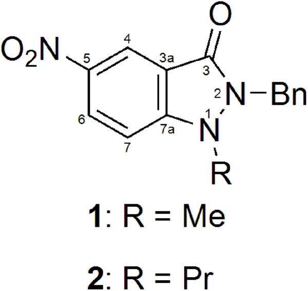


 Open access

