Introduction
Mammalian sperm undergo biochemical and physiological changes in the female reproductive tract for some period of time (Austin, Reference Austin1951; Chang, Reference Chang1951). The changes that render the spermatozoa competent to fertilize are collectively referred to as ‘capacitation’. Capacitation has been correlated with changes in sperm plasma membrane fluidity, intracellular ion concentrations, metabolism and motility (Yanagimachi, Reference Yanagimachi, Knobil and Neill1994). During capacitation, loss of cholesterol from the surface membrane of spermatozoa has been shown to be one of the molecular events of capacitation in vitro (Shadan et al., 2004; Witte et al., 2007). Cholesterol is a major constituent of the cell membrane and is known to regulate the fluidity of membrane lipid bilayers and the permeability of the membrane (Chapman et al., Reference Chapman and Chapman1982; Yeagle et al., Reference Yeagle1985). It has been suggested that removal of membrane cholesterol and the resulting decrease in the cholesterol/phospholipid (C/PL) ratio in the membrane constitutes an important step in sperm capacitation (Davis, Reference Davis and Kabara1978, Reference Davis1981). The requirement for bovine serum albumin (BSA) in regulating capacitation is thought to be due to its ability to remove cholesterol from the sperm plasma membrane (Jha, 2006). However, BSA is contaminated by other serum components including enzymes, hormones, lipids and organic and inorganic ions. Thus, the molecular mechanisms of BSA-induced changes have yet to be determined clearly (Maurer et al., Reference Maurer and Freshney1992).
Cyclodextrins are cyclic oligosaccharides derived from corn by a bacterial process that links six, seven or eight glucose units together in a ring-shaped structure (Valentino, 2008). Cyclodextrins form molecular complexes with lipophiles, a process that usually enhances the solubility of lipophiles in water (Valentino, 2008). Cyclodextrin has also been shown to stimulate the efflux of cholesterol from cultured cells with high efficiency. The β-cyclodextrins, when compared with α- and γ-cyclodextrins, possess a suitable structure to encase natural and synthetic molecules such as hormones, vitamins and lipids components and have the highest affinity for encapsulating sterols, in particular cholesterol (Schonfelder et al., 2009). Recent studies have indicated the abilities of β-cyclodextrins, 2-hydroxypropyl-β-cyclodextrins (2OHpβ CDs) and methyl-β-cyclodextrins (MBCD) to promote cholesterol efflux from mouse L-cell fibroblasts (Kilsdonk et al., Reference Kilsdonk, Yancey, Stoudt, Bangerter, Johnson, Phillips and Rothblat1995). The order of efficiency in accepting cholesterol was found to be MBCD > 2OHp β CDs > β-cyclodextrin (Patricia et al., 1996). Therefore, MBCD may represent a useful tool for studying the mechanism of capacitation, especially in the relationship between the process of removing cholesterol and capacitation. The other authors reported that MBCD treatment removed cholesterol from the mouse and rat sperm membrane and resulted in capacitation (Takeo et al., Reference Takeo, Hoshii, Kondo, Toyodome, Arima, Yamamura, Irie and Nakagata2008; Seita et al., 2009). Exposure to MBCD could induce the acrosome reaction in frozen–thawed bovine sperm cultured in a chemically defined medium (Dinkins et al., Reference Dinkins and Brackett2000). However, the effect of MBCD upon the fertilization of bovine oocytes and their subsequent development has yet to be demonstrated in detail. In the present study, we examined the effects of MBCD during the in vitro fertilization of bovine oocytes in chemically defined conditions, with particular emphasis upon fertilization and subsequent embryonic development.
Materials and Methods
Bovine oocyte collection and in vitro maturation
Oocyte collection and in vitro maturation of oocytes were carried out as previously described by Nagao et al. (Reference Nagao, Iijima and Saeki2008), with some modifications. In short, bovine oocytes were obtained from small antral follicles (2–5 mm in diameter) in slaughterhouse ovaries and the oocytes with intact cumulus cells and evenly granulated cytoplasm were selected for experimental use. Selected oocytes were washed three times and cultured in modified TCM-199 (m-TCM199) for 22–25 h at 39°C under 5% CO2 in air with high humidity. The m-TCM199 consisted of HEPES-buffered medium 199 (Gibco Laboratories) supplemented with 0.1% (w/v) polyvinyl alcohol (Sigma Chemical Co.; PVA), 0.5 mM sodium pyruvate (Nacalai Tesque Inc.), 1% antibiotic–antimycotic (Gibco, AB), 0.02 AU/ml FSH (Antrin) and 1 μg/ml estradiol-17β (Sigma).
Pre-incubation of spermatozoa (PI)
Spermatozoa were pre-incubated in defined medium (Brackett et al., Reference Brackett and Oliphant1975) modified by excluding BSA (Sigma) and supplementing with 0.1% PVA (m-BO) and MBCD (0–3.0 mM, Sigma) in a plastic dish (Falcon) covered with mineral oil for 0–8 h at 39°C in 5% CO2 in air at high humidity. After pre-incubation, the spermatozoa were used for insemination. Frozen–thawed semen from five different sires were used for experiments (sire A for Experiment 1, 2, 4 and 5, sire B, C, D and E for Experiment 3).
In vitro fertilization
In vitro fertilization was conducted as described by Nagao et al. (Reference Nagao, Iijima and Saeki2008). Briefly, matured COCs were inseminated with frozen–thawed spermatozoa (2.0 × 10⁶ cells/ml) immediately after Percoll washing or following pre-incubation in m-BO (10 COCs/100 μl droplets). Oocytes and spermatozoa were co-incubated for 18 h at 39°C in 5% CO2 in air with high humidity. Following co-incubation, the oocytes were freed from spermatozoa and cumulus cells by vortex agitation. Some oocytes were fixed on a slide with 3:1 acetic acid: ethanol and stained with 1% orcein following insemination for assessment of fertilization. Remaining oocytes were cultured to the blastocyst stage.
In vitro culture
In vitro culture of IVF embryos was carried out as previously described Nagao et al. (Reference Nagao, Iijima and Saeki2008). In short, 18–20 h after IVF, IVF embryos were removed from the fertilization medium, stripped of cumulus cells by vortexing and washed twice in modified-SOF supplemented with 3 mg/ml BSA, 0.05% (w/v) PVA and 1% AB (SOF + BSA). Then, 15–50 of these IVF embryos were placed in SOF + BSA and cultured at 39°C under 5% CO2, 5% O2 and 90% N2 at high humidity (20 to30 embryos/50 μl droplet) for 7–8 days.
Assessment of sperm stain
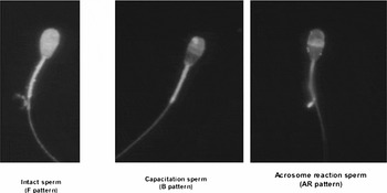
Spermatozoa were stained with chlortetracycline (CTC) stain to evaluate cultured sperm as reported by Fraser et al. (Reference Fraser, Abeydeera and Niwa1995). Briefly, the suspended spermatozoa were stained with 1 μg/ml bis-benzamide 33258 (Sigma) in BO supplement with PVA (BO + PVA). The suspensions were centrifuged at 600 g for 6 min. The precipitated spermatozoa were mixed thoroughly with 750 μM CTC solution (Sigma), 5 mM cysteine (Sigma), 130 mM NaCl (Kanto Chemicals), 20 mM Tris (Sigma: pH 7.8) and then fixed with 12.5% (w/v) paraformaldehyde in 0.5 M Tris–HCl (pH 7.4). At least 100 spermatozoa were observed under a fluorescence microscope (BX-51, Olympus) for bis-benzamide 33285 staining to determine whether they were living or dead using a 330–385 nm filter and a DM 400 dichroic mirror (U-MWU2: Olympus). At least 50 spermatozoa judged as living (Hoechst stain negative) were examined for CTC staining using a 400–440 nm filter and a DM 455 dichroic mirror (U-MWBV2: Olympus). The CTC-staining patterns could be classified into three types according to the method described by Fraser et al.: F (uncapacitated) pattern, bright fluorescence detected over the whole region of the sperm head; B (capacitated) pattern, fluorescence detected on the sperm head but not over the post-acrosomal region; AR (acrosome-reacted) pattern, weak fluorescence observed over the sperm head with a bright band sometimes present in the equatorial region (Fig. 1).
F, B and AR patterns were shown below. F (uncapacitated) pattern, bright fluorescence detected over the whole region of the sperm head; B (capacitated) pattern, fluorescence detected on the sperm head but not over the post-acrosomal region; AR (acrosome-reacted) pattern, weak fluorescence observed over the sperm head with a bright band sometimes present in the equatorial region.

Assessment of fertilization and early development
To evaluate the pronuclear stage during fertilization, oocytes considered to be fertilized were classified in three groups: ‘2PN’ (female pronucleus, male pronucleus and sperm tail; normal fertilization); ‘polyspermy’ (two or more sperm tails in the cytoplasm with condensed heads, or two or more decondensed heads in the cytoplasm); and ‘asynchrony’ with a condensed sperm head (female pronucleus and a condensed sperm head). The percentage of each category of fertilization was calculated from the total number of oocytes inseminated. Early development was examined with a stereomicroscope (×60) for cleavage and development to the blastocyst stage 7 days (168 h) after insemination. Cleaved embryos included embryos from the 2-cell stage to the blastocyst stage. Only embryos with a clear blastocoel and an inner cell mass were counted as blastocysts.
Evaluation of chromosomal integrity at the blastocyst stage
Chromosomes were counted as previously described (Yoshizawa et al., Reference Yoshizawa, Konno, Zhu, Kagegama, Fukui, Muramatsu, Kim and Araki1999). Briefly, some blastocysts were washed and placed in 1% sodium citrate solution for 15 min and fixed by pouring 20–30 μl of acetic–alcohol fixative (acetic acid 1: ethanol 1) into 0.4 ml of sodium citrate solution. Blastocysts were placed onto a glass slide, immediately covered with a very small droplet of acetic acid to separate each blastomere and then immediately re-fixed with several drops of the initial fixative. After drying completely, chromosome samples of blastocysts were stained with 2% Giemsa solution for 10 min. The numbers of chromosomes were counted as ‘normal’, 60 (2n), or ‘abnormal,’ 30 (n) and 90 (3n).
Vitrification, warming and transfer of blastocysts to recipient heifers
To examine the developmental ability to offspring of the blastocysts derived from IVF with PI sperm with CD, some blastocysts were vitrified, warning and transferred to recipients. Vitrification was carried out as previously described. The vitrification solution used (VSED) was PBS (Sigma) containing 4.5 M ethylene glycol (Wako Pure Chemical Industries) and 3.5 M dimethyl sulphoxide (Wako Pure Chemical Industries) as cryoprotectants. Blastocysts were equilibrated with 50% VSED and then vitrified with VSED in 0.25 ml straw (FHK). After storage in the liquid nitrogen, the straws were warmed in a 20°C water bath. The contents of the straws were expelled onto a petri dish and recovered blastocysts were transferred into 1 ml of PBS with 0.5 M sucrose. Five minutes later, blastocysts was washed with m199 supplemented with 10% FCS (Equitech Bio.) and transferred non-surgically to recipient heifers 7 days after estrus (1 embryo per recipient). Diagnosis of pregnancy was carried out by ultrasonography on day 30 following transfer.
Experimental design
Experiment 1
To determine the effect of MBCD on sperm viability, capacitation and acrosome reaction, spermatozoa were stained using the CTC method described earlier after 0, 2, 4, 6 and 8 h of incubation in BO + PVA supplemented with 1.5 mM MBCD (MBCD) or BO + PVA (control).
Experiment 2
We also examined the effects of MBCD concentration and length of pre-incubation period of spermatozoa upon fertilization. Frozen–thawed spermatozoa were incubated with 0, 0.25, 0.5, 1, 1.5, 2 and 3 mM MBCD for 4 h, or with 1.5 mM MBCD for 0, 3 and 10 min and 0.5, 1, 2, 4 and 8 h (2.0 × 10⁶ cells/ml). Spermatozoa were then co-incubated with matured oocytes.
Experiment 3
To investigate for characteristic differences in the sensitivity to MBCD, frozen–thawed spermatozoa from four different sires (B, C, D and E) were pre-incubated with 0, 0.5, 1.5 and 3 mM MBCD for 4 h and then the spermatozoa were co-incubated with matured oocytes.
Experiment 4
Examination was made of the effects of MBCD concentration and the length of pre-incubation period of spermatozoa on subsequent early development following in vitro fertilization. Fertilized embryos derived from co-incubation of matured oocytes in the frozen–thawed spermatozoa were pre-incubated with 0.5 and 1.5 mM MBCD for 0.5, 2 and 4 h were cultured for 7 days.
Experiment 5
Three blastocysts derived from oocytes fertilized with sperm pre-incubated with MBCD were transferred to three recipient heifers to investigate subsequent viability of the blastocyst. Remaining blastocysts were examined in order to evaluate chromosome integrity.
Statistical analysis
The percentage data obtained from each experiment were analysed by analysis of variance (ANOVA) followed by Fisher's protected least significant difference (PLSD) test. Differences of p < 0.05 were considered significant.
Results
Experiment 1
After 0, 2, 4 and 8 h of incubation in BO + PVA supplemented with 1.5 mM MBCD, the proportion of live sperm decreased with the length of the incubation time (Table 1, p < 0.05). However, that in BO + PVA without MBCD did not decrease in the incubation (Table 1; p > 0.05).
Effect of MBCD on sperm viability in bovine sperm (n = 3–12)

n = Number of replicated experiments.
a,bValues differ significantly (p < 0.05).
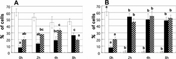
The proportion of live sperm displaying the B (capacitated) pattern at 2, 4 and 8 h of incubation in BO + PVA supplemented with 1.5 mM MBCD, were higher than that at 0 h (Fig. 2, p < 0.05). On the other hand, that in BO + PVA without MBCD was not different with the incubation time (Fig. 2, p > 0.05).
Effect of MBCD on capacitation and the acrosome reaction in bovine sperm. Uncapactitated bovine sperm suspension was incubated for 0, 2, 4 and 8 h in the absence (A) or presence (B) of 1.5 mM MBCD in modified BO + PVA and then analysed using CTC stain. Experiments were repeated three to eight times. Data are percentage of cells expressing the F pattern (white), the B pattern (black) and AR pattern (chequered) of CTC fluorescence. a–c, letters within same content indicate significant differences (ANOVA, p < 0.05).

Experiment 2
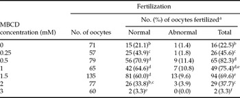
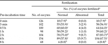
As shown in Table 2, the frequencies of normal fertilization at 0.5, 1.0 and 1.5 mM MBCD were higher than that of other concentrations (p < 0.05). As shown in Table 3, the frequencies of normal fertilization at 3 min, 10 min, 0.5, 1, 1.5, 2 and 4 h were higher than that of other periods (p < 0.05) and the highest rate of normal fertilization was evident in the 2-h group (p < 0.05).
Effects of MBCD concentration in sperm preincubation for 4 h on in vitro fertilization of bovine oocytes

aPercentage of inseminated oocytes.
b–fValues differ significantly in the same column (p < 0.05).
Effects of time in sperm preincubation with 1.5 mM MBCD on in vitro fertilization on bovine oocytes

aPercentage of inseminated oocytes.
b–dValues differ significantly in the same column (p < 0.05).
Experiment 3
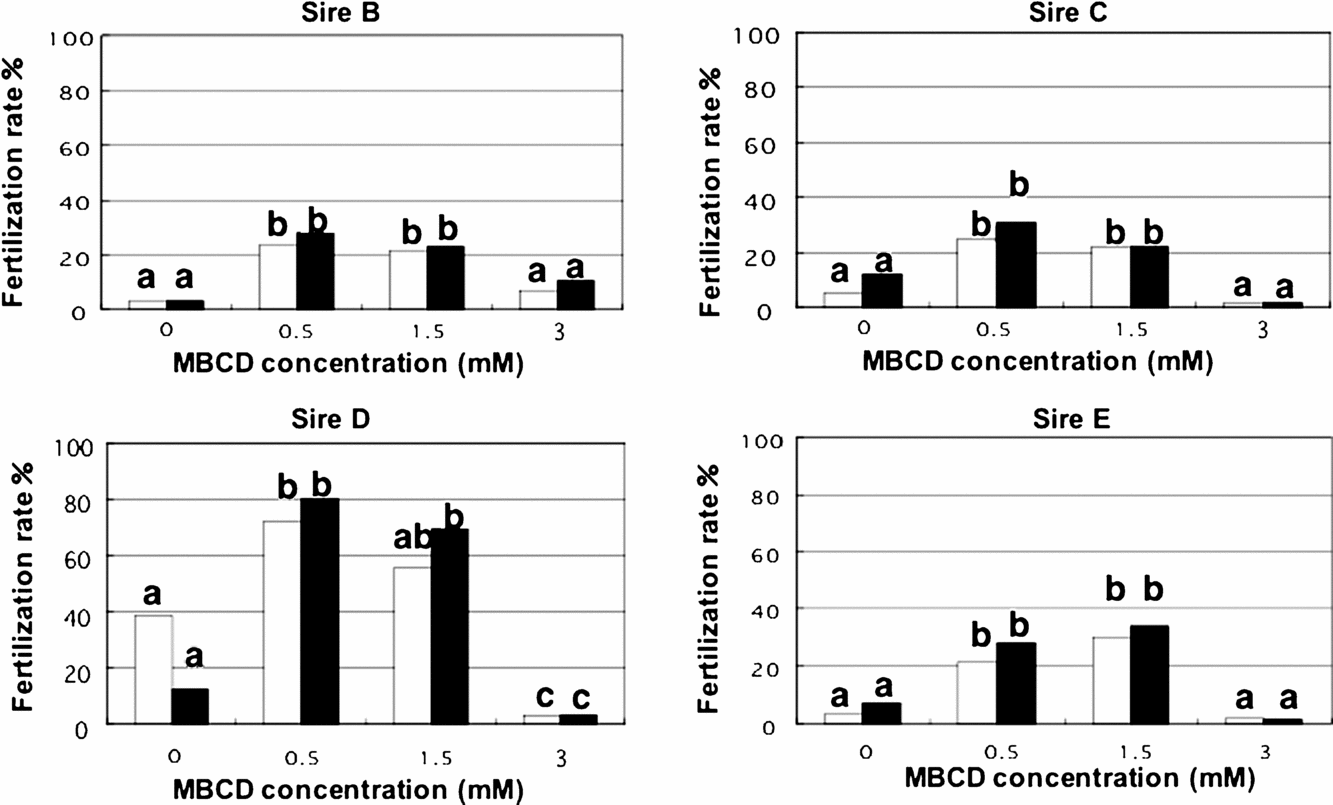
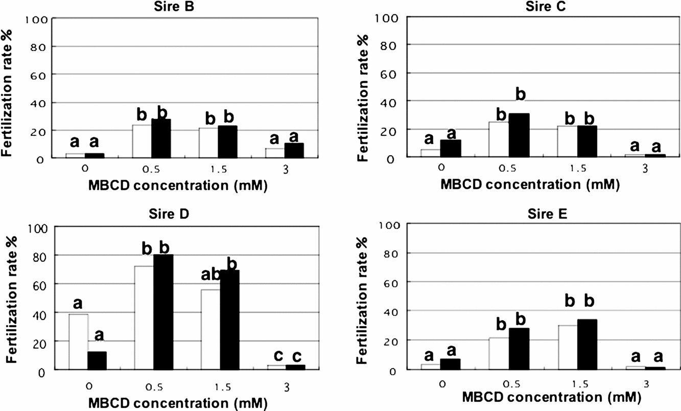
As shown in Figure 3, the frequencies of normal fertilization of sperm from 4 different sires exhibited similar tendencies which were dependent upon the concentration of MBCD and pre-incubation with 0.5 or 1.5 mM MBCD for 4 h was optimum for promoting fertilization in vitro (p < 0.05).
Sensibility to MBCD of sperm derived form different sires in sperm pre-incubation with various MBCD concentrations for 4 h on in vitro fertilization of bovine oocytes. Normal: embryos with two pronuclei and one sperm tail. Total: all penetrated oocytes. Data are percentage of fertilization expressing the normal (white) and total (black). a–c, letters within same content indicate significant differences (ANOVA, p < 0.05).
Experiment 4
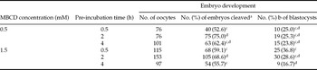
As shown in Table 4, the frequencies of cleavage were the same as that of normal fertilization in Experiment 2. The frequencies of development to the blastocyst stage were influenced by both the concentration of MBCD and the pre-incubation period (p < 0.05) and were highest in the 1.5 mM and 0.5 h group (p < 0.05).
In vitro development of bovine oocytes fertilized by spermatozoa for various time preincubated with various MBCD concentration

aPercentage of cultured oocytes.
bPercentage of cleaved oocytes.
c,dValues differ significantly in the same column (p < 0.05).
Experiment 5
We examined chromosomal integrity of blastocysts derived from oocytes fertilized with spermatozoa pre-incubated with MBCD (63.6%: 21/33). One recipient heifer became pregnant (pregnancy rate: 1/3, 33.3%) and delivered a live calf.
Discussion
In Experiment 1, sperm cultured in BO + PVA with MBCD demonstrated decreased rate of live sperm when compared with sperm cultured in BO + PVA (Table 1). Cyclodextrins caused the release of some membrane components such as cholesterol, phospholipids and proteins from erythrocytes (Irie et al., Reference Irie, Otagiri, Sunada, Uekama, Ohtani, Yamada and Sugiyama1982). These changes of the membrane components may affect sperm viability, resulted in decreasing the viability of the sperm.
On the other hand, sperm cultured in BO + PVA supplemented with MBCD exhibited higher capacitation and acrosome reaction rates in vitro compared with sperm cultured in BO + PVA (Fig. 2). This finding shows that MBCD in the pre-incubation made the sperm fertilizable resulted in a significant enhancement in the rate of sperm incorporation into oocytes (Tables 2 and 3; Fig. 3). MBCD has been shown to be able to enhance capacitation, acrosome reaction and fertility (Choi & Toyoda, Reference Choi and Toyoda1998). Cholesterol effluxed from sperm membrane by MBCD (David et al., Reference Davis1981; Irie et al., Reference Irie, Otagiri, Sunada, Uekama, Ohtani, Yamada and Sugiyama1982). Loss of cholesterol induces a change in the stability and distribution of lipid rafts (Simons et al., 1997). A change of the architecture of lipid rafts may regulate an influx of Ca2+ ion coupled with the release of intracellular Ca2+ leading to the opening of store-operated channels in the plasma membrane in mouse sperm (Trevino et al., 2001). While numerous events are known to occur during capacitation, it appears that the regulation of the intracellular concentration of Ca2+ is one of the most important for capacitation and acrosome reaction. Therefore, we inferred that MBCD may reduce cholesterol from sperm membrane and increase Ca2+ concentration in the sperm and localized around the oocytes resulted in enhancement of the rate of the fertilization.
In Experiment 2, we examined the effects of pre-incubating spermatozoa in m-BO with MBCD prior to insemination on subsequent fertilization and embryo development. The frequencies of normal fertilization at pre-incubation with 0.5, 1.0 and 1.5 mM MBCD for 4 h (Table 2) and pre-incubation for 3, 10 and 30 min and 1, 2 and 4 h at 1.5 mM MBCD (Table 3) were higher than that of the other concentrations and pre-incubation periods. The effects of MBCD upon fertilization depended on the concentration of MBCD and the incubation period, as reported previously in mice (Choi & Toyoda, Reference Choi and Toyoda1998). MBCD has been shown to enhance enhanced sperm capacitation and fertilization on mouse, rat and bovine (Dinkins et al., Reference Dinkins and Brackett2000; Seita, et al., 2008; Takeo, et al., Reference Takeo, Hoshii, Kondo, Toyodome, Arima, Yamamura, Irie and Nakagata2008) and increase Ca2+ concentration in sperm (Takeo et al., Reference Takeo, Hoshii, Kondo, Toyodome, Arima, Yamamura, Irie and Nakagata2008). Increasing of Ca2+-induced capacitation, hyperactivation and fertilization. There is evidence that Ca2+ was involved in signalling the onset of hyperactivation. Increased intracellular Ca2+ has been detected in hyperactivated hamster and bull sperm (Suarez et al., 1993; Ho & Suarez, 2001). Ca2+ increased cAMP in sperm cell along with pH increases, have been correlated with onset of hyperactivated motility during capacitation of sperm in vitro (White & Aitken, 1989; Yanagimachi, Reference Yanagimachi, Knobil and Neill1994). The hyperactive state associated with the acrosome reaction allows sperm to penetrate the cumulus and zona pellucida surrounding the egg (Suarez et al., 2007). Our results, combined with these previous reports indicate that the pre-incubation of bovine spermatozoa with MBCD prior to insemination may have promotional effects upon the capacitation of sperm, resulting in enhanced subsequent fertilization under defined conditions.
Examination was made of the sire variation of sensibility to MBCD in pre-incubation using spermatozoa derived from four different sires in Experiment 3. Fertilization rate was different among each of the four sires, although the frequencies of normal fertilization at 0.5 and 1.5 mM MBCD was higher than that at 0 and 3 mM MBCD with all spermatozoa. In fact, it is thought that the efflux of cholesterol from the sperm membrane relates closely to the process of capacitation. However, each of the fertilization rates was distinct presumably reflecting differences in the spermatozoa. According to Hoshi et al. (Reference Hoshi, Aita, Yanagida, Yoshimatu and Sato1990), who examined the concentration of cholesterol (C) and phospholipids (PL) in spermatozoa found that the C/PL ratio varied considerably from man to man. Lower C/PL ratios have been shown to relate to faster sperm penetration and capacitation. Capacitation status and fertilization rates have been shown to be difference in in vitro (Suzuki et al., 2003). These results suggest that the effects of MBCD on sperm might be universal, however there is variation of sensitivity to MBCD among various sperm from various sires.
In Experiment 4, we found that embryos treated under in these experimental conditions developed to the blastocyst stage normally (Table 4). Such blastocysts exhibited normal chromosomal integrity and normal developmental ability to term (Experiment 5). Chromosomal normality has been shown to be around in 60% (20/33) of the embryos (Yoshizawa et al., Reference Yoshizawa, Konno, Zhu, Kagegama, Fukui, Muramatsu, Kim and Araki1999). Birth rates of calves following the transfer of in vitro produced embryos were has been shown to be 38.8–57.1% (Numabe et al., 2001). In addition, it was also reported that live offspring were born when mouse eggs fertilized by spermatozoa treated with MBCD were transferred to recipients (Choi & Toyoda, Reference Choi and Toyoda1998, Takeo et al., Reference Takeo, Hoshii, Kondo, Toyodome, Arima, Yamamura, Irie and Nakagata2008). Our results showed that bovine embryos fertilized with MBCD-treated spermatozoa in protein-free medium exhibit developmental ability to term firstly. These finding showed that it is effective to be MBCD as a means of in vitro embryo pregnant.
We conclude that MBCD induces the capacitation of bovine sperm in a protein-free medium, resulting in the effective and normal fertilization of bovine oocytes. Oocytes fertilized with spermatozoa pre-incubated with MBCD developed to the blastocyst stage normally and exhibited normal developmental ability to term.
Acknowledgements
The authors thank Dr Yutaka Toyoda, Obihiro University of Agriculture and Veterinary Medicine, for critical reading of the manuscript, Miyagi Prefectural Livestock Experiment Station for providing the frozen semen and Chikusei Meat Center and Nakao Chikusan Co. Ltd for providing the ovaries, used in this study.