INTRODUCTION
Research on calcareous sponges of Greenland dates back to the 18th Century, the first specimens being described by Fabricius (Reference Fabricius1780). Until the first part of the 20th Century several papers on the Calcarea of Greenland were published, and about 30 species are reported from Greenlandic waters (Schmidt, Reference Schmidt1869, Reference Schmidt1870; Haeckel, Reference Haeckel1870, 1872, 1874; Fristedt, Reference Fristedt1887; Breitfuss, Reference Breitfuss1897, Reference Breitfuss1898c, Reference Breitfuss1933; Vanhöffen, Reference Vanhöffen1897; Lundbeck, Reference Lundbeck1909; Brøndsted, Reference Brøndsted1914, Reference Brøndsted1916, Reference Brøndsted1933a, b; Burton, Reference Burton1934; Laubenfels, Reference Laubenfels1942; Koltun, Reference Koltun1964; Tendal, Reference Tendal1970; Thorbjørn & Petersen, Reference Thorbjørn and Petersen2003; Rapp, Reference Rapp2004a, Reference Rappb), and in the early works of Fristedt, Schmidt and Haeckel several new species were described from Greenland, species that later have been considered as widely distributed in the Atlantic Ocean.
During the last 30 years there has been extensive sampling of benthic invertebrates in the Thule area on the north-western coast, along Peary Land, Jørgen Brønlund Fjord and Independence Fjord on the north-eastern coast, in the Kap Farvel area on the southernmost coast, and on the banks along the south-western coast of Greenland. This new and voluminous material has been of great value for this revision. Brøndsted (Reference Brøndsted1914) made a catalogue of the species known in the area at that time, but no comprehensive revisions including full description of species have until now been made.
The main aim of this publication is, therefore, to provide a faunal inventory, a taxonomic revision of the species, and an identification key. In order to make the publication as independent as possible from older publications, all encountered species are described and depicted wherever possible. Of the 28 species of Calcarea described here, six are new records for Greenland, two are resurrected and one is new to science.
MATERIALS AND METHODS
The present study is mainly based on sponge specimens from the collections in the Zoological Museum of Copenhagen, with some additions from the Zoological Museum, University of Oslo (The Norwegian Expeditions to East Greenland 1930, 1931 and 1933, marked Hoel's stations). Specimens from the inter-Nordic project BIOICE (benthic invertebrates from Icelandic waters) have been included for comparison and complementary illustration of particular species. Type material has been studied when available. Wherever possible the species are fully described and depicted.
Study area
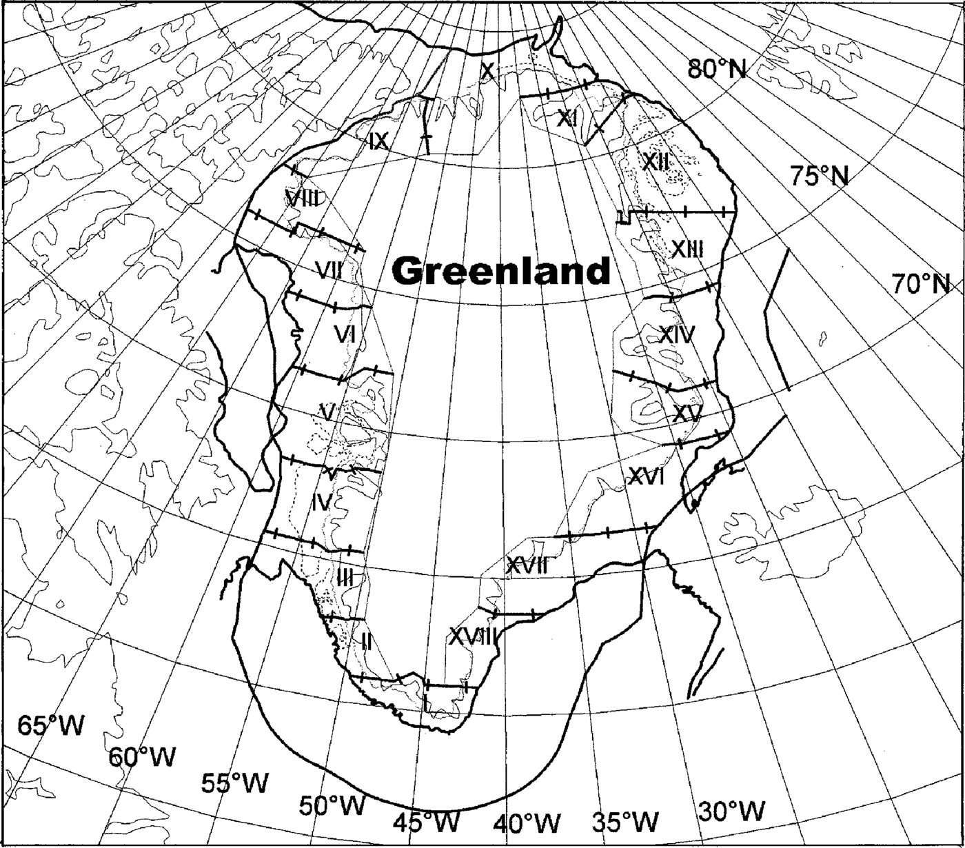
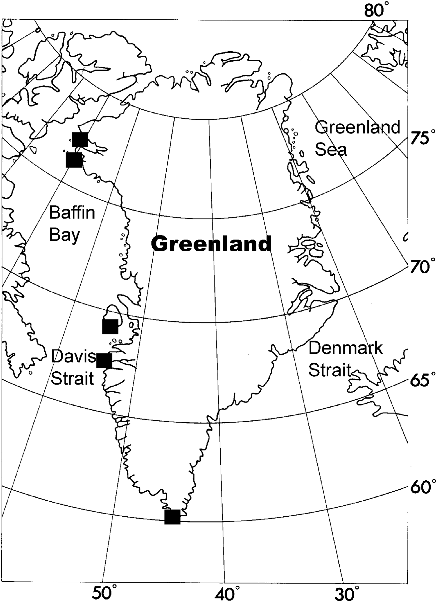
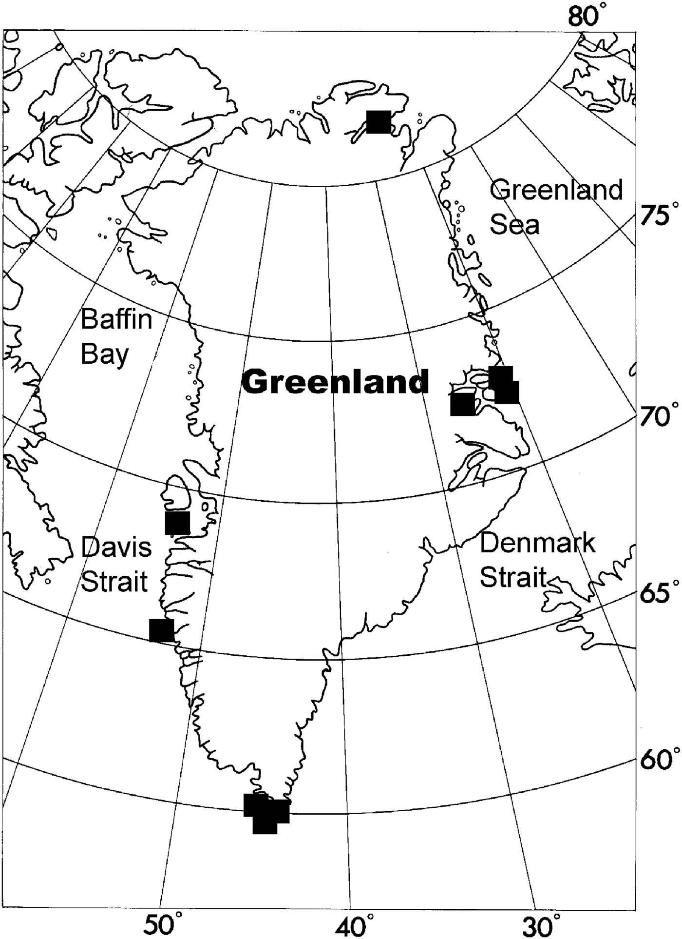
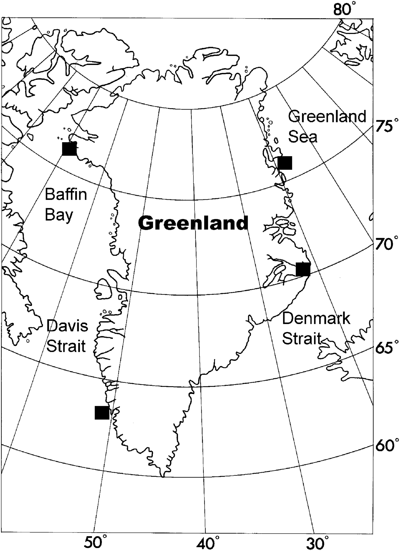
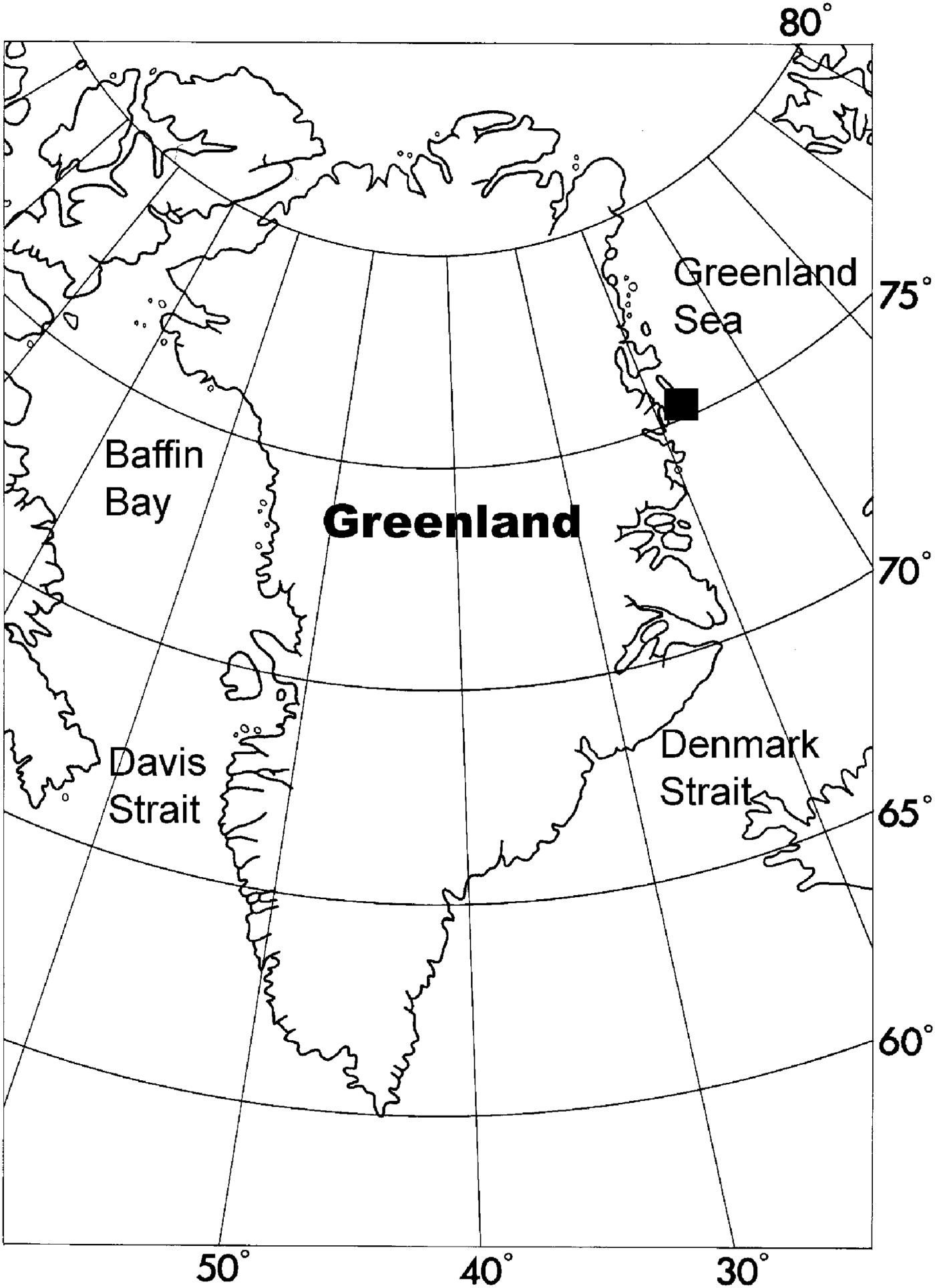
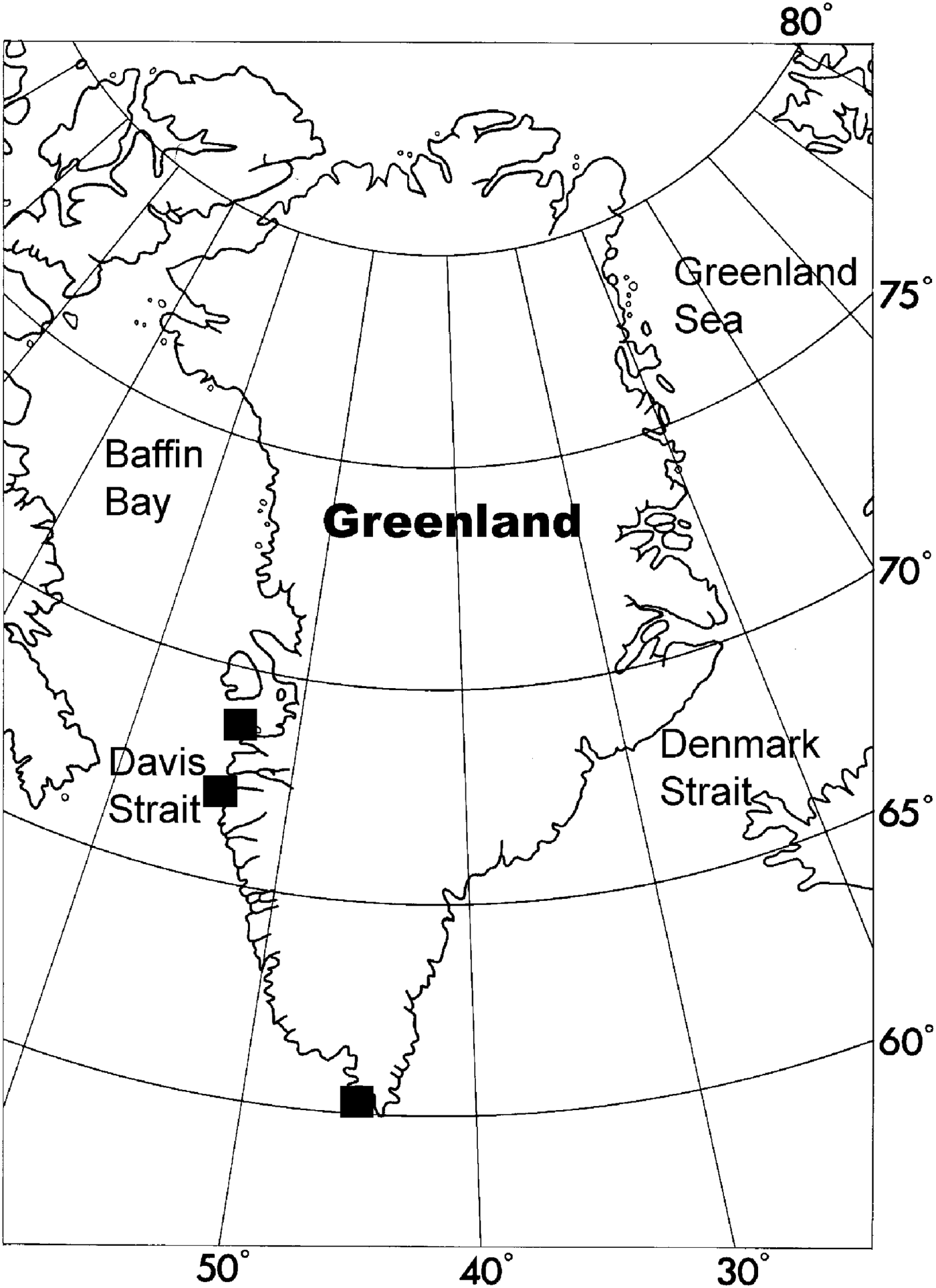
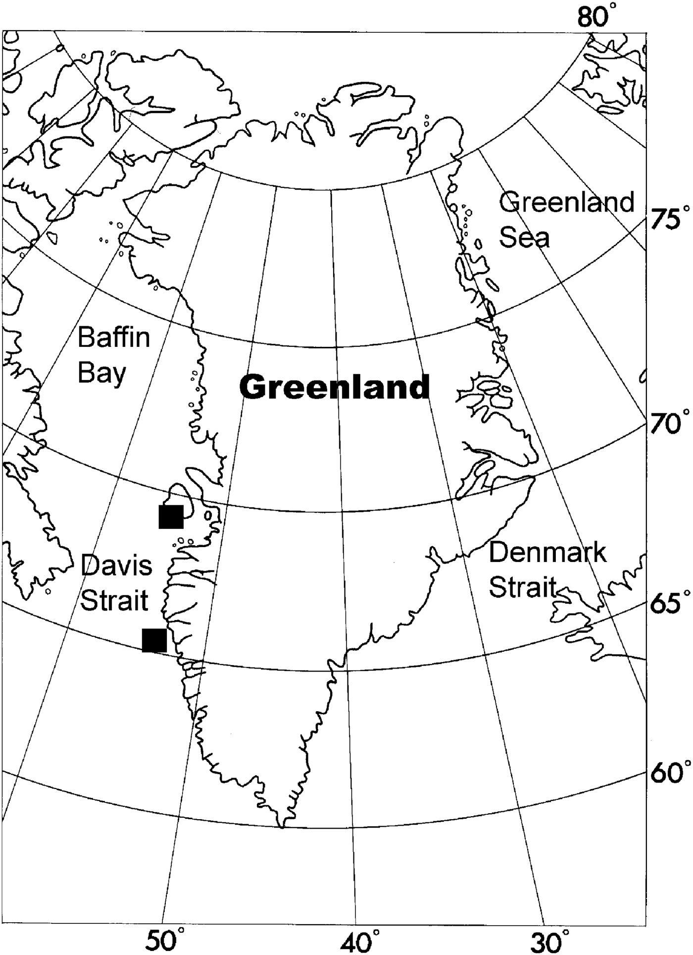
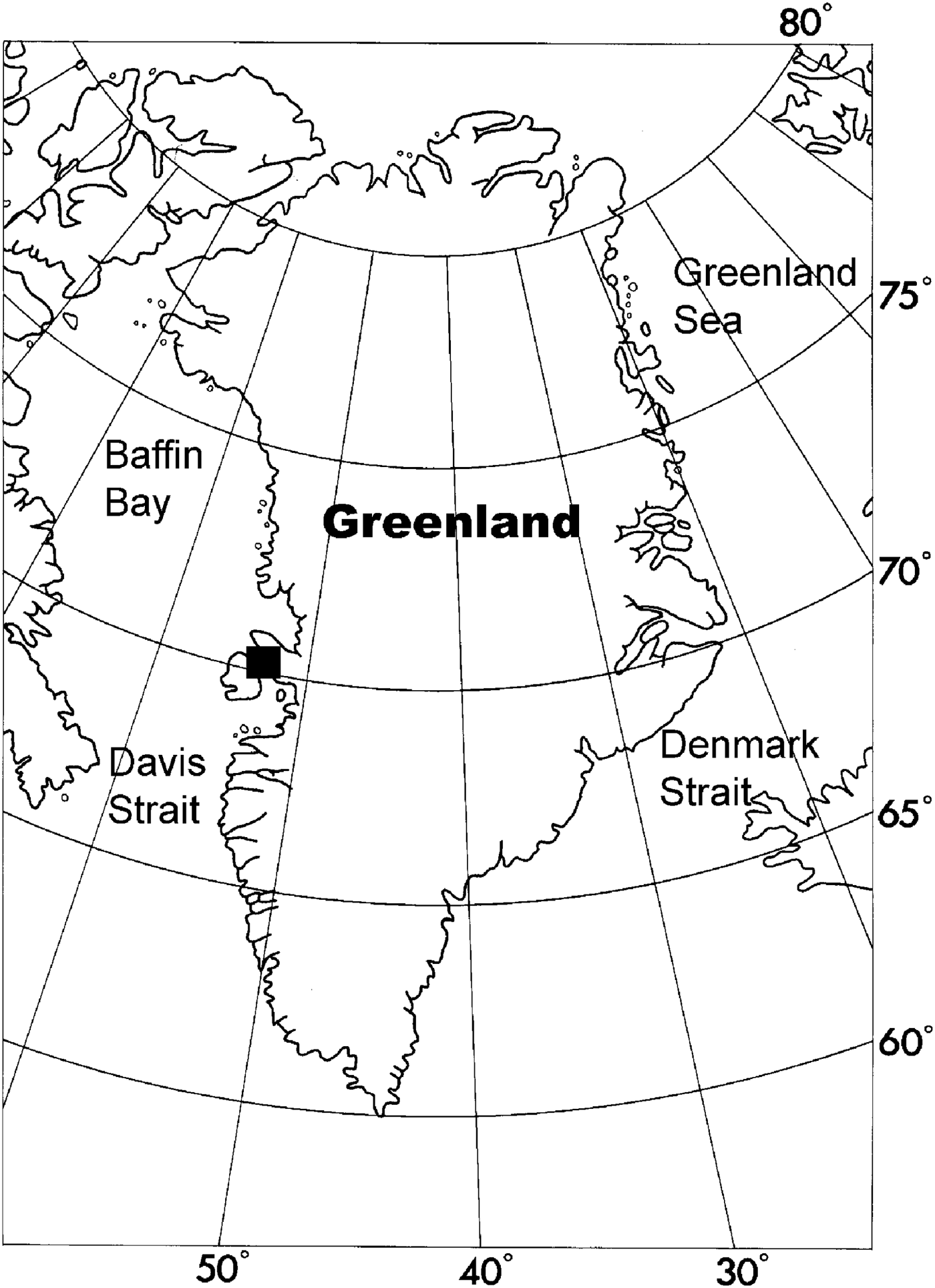
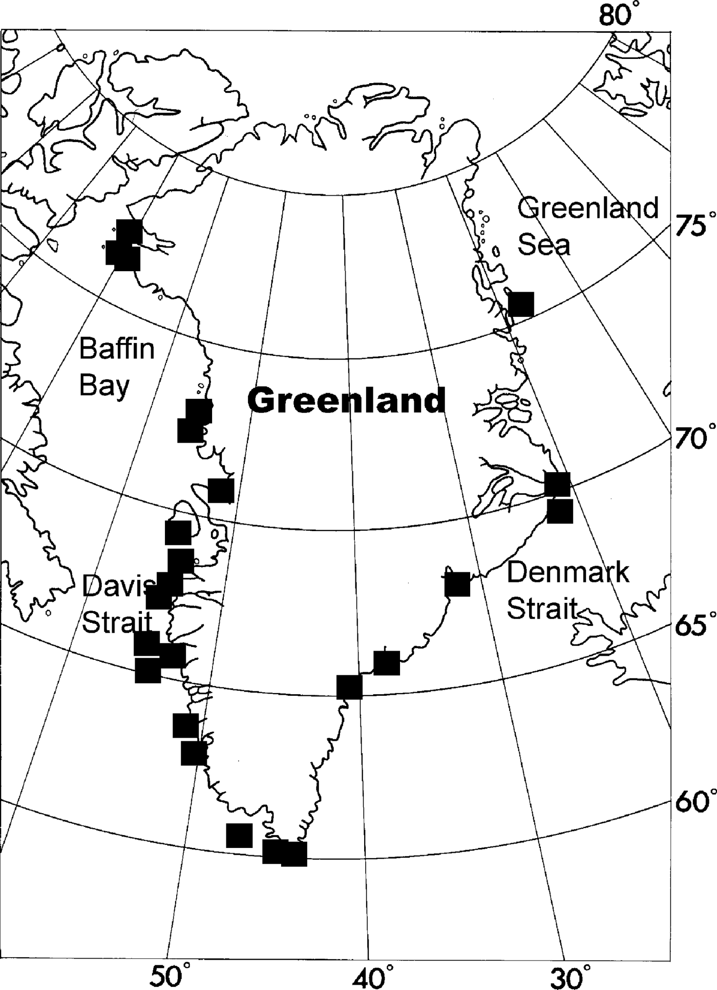
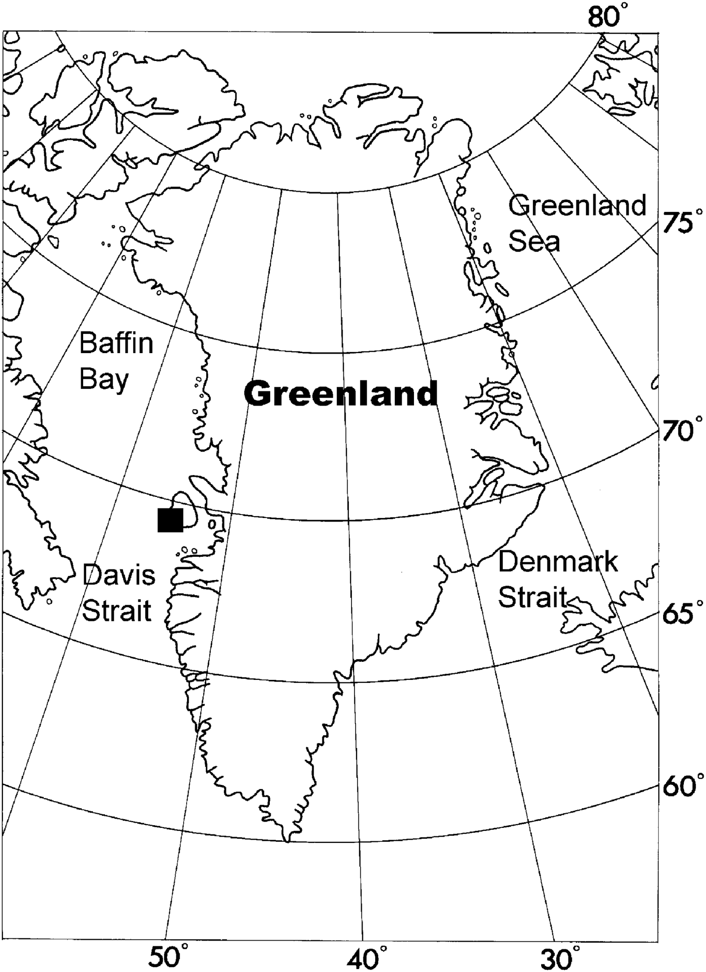
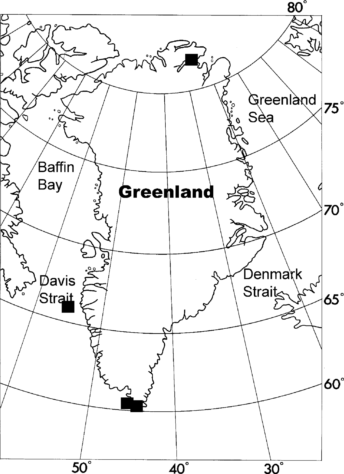
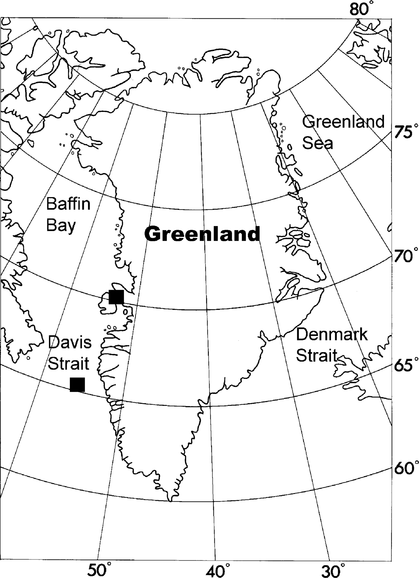
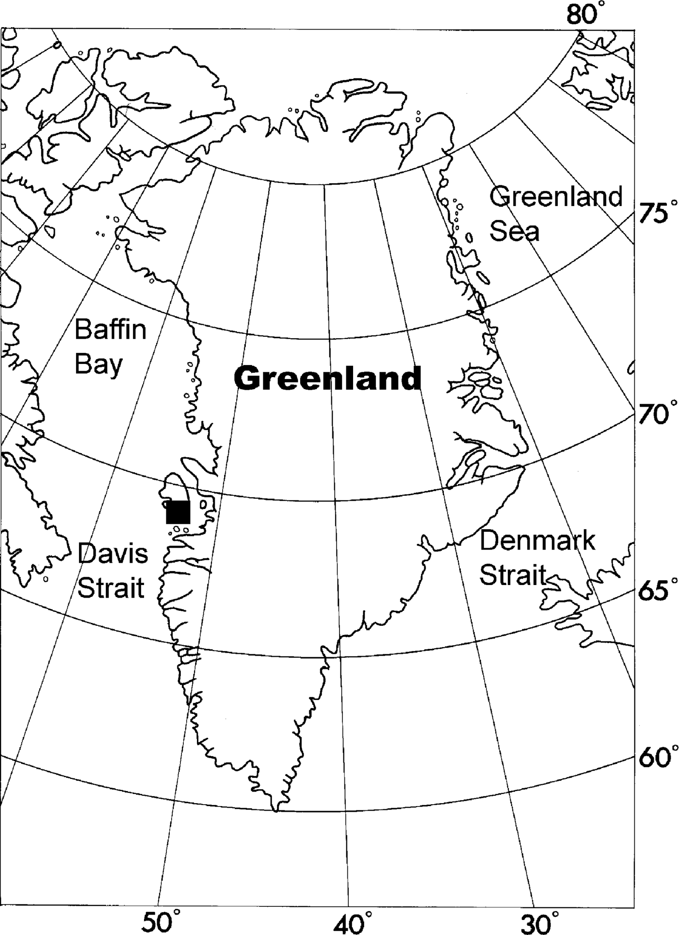
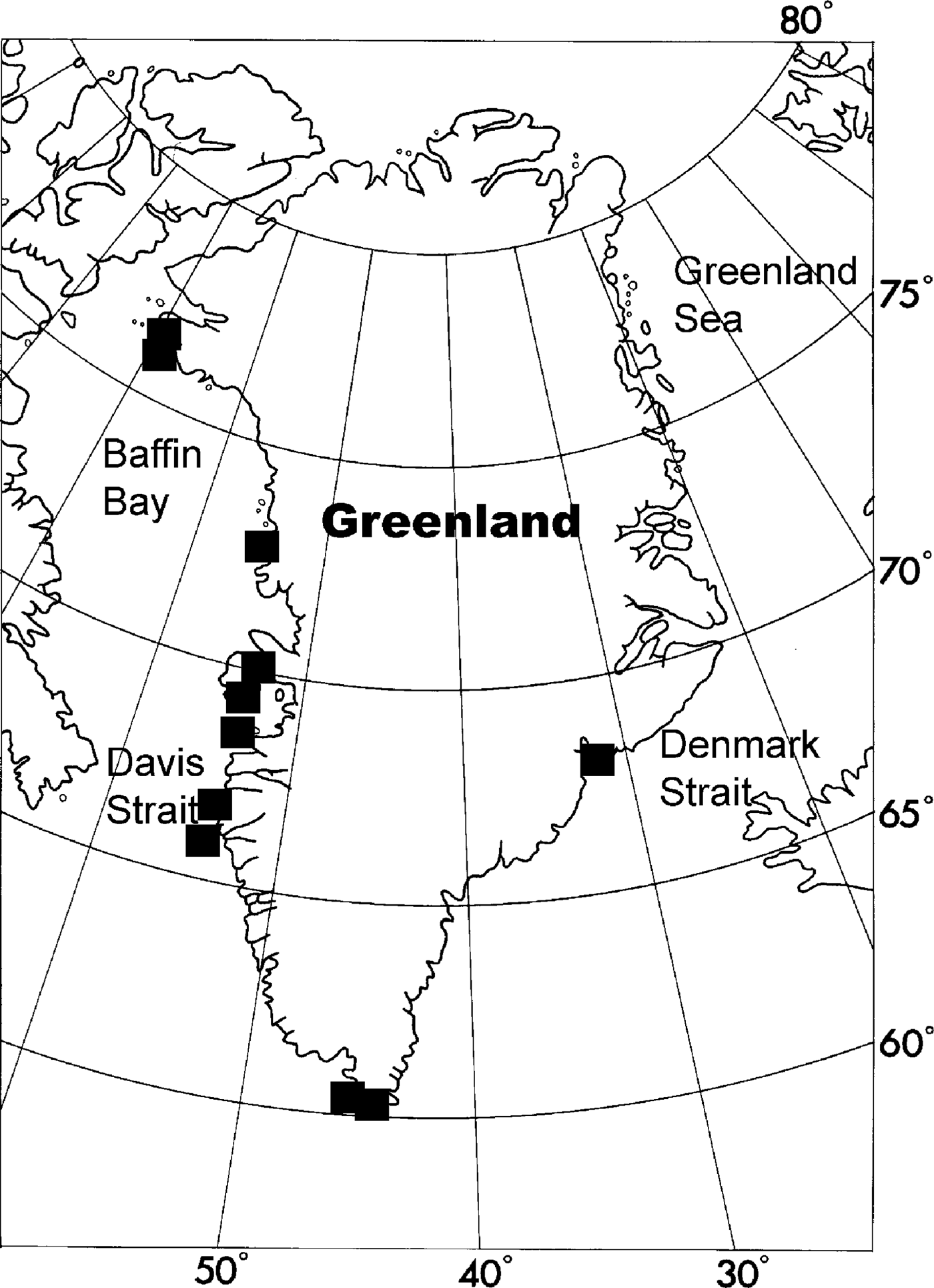
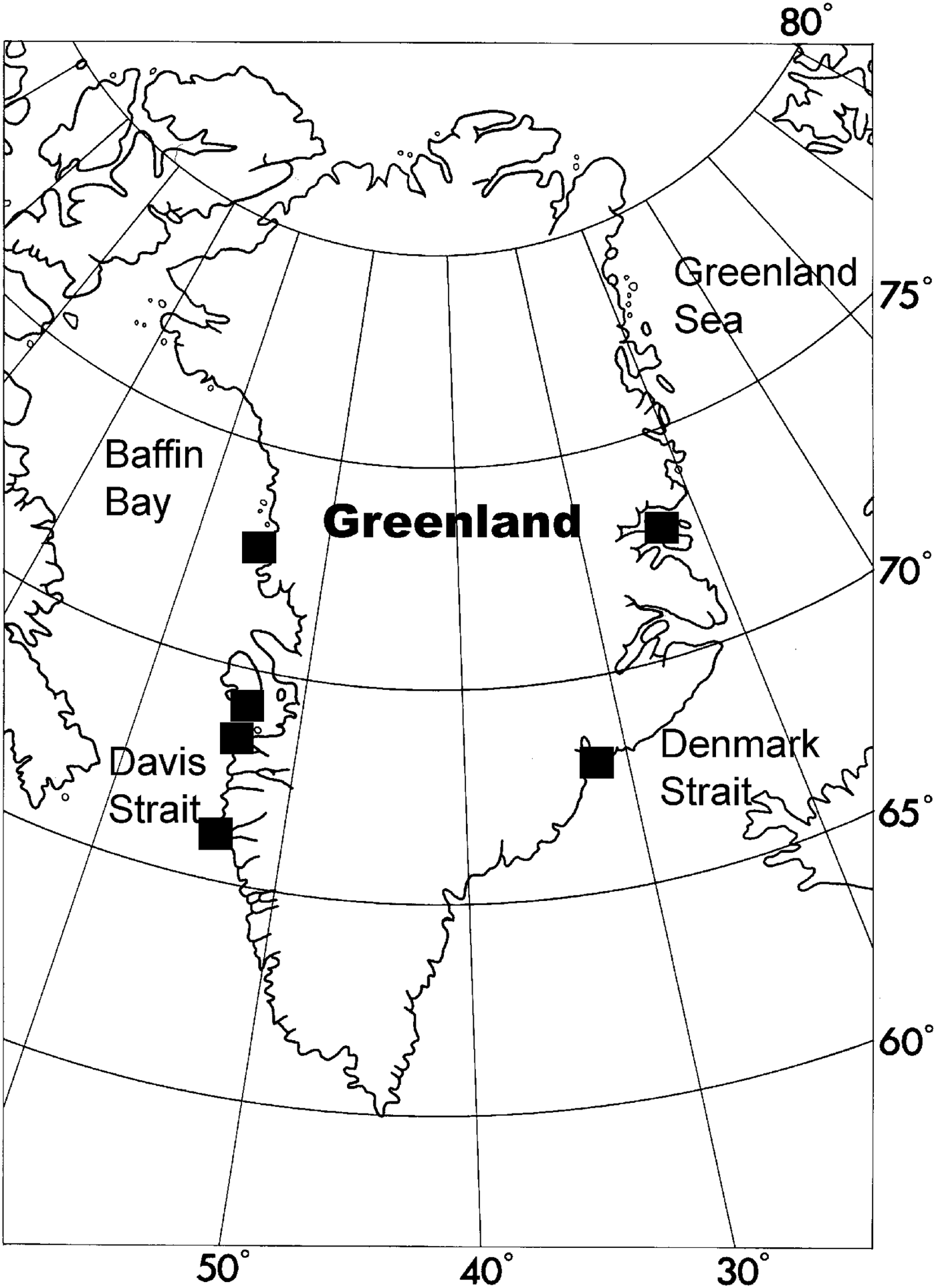
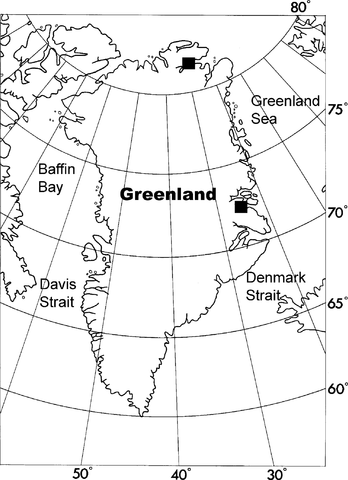
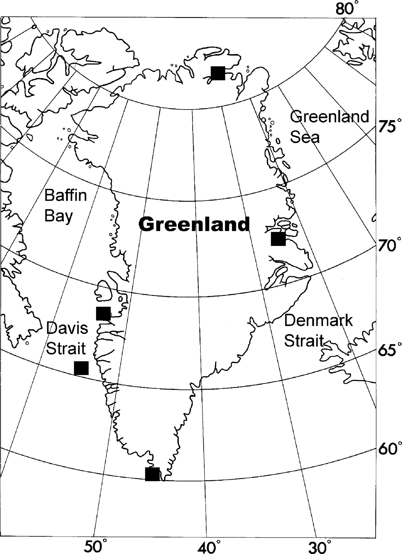
The present study area comprises the entire Greenlandic coast from the southern tip of Kap Farvel (about 59°60′N) to the northernmost coast of Peary Land at about 83°N. The study comprises the area that is within the scope for the marine benthic fauna of Greenland, including the coast and shelf areas down to 1000 m depth (Tendal & Schiøtte, Reference Tendal and Schiøtte2001) (Figure 1). The coasts of Greenland have many types of marine habitats, ranging from fjords heavily influenced by glaciers to a highly exposed and topographically diverse outer coastline. Throughout the Greenlandic coast there are distinct gradients in temperature, salinity, topography and ice cover, resulting in a diverse and varied marine fauna and flora. The southern coast is influenced by Atlantic water with quite high average temperature (about 5–6°C) and high salinity (about 34.3S). The West Greenland Current and the Labrador Sea Current dominate the western coast, characterized by Arctic cold (−1.3–0.8°C) and low salinity (32.5–33.9S) water (Thorson & Ussing, Reference Thorson and Ussing1934; Dinter, Reference Dinter2001). The area north and north-east of Greenland is dominated by polar water with a temperature lower than 0°C and salinity lower than 34.4S. In general these waters are ice-covered throughout the year (Wadhams, Reference Wadhams1981) and thus belong to the permanent pack ice zone (Hempel, Reference Hempel, Gray and Christiansen1985). Along the eastern coast approaching the Denmark Strait the surface water is still highly influenced by the East Greenland Current carrying cold polar water, but in deeper layers warmer and more saline Atlantic water becomes more dominant (Coachman & Aagaard, Reference Coachman, Aagaard and Herman1974). The north-eastern part of the Denmark Strait is influenced by Arctic water from the Iceland Current, the western part by a cyclonic gyre located between the Iceland–Faroe Ridge and the Jan Mayen Ridge, a water mass with a temperature–salinity (TS) signature indicating an intermediate between polar water and Atlantic water (Hopkins, Reference Hopkins1991; Hansen & Østerhus, Reference Hansen and Østerhus2000). Further south the western branch of the Irminger Current with Atlantic origin joins the East Greenland Current, and temperature and salinity are increasing heading towards the southern coast.
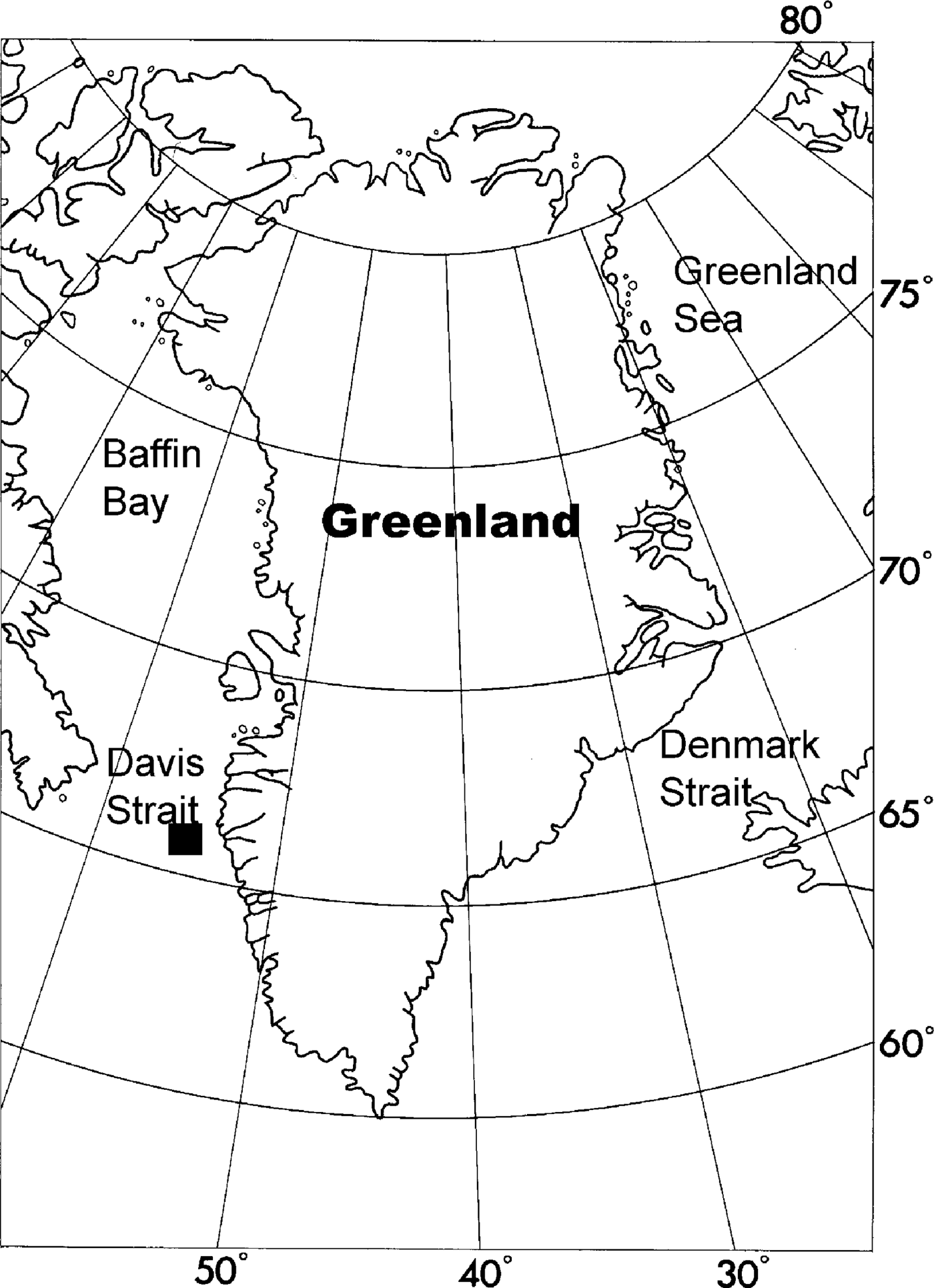
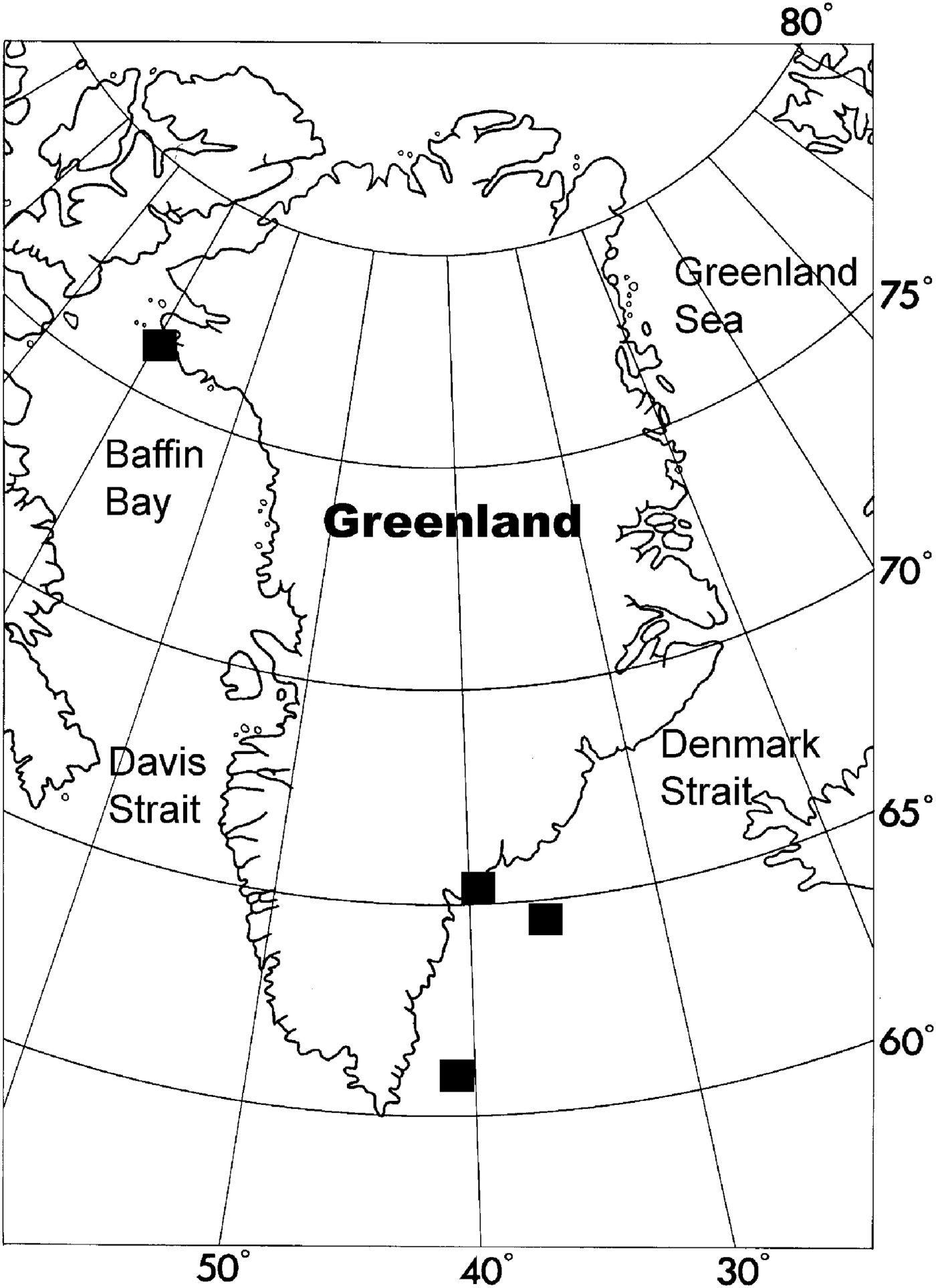
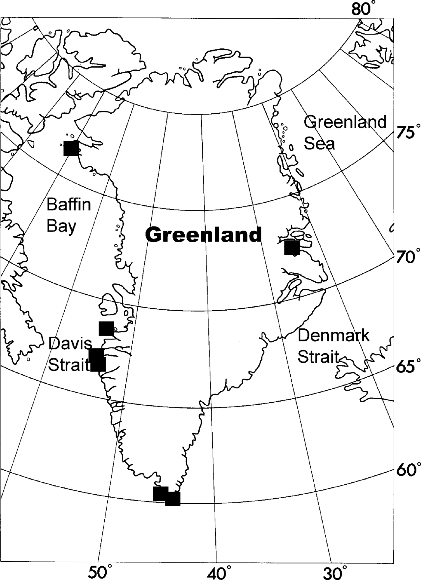
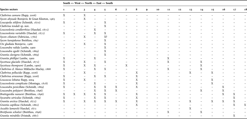
Map of Greenland. This map where the coastline of Greenland is divided into 18 zones was constructed as a tool to map the distribution of marine invertebrates in Greenlandic waters (Tendal & Schiøtte, Reference Tendal and Schiøtte2001) (Zoological Museum, SNM, University of Copenhagen). Distribution of Calcarea in the area is given with reference to these zones.

Description of species
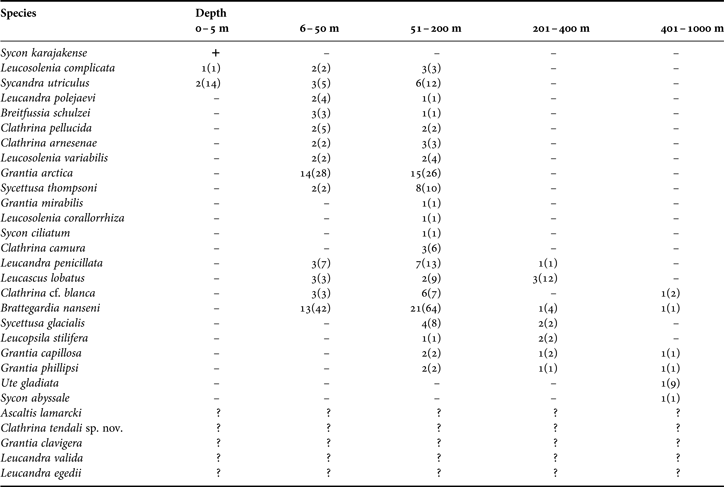
For each species the following information is given. Synonyms and citations, including original allocation and other names used in the north-east Atlantic and Arctic regions. Type locality and deposition of type material (when known). Previous records from the Greenlandic coast. Overview of material examined, given as follows: expedition, station number, name of locality, coordinates in decimal degrees, collection date, depth interval and number of specimens. Where nothing else is stated, the material is deposited in the Zoological Museum of Copenhagen. Etymology (for new species). A full description, including spicule measurements. Occurrence along the Greenlandic coast is given with reference to the different sectors introduced in the Fauna Groenlandica project (Tendal & Schiøtte (Reference Tendal and Schiøtte2001), Zoological Museum (SNF), University of Copenhagen) (Figure 1), in addition to comments on the total distribution range. Distribution maps are based on examined material, but also type locality is included when known. Misidentifications of calcareous sponges are numerous in the literature, hence the inclusion of such records could be a considerable source of error. Selected and validated literature records are provided in the text. Descriptions and photographs are based on examined material unless mentioned specifically in the text. For species considered valid and with type locality from Greenland or adjacent waters, and where no material has been available for examination, a short description based on the original description and additional scources is provided. For the figures typical specimens were chosen or the extent of variation is also depicted. Further information and discussions are presented in a Remarks section for each species.
Spicule preparations
Descriptions of spicules were based on examination of different regions of the sponge. Permanent spicule preparations were made following standard protocols for the sodium hypochloride method (e.g. Rapp et al., Reference Rapp, Klautau and Valentine2001; Rapp, Reference Rapp2006; Janussen & Rapp, Reference Janussen and Rapp2011) and using Euparal as embedding medium. Measurements of thirty spicules of each type were made using a micrometric ocular.
Histological sections
Unfortunately a major part of the material was not fixed for proper fine scale histological investigations of the soft parts. Therefore the main effort is put into describing the skeleton arrangement and the aquiferous system. Small pieces of sponge tissue were stained in a saturated solution of acid fuchsin in 70% ethanol for 60 min, washed in 75% ethanol (2 × 30 min), transferred to 90% ethanol (30 min), and to two changes of absolute ethanol (2 × 30 min). Thin-walled sponges like Leucosolenia and Clathrina were only rinsed in absolute alcohol to get rid of excess colour. For clearing the sponge tissue was transferred into xylene (2 × 30 min). For infiltration tissue was transferred to a 1:1 mixture of xylene and melted paraffin wax in an oven (60°C) for 60 min, followed by two changes of paraffin wax at 60°C (60 min each). The tissue was embedded in paraffin, and when partly set, the blocks were put into the refrigerator to avoid crystallization of the wax. The blocks were cut by hand at various thicknesses (~30–100 μm) under a dissection microscope. Microscopic slides were prepared in a 2% solution of silane (3-aminopropyltriethoxy-silane) in 100% acetone for 1 min, washed for a few seconds in acetone, rinsed in distilled water, and allowed to dry. The sections were placed on preheated microscopic slides to allow the paraffin to melt and to flatten out the section. Excess paraffin was soaked up by porous paper. Slides were kept at 60°C for 12 h to make the section stick to the slide. Thick and heavy sections containing many spicules were glued to the slide by means of a 1:1 solution of glycerol and the filtered white of an egg, and carefully heated just to allow the paraffin to melt and flatten out the section. The slides were allowed to re-harden, and were de-paraffinized in xylene (60 min). The sections were mounted using Euparal or Histokitt and covered by a cover slip. Sections mounted with Euparal were kept at 50°C for 48 h to let the Euparal polymerize, while Histokitt polymerizes after a few hours at room temperature (modified from Hoffmann et al., Reference Hoffmann, Rapp, Zöller and Reitner2003; Rapp, Reference Rapp2004a, Reference Rapp2006). Additional thick sections were produced from epoxy embeddings, following the standard procedures by Boury-Esnault & Bézac (Reference Boury-Esnault, Bézac, Custódio, Lôbo-Hajdu, Hajdu and Muricy2007) and Plotkin et al. (Reference Plotkin, Gerasimova and Rapp2012). Specimens were stained with toloudine blue after sectioning.
Taxonomy
If not stated otherwise, the scopes of the supraspecific taxa used here correspond to Borojevic et al. (Reference Borojevic, Boury-Esnault and Vacelet1990, Reference Borojevic, Boury-Esnault, Manuel, Vacelet, Hooper and van Soest2002a, b, c), Manuel et al. (Reference Manuel, Borojevic, Boury-Esnault, Vacelet, Hooper and van Soest2002), Rossi et al. (Reference Rossi, Russo, Solé-Cava, Rapp and Klautau2011), Cavalcanti et al. (Reference Cavalcanti, Rapp and Klautau2013) and Klautau et al. (Reference Klautau, Azevedo, Cóndor-Luján, Rapp, Collins and Russo2013). Definitions and technical terms used in this study can be found in Boury-Esnault & Rützler (Reference Boury-Esnault and Rützler1997) and Manuel et al. (Reference Manuel, Borojevic, Boury-Esnault, Vacelet, Hooper and van Soest2002).
Institutional abbreviations
- BMNH
—Natural History Museum, London, Great Britain.
- MNHN
—Muséum National d'Histoire Naturelle, Paris, France.
- SMNH
—Swedish Museum of Natural History, Stockholm, Sweden.
- TMU
—Tromsø Museum, University of Tromsø, Norway.
- NTNU-VM
—Museum of Natural History and Archaeology, the Norwegian University of Science and Technology (NTNU), Trondheim, Norway.
- ZIL
—Zoological Institute of the Russian Academy of Sciences, St Petersburg, Russia.
- ZMO
—Zoological Museum, University of Oslo, Norway.
- ZMB
—Museum für Naturkunde, Berlin, Germany.
- ZMBN
—Bergen University Museum, University of Bergen, Norway.
- ZMUC
—Zoological Museum, SNM, University of Copenhagen, Denmark.
SYSTEMATICS
Systematic index
Description of taxa
Exclusively marine Porifera in which the mineral skeleton is composed entirely of calcium carbonate. Spicules are diactines, triactines and tetractines. All Calcarea are viviparous.
KEY TO HIGHER TAXA OF THE CALCAREA OF GREENLAND
1. Regular (equiangular and equiradiate) triactines and tetractines, choanocytes are basinucleate (Calcinea)………2
— Sagittal triactines and tetractines, choanocytes apinucleate (Calcaronea)………5
2. Cormus formed by anastomosed tubes, with or without a peduncle, without a common cortex surrounding the whole cormus………3
— Cormus formed of anastomosed tubes, with a common cortex and/or a cortical membrane surrounding the whole cormus………4
3. Skeleton solely composed of triactines.………Clathrina
— Skeleton composed of tri- and tetractines.………Brattegardia
4. No exhalant aquiferous system; a pseudo-atrium may be exceptionally formed through the calyciform growth of the sponge………Ascaltis
— Well developed exhalant aquiferous system represented by a central atrial cavity surrounded by a pinacoderm with a proper skeleton………Leucascus
5. Skeleton either composed exclusively of microdiactines or in which microdiactines constitute exclusively or predominantly a specific sector of the skeleton. Large or giant spicules frequently present in the cortical skeleton. Dagger-shaped small tetractines (pugioles) frequently the sole skeleton of the exhalant aquiferous system. Aquiferous system leuconoid (Baerida).………Leucopsila
— Free spicules diactines, sagittal triactines and/or tetractines. Aquiferous system asconoid, syconoid, sylleibid or leuconoid (Leucosolenida)………6
6. Homocoel organization, all the internal cavities are lined by choanocytes………Leucosolenia
— Heterocoel organization, with separate choanocytes chambers and an atrium devoid of choanocytes, with or without a separate inhalant system ……… 7
7. Sponge body composed of a central atrial tube bearing, at least in its median region, radial tubes lined with choanoderm and ending in conspicuous distal cones that are frequently crowned by tufts of radial diactines ……… 8
— Sponge body covered by a continuous cortex supported by tangential spicules ……… 9
8. The atrial cavity contains an internal tissue network supported by a skeleton of parallel bundles of diactines ……… Sycandra
— No network inside the atrial cavity ……… Sycon
9. Presence of a distinct layer of subcortical pseudosagittal spicules ……… Sycettusa
— Absence of a distinct layer of subcortical pseudosagittal spicules ……… 10
10. Inarticulated choanoskeleton composed of the unpaired actine of subatrial triactines and radially arranged diactines ……… Breitfussia
— Articulated choanoskeleton, with at least some trace of the tube organization, the majority of spicules have the unpaired actine pointing towards the outer surface of the sponge ……… 11
11. Presence of longitudinal diactines in the cortical skeleton ……… Ute
— Absence of longitudinal diactines in the cortical skeleton ……… 12
12. Aquiferous system syconoid ……… Grantia
— Aquiferous system leuconoid ……… Leucandra
Calcarea with regular (equiangular and equiradial), or exceptionally parasagittal or sagittal triactines, and/or a basal system of tetractines. In addition to the free spicules, there may be a non-spicular basal calcareous skeleton. In terms of ontogeny, triactines are the first spicules to be secreted. Choanocytes are basinucleate with spherical nuclei. The basal body of the flagellum is not adjacent to the nucleus. Calcinea incubate calciblastula larvae.
Calcinea with a skeleton composed exclusively of free spicules, without hypercalcified non-spicular reinforcements, spicule tracts, calcareous scales or plates.
Clathrinidae with an essentially tubular organization. The skeleton is formed by tangential triactines, to which tripods, tetractines and diactines may be added. A continuous choanoderm lines all the internal cavities. The water crosses the wall through pores, delimited by porocytes. The young sponges have an olynthus form that grows through longitudinal median division, budding and anastomosis of individual tubes to form the large units, called the cormus. There is neither a common cortex nor a well-defined inhalant or exhalant aquiferous system.
TYPE SPECIES
Brattegardia nanseni (Breitfuss, Reference Breitfuss1896). Calcinea in which the cormus is formed by anastomosed tubes covered by a thin membranous layer, at least in young specimens. Cormus is massive/globular with or without a stalk. The skeleton contains regular (equiangular and equiradiate) triactines and tetractines, but parasagittal triactines may be present. Triactines are the most numerous spicules. Aquiferous system asconoid.
KEY TO SPECIES OF BRATTEGARDIA
Only one species is known in the area.
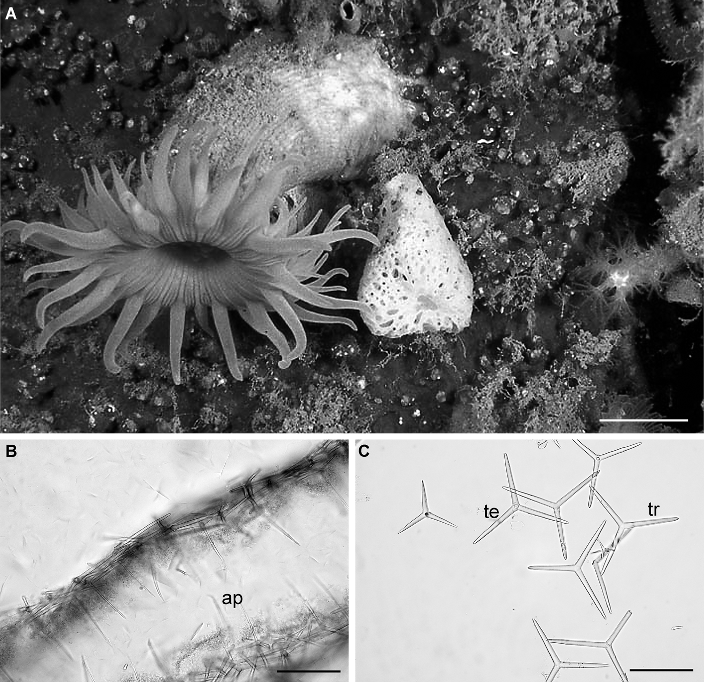
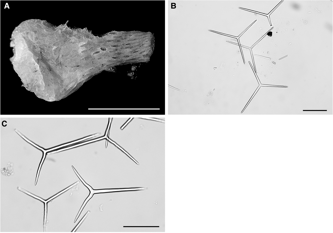
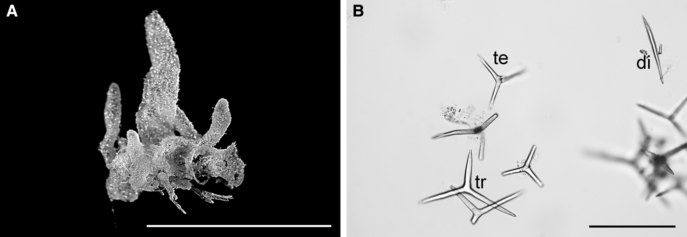
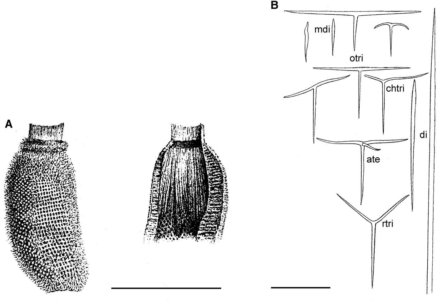
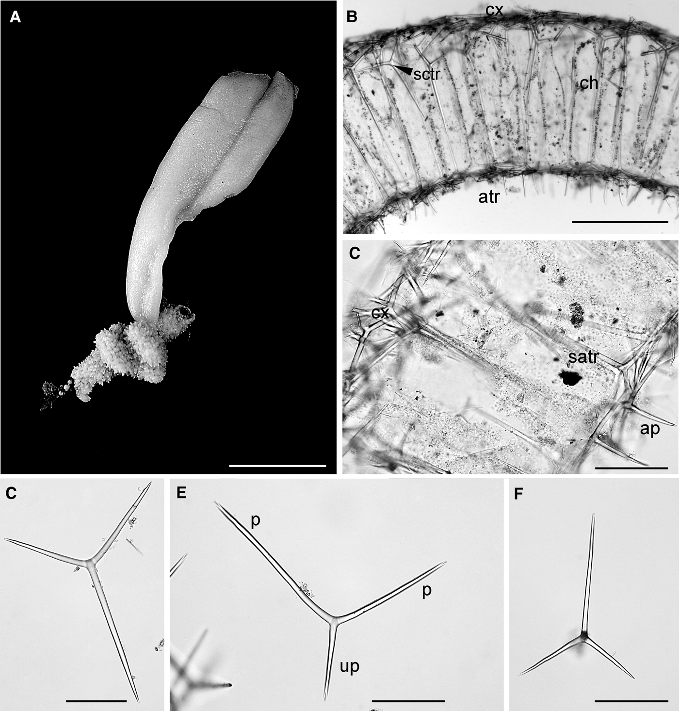
Brattegardia nanseni: (A) specimen photographed in situ at about 5 m depth (from Spitsbergen; (B) section of a asconoid tube. Note the apical actines (ap) projecting into the atrium; (C) regular tri- and tetractines: tr, triactines; te, tetractine; Scale bars: A, 1 cm; B, C, 100 µm.
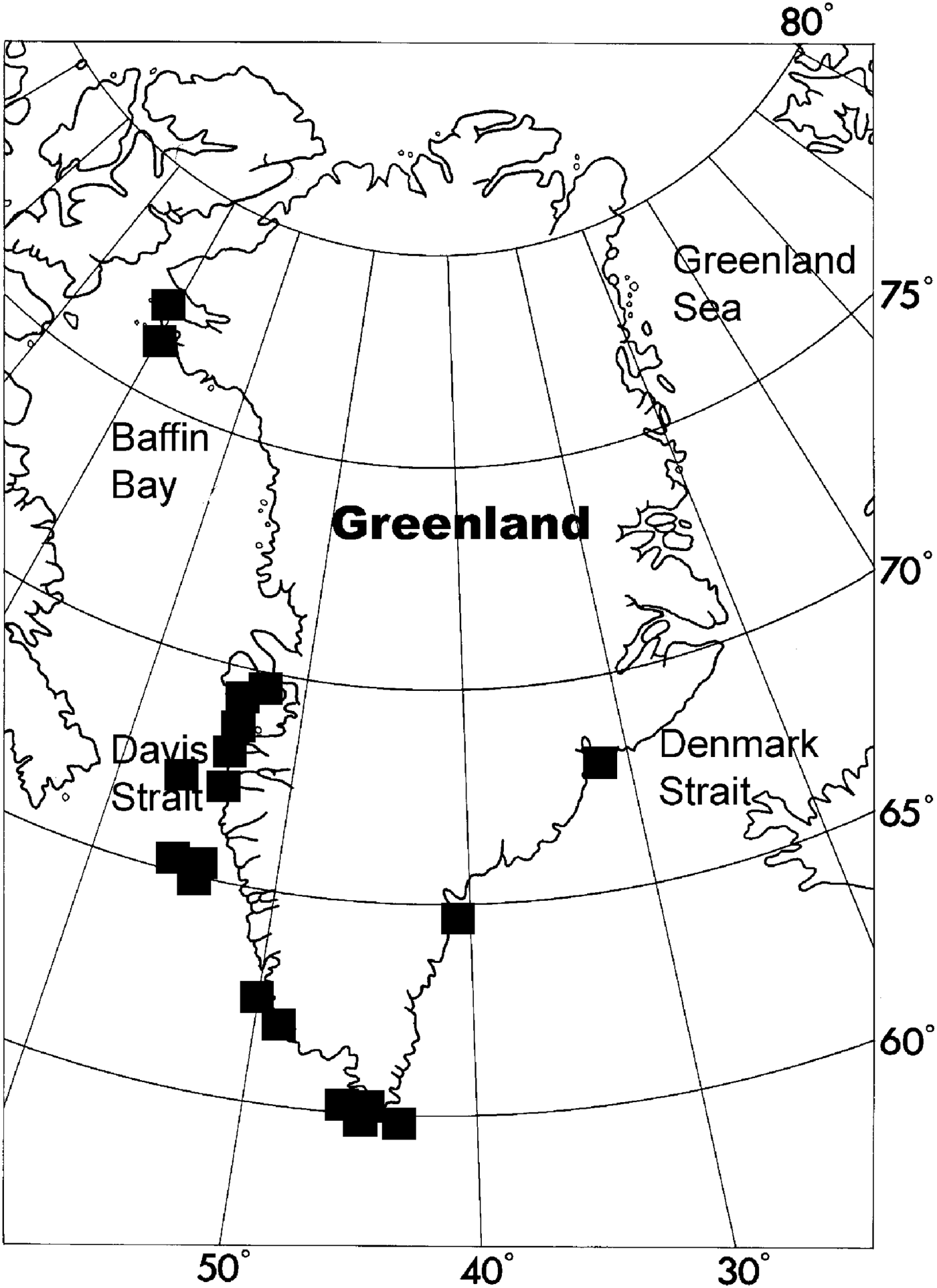
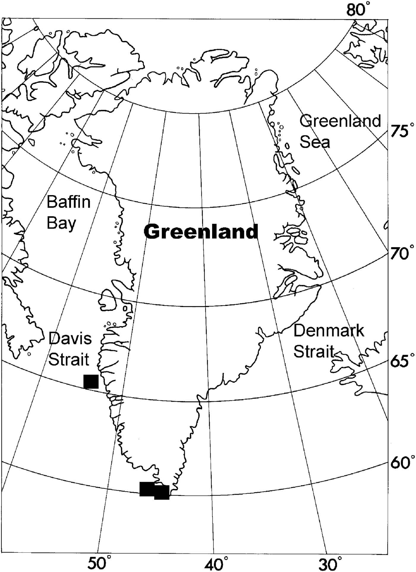
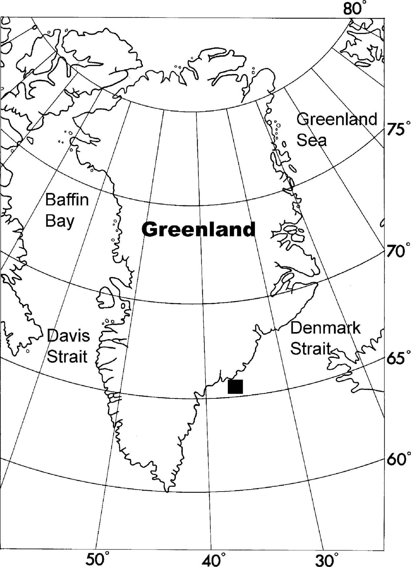
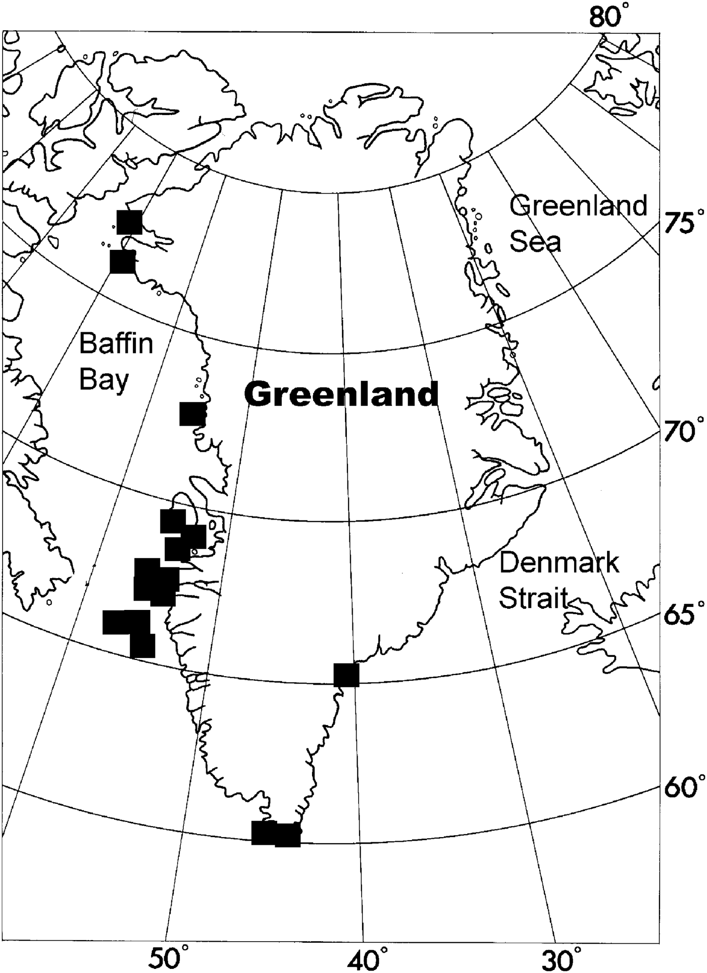
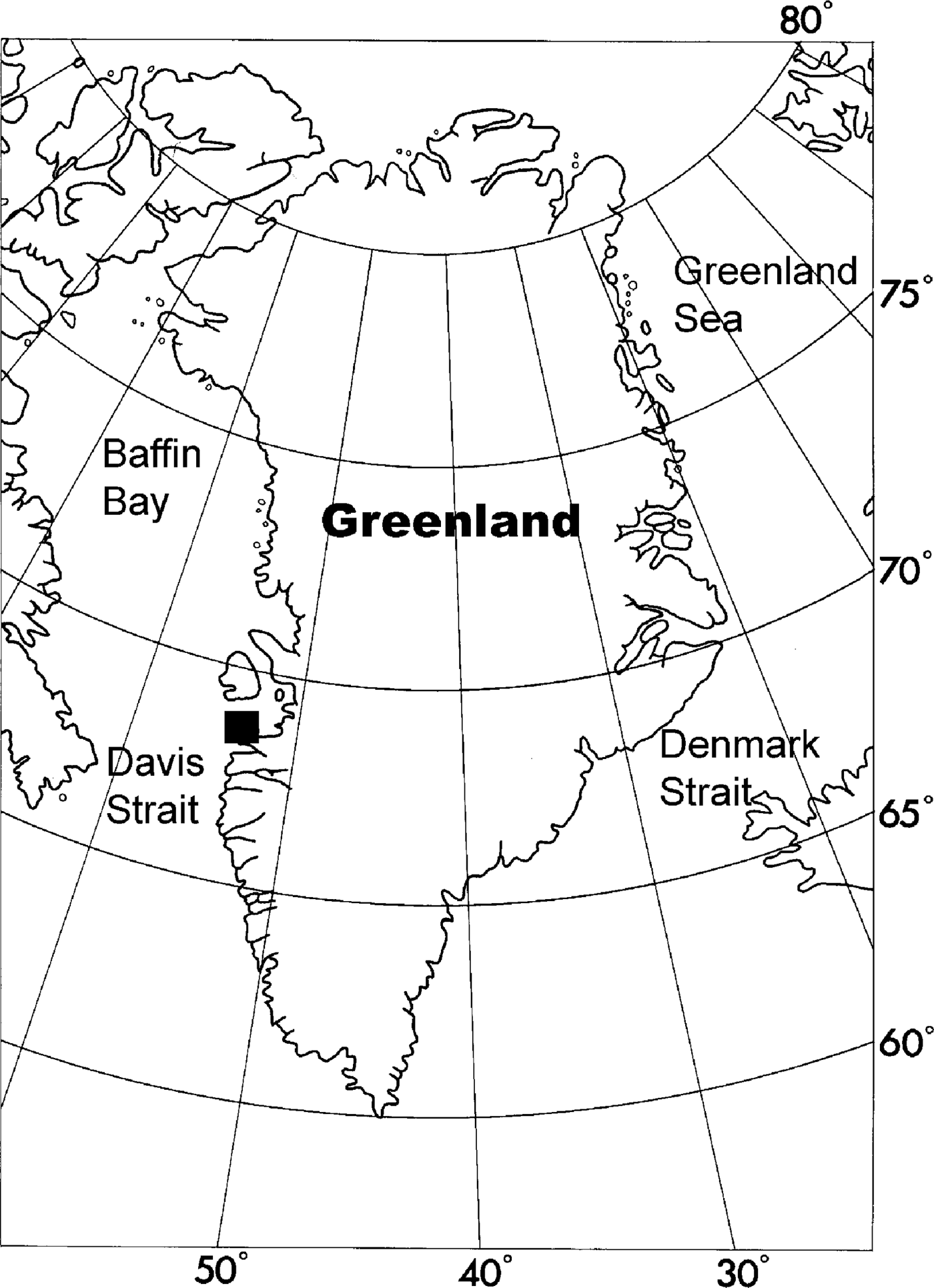
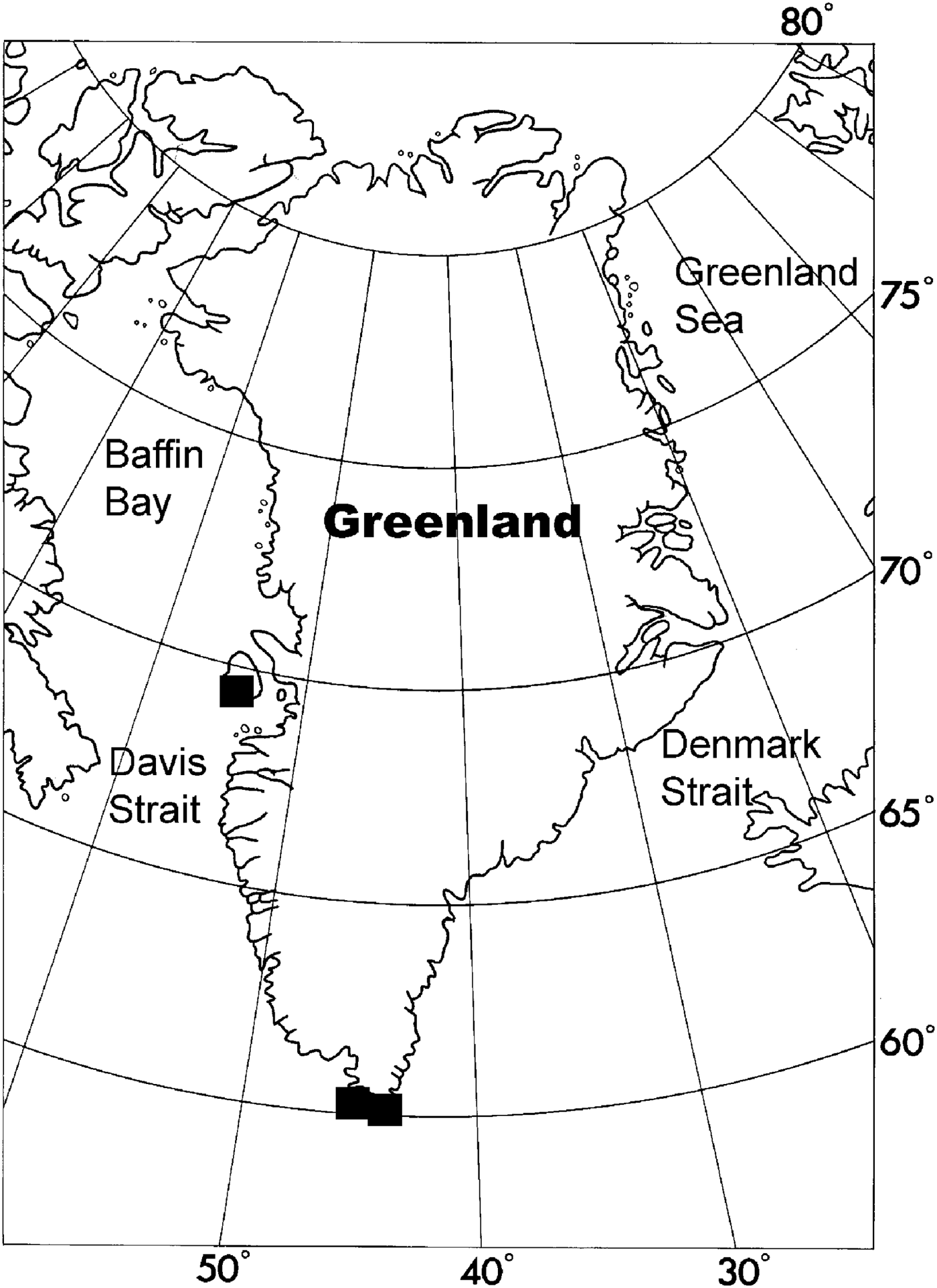
Distribution of Brattegardia nanseni along the coast of Greenland.
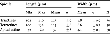
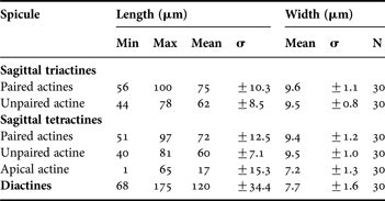
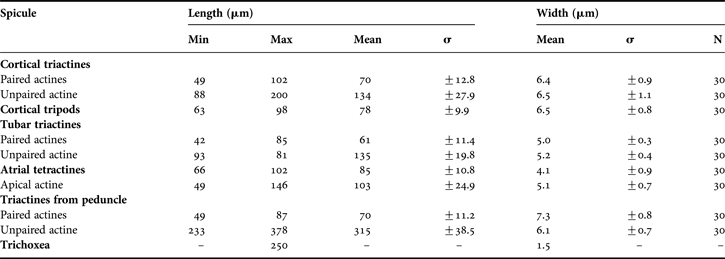
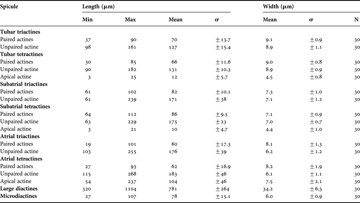
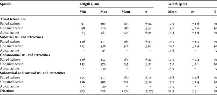
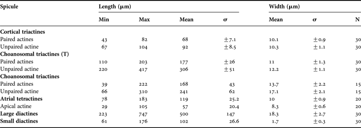
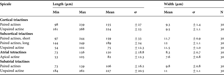
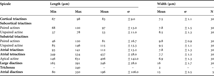
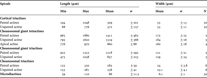
Spicule measurements of Brattegardia nanseni (Breitfuss, Reference Breitfuss1896) from Greenland.
Original description: Leucosolenia nanseni Breitfuss, Reference Breitfuss1896: 427–428.
SYNONYMS AND CITATIONS
Ascandra reticulum (Haeckel, Reference Haeckel1872; Lütken, Reference Lütken and Jones1875; Breitfuss, Reference Breitfuss1897, Reference Breitfuss1898b, Reference Breitfussc; Vanhöffen, Reference Vanhöffen1897; Brøndsted, Reference Brøndsted1914).
Brattegardia nanseni (Klautau et al., 2013).
Clathrina nanseni (Rapp et al., Reference Rapp, Klautau and Valentine2001; Klautau & Valentine, Reference Klautau and Valentine2003; Rapp, Reference Rapp2006).
Leucosolenia nanseni (Breitfuss Reference Breitfuss1898a, Reference Breitfussc, Reference Breitfuss1933; Lundbeck, Reference Lundbeck1909; Brøndsted, Reference Brøndsted1914, Reference Brøndsted1933b).
Nardoa reticulum (Schmidt, Reference Schmidt1869 pars; 1870 pars).
TYPE LOCALITY
Eastern Spitsbergen (Breitfuss, Reference Breitfuss1896).
TYPE MATERIAL
ZIL, Breitfuss collection No. 1 (lectotype) and No. 2 (paralectotype) (Rapp, Reference Rapp2006).
PREVIOUS RECORDS
Angmagsalik (Lundbeck, Reference Lundbeck1909) and Lille Karajakfjord (Brøndsted, Reference Brøndsted1914).
MATERIAL EXAMINED
152 specimens. Davis Strait, 1884 (1). Davis Strait, 09.07.1884, 188 m (15). Frederikshåb, 17.08.1886, 50 m (1). Egedesminde, 20.10.1890 (1). Egedesminde 11.11.1892 (11). Egedesminde, 1892 (4). Godhavn, 04.07.1892 (1). Amdrup Expedition, Tasiusak, 25.05.1899, 40–60 m (1). Tasiusak, 01.06.1899, 50–60 m (1). Tasiusak, 65.35°N 22.08.1902, 60–100 m (3). Angmagsalik Fjord, 27.11.1902, 20–50 m (1). 63.48°N 52.23°W, 10.06.1908, 194 m (1). 65.06°N 54.19°W, 07.06.1909, 85 m (4). Nordre Strømfjord, 25.06.1911, 200–240 m (3). Nordre Strømfjord, 29.07.1911, 65–98 m (1). Nordre Strømfjord, 06.07.1911, 44–64 m (2). Godthaab Expedition Station 47, 69.5°N 54°W, 13.07.1928, 70 m (1). Kangerdlugssuak, 11.08.1932, 70 m (1). Thule Expedition Station 7, 20.07.1933, 20–30 m (7). Discofjord, 05.08.1988, 17 m (11). 65.22°N 54.02°W, 125 m (8). Ingolf Station 29, 65.34°N 54.31°W, 05.09.1895, 128 m (1). Station 34, 65.17°N 54.17°W, 18.07.1895, 104 m (6). Just and Vibe Station 20, Bylot Sound, 76.34.5°N 69.24.5°W, 14.08.1968, 35 m (2). Station 67, Murchison Sound, 77.31.6°N 70.40°W, 24.08.1968, 50 m (10). Kap Farvel Station 11, Ilua, 60.09.4°N 44.14.9°W, 13.07.1970, 60–70 m (6). Station 22, 60.09.4°N 44.14.9°W, 17.07.1970, 100–120 m (2). Station 45, 60.097°N 44.16.9°W, 24.07.1970, 25–35 m (1). Station 54, 60.08.3°N 44.18.7°W, 30.09.1970, 30–40 m (1). Station 59, 60.08.3°N 44.18.7°W, 03.08.1970, 40 m (1). Station 60, 60.08.5°N 44.17.4°W, 03.08.1970, 40 m (1). Station 62, 60.08.5°N 44.17.4°W, 03.08.1970, 50–60 m (2). Station 74, 60.05.3°N 44.14.5°W, 25–30 m (1). Station 75, 60.05.3°N 44.14.5°W, 05.08.1970, 40–50 m (2). Station 87, 59.53.3°N 44.22.2°W, 09.08.1970, 70 m (3). Station 90, 60.09°N 44.10°W, 12.08.1970, 40–60 m (1). Station 93, Akuliaruseq, 60.09°N 44.10°W, 12.08.1970, 140 m (3). Station 95, 59.91°N 44.43°W, 13.08.1970, 80–100 m (4). Station 101, 60.10.1°N 44.13°W, 17.08.1970, 40 m (2). Station 132, 59.55.5°N 44.23.0°W, 23.08.1870, 400 500 m (1). Station 142, 60.03.8°N 43.09°W, 26.08.1970, 120 m (2). Station 148, 60.07°N 43.20°W, 28.08.1970, 50 m (1). Bankeundersøkelser Station 3B, 28.07.1975 (8). Station 64A (3). Station 5298-5, 67.05°N 54.67°W, 23.08.1976, 70 m (4). Station 5299-5, 67.08°N 54.12°W, 23.08.1976, 80–95 m (1).
DESCRIPTION
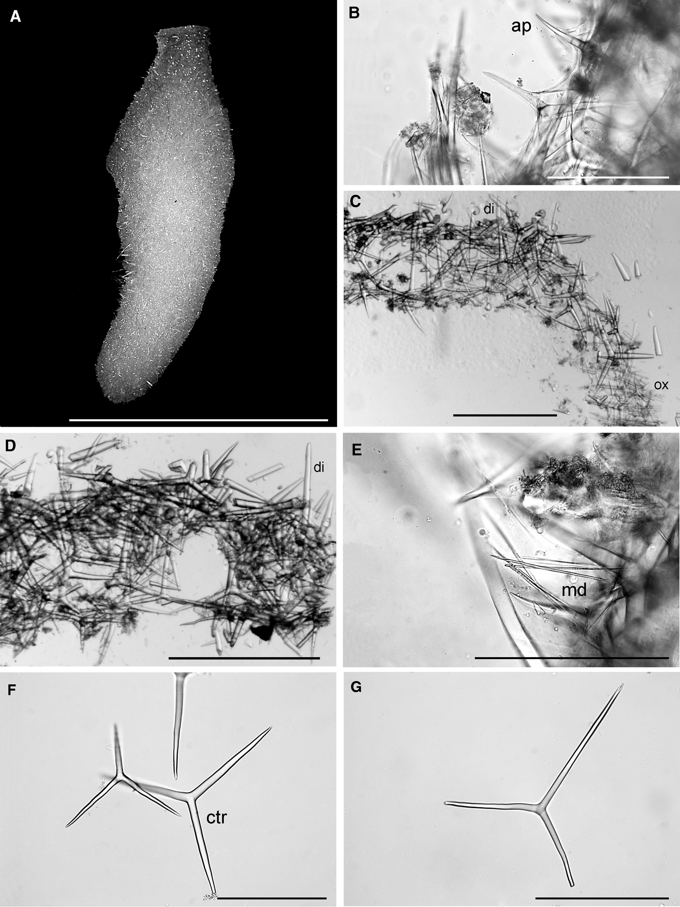
The cormus of this species is composed of regularly and tightly anastomosing tubes forming a massive and more or less globular or pear-shaped cormus. The cormus is up to 5 cm in diameter and is often wider in the top than at the base (Figure 2A). Some specimens have a poorly developed stalk composed of normal tubes with choanoderm. Water-collecting tubes converge at the centre of the sponge, ending in one or several large oscula, normally surrounded by a membranous rim. Texture is soft. Surface is smooth. The colour white to greyish-white in life and greyish-white to light-brown in alcohol. A thin membrane is surrounding parts of, or the entire cormus in young specimens. No cells with brown inclusions in the mesohyl.
The skeleton has no special organization, and it is composed of regular and equiangular triactines of one type, and two types of tetractines (Figure 2C). Slightly parasagittal triactines are sometimes present in the base of the sponge. The apical actine of the tetractines is projected into the interior of the tubes (Figure 2B). Actines are cylindrical to slightly conical with short points. In the tetractines the apical actine is thinner than the basal ones, cylindrical, slightly curved, and sharply pointed. In some of the tetractines the apical actine is rudimentary, forming only a small projection. All tetractines are of equal size.
OCCURRENCE
The species was found in sectors 1–5, 8, 16 and 17 (Figure 3). Depth 17–500 m.
GEOGRAPHICAL DISTRIBUTION
Widely distributed in the Arctic, north-east Canada, Spitsbergen, Barents Sea, Murman coast and northern Norway.
REMARKS
Brattegardia nanseni is very similar to Clathrina septentrionalis Rapp et al., Reference Rapp, Klautau and Valentine2001, but can be distinguished from C. septentrionalis by the presence of tetractines with a rudimentary apical actine, and absence of parasagittal spicules and special cells with brown inclusions (Rapp et al., Reference Rapp, Klautau and Valentine2001; Klautau & Valentine, Reference Klautau and Valentine2003; Rapp, Reference Rapp2006). Brattegardia nanseni also has similarities with Clathrina canariensis (Miklucho-Maclay, Reference Miklucho-Maclay1868) in respect of shape and types of spicules. However, the spicules are about two times bigger in B. nanseni than in C. canariensis. The latter species has been reported from the Arctic several times (Hentschel, Reference Hentschel1916; Breitfuss, Reference Breitfuss1933), but according to Klautau & Valentine (Reference Klautau and Valentine2003), the distribution is restricted to the Atlantic coasts of Europe, and perhaps, the Mediterranean Sea. Breitfuss (Reference Breitfuss1933) claimed that it is very difficult to recognize one species from the other, and it is possible that his records were based on specimens of Brattegardia nanseni with slightly smaller spicules than in his type material from Spitsbergen. Most specimens of B. nanseni from the northern Norwegian coast also have spicules differing slightly in size from the original material (Rapp, Reference Rapp2006, as Clathrina nanseni). Clathrina cancellata (Verrill, Reference Verrill1874) described from Casco Bay, Maine, is also very similar to B. nanseni. The description of Verrill (Reference Verrill1874) is not very detailed, but the shape of the cormus, and the size and shape of spicules are within the range of what can be found in B. nanseni. Lambe (Reference Lambe1900c) reported C. cancellata from several localities along the north-eastern coast of Canada. As I have not been able to re-examine the type specimen of C. cancellata, I have examined one of Lambe's specimens from Strait of Belle Isle, Labrador, deposited in the ZMUC. The specimen revealed to be identical with a typical Brattegardia nanseni, and it is therefore questioned if C. cancellata and B. nanseni are synonymous. If proven synonymous B. nanseni should be regarded as a junior synonym of C. cancellata, and the Arctic and amphiatlantic distribution of the species would follow the same pattern as has been shown in other sponges, e.g. the Geodiidae (Cardenas et al., Reference Cardenas, Rapp, Klitgaard, Best, Thollesson and Tendal2013; Hestetun et al., Reference Hestetun, Fourt, Vacelet, Boury-Esnault and Rapp2013). However, I want to keep the species separated until the type material has been found and re-examined. According to Rossi et al. (Reference Rossi, Russo, Solé-Cava, Rapp and Klautau2011) and Klautau et al. (Reference Klautau, Azevedo, Cóndor-Luján, Rapp, Collins and Russo2013) only ‘Clathrina’ with a skeleton consisting of solely triactines are true Clathrina. Their phylogenetic analyses showed that the genus is not monophyletic and that Brattegardia nanseni forms a well-supported clade separated from the true Clathrina.
TYPE SPECIES
Clathrina clathrus (Schmidt, 1864). Calcinea in which the cormus comprises anastomosed tubes. A stalk may be present. The skeleton contains regular (equiangular and equiradiate) and/or parasagittal triactines, to which diactines and tripods may be added. Asconoid aquiferous system (Klautau et al., Reference Klautau, Azevedo, Cóndor-Luján, Rapp, Collins and Russo2013).
REMARKS
According to Rossi et al. (Reference Rossi, Russo, Solé-Cava, Rapp and Klautau2011) and Klautau et al. (Reference Klautau, Azevedo, Cóndor-Luján, Rapp, Collins and Russo2013) the genus Clathrina should now also include former Guancha species with a skeleton composed of regular and/or parasagittal triactines. The diagnosis of the genus is here updated accordingly.
KEY TO SPECIES OF CLATHRINA
1. Spicules are regular triactines, with additional parasagittal spicules, at least in the peduncle ……… 2
— Spicules are only regular triactines ……… C. tendali sp. nov.
— Spicules are all parasagittal triactines ……… C. arnesenae
2. — Spicules are undulated or ‘horn-shaped’ ……… 3
— All spicules have straight actines ……… C. cf. blanca
3. All spicules have undulated actines ……… C. pellucida
— Peduncular parasagittal triactines have ‘horn-shaped’ paired actines ……… C. camura
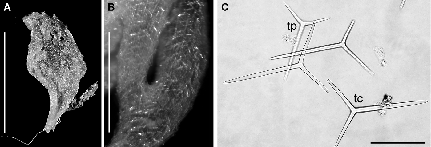
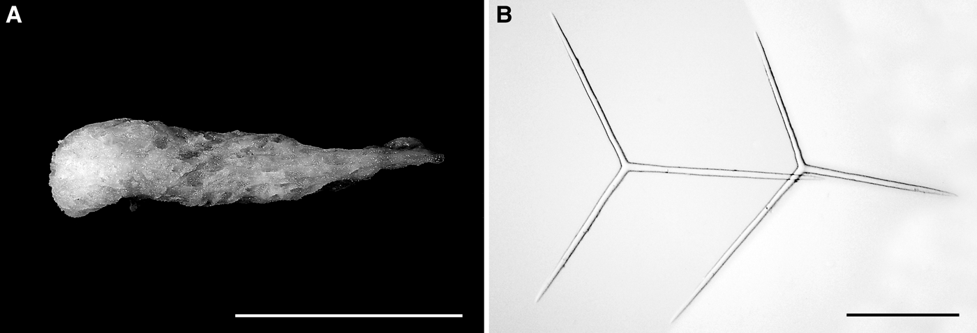
Clathrina arnesenae: (A) preserved specimen; (B) close-up of asconoid tubes. Note that unpaired actines are arranged in parallel, pointing towards the base of the sponge; (C) spicules; tc, parasagittal triactine from cormus; tp, parasagittal triactine from peduncle. Scale bars: A, 0.5 cm; B, 0.7 mm; C, 100 µm.
Distribution of Clathrina arnesenae along the coast of Greenland.
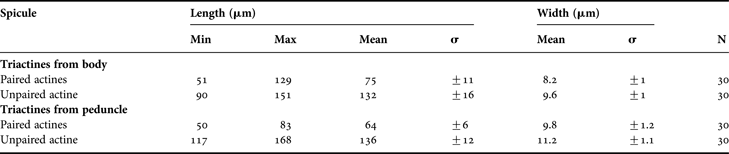
Spicule measurements of Clathrina arnesenae (Rapp, Reference Rapp2006) from Greenland.
Original description: Guancha arnesenae Rapp, Reference Rapp2006: 349–352.
SYNONYMS AND CITATIONS
Guancha arnesenae (Rapp, Reference Rapp2006; Ereskovsky & Willenz, Reference Ereskovsky and Willenz2008).
Guancha sagittaria (Rapp, Reference Rapp2004b).
Leucosolenia sagittaria (Merejkowsky, Reference Merejkowsky1878; Breitfuss, Reference Breitfuss1898c, Reference Breitfuss1927, Reference Breitfuss1933).
TYPE LOCALITY
Evenskjer, Norway, 68°35′N 16°35′E, 25 m.
TYPE MATERIAL
Holotype: VM-398, Evenskjer, Norway, 68°35′N 16°35′E, 25 m, 18.08.1919, 1 specimen.
Paratype: TMU-183, Ramfjorden, 10–20 m, 14.07.1921, 1 specimen.
PREVIOUS RECORDS
As Leucosolenia primordialis, from northern Strømfjord inlet (Brøndsted, Reference Brøndsted1933a).
MATERIAL EXAMINED
Seven specimens. Holsteinsborg, 17.05.1892, 75 m (1). Godhavn, 04.07.1892 (2). Northern Strømfjord inlet, 27.06.1911, 51–54 m (1). Kap Farvel Station 63, 60.08.5°N 44.18.5°W, 03.08.1970, 30–40 m (1). Just and Vibe Station 16, 76.30.9°N 69.28.6°W, 14.08.1968, 94 m (1). Station 67, 77.31°N 70.4°W, 24.08.1968, 50 m (1).
DESCRIPTION
Clathrinidae with a cormus normally composed of a clathroid cormus of irregularly and loosely anastomosing tubes and a peduncle. Size is ranging from 4 to 10 mm in height. Several true tubes with a normal choanoderm form the peduncle. The peduncular tubes run in parallel, free, or slightly anastomosed. In specimens without a proper peduncle the cormus is narrower at the base than in the apical region. Water-collecting tubes converge at the centre of the sponge, ending in one or several apical oscula. The single tubes are about 0.5 mm in diameter. The colour is light beige in alcohol and dried. Texture is soft. The skeleton of the cormus is solely composed of tripodic, parasagittal triactines (Figure 4C). The spicules are irregularly oriented in several layers in the walls of the tubes in the main body, resulting in a wall thickness of about 30–50 μm. In the peduncle the paired actines are shorter than in the clathroid body. Spicules in the peduncle with the unpaired actine basipetally oriented. All the actines are straight, cylindrical and with blunt tips.
OCCURRENCE
The species was found in sectors 1, 4, 5, and 8 (Figure 5). Depth 30–94 m.
GEOGRAPHICAL DISTRIBUTION
Clathrina arnesenae has also been found in the northern part of Norway at 10–90 m depth (Rapp, Reference Rapp2006), the White Sea (Merejkowsky, Reference Merejkowsky1878; Derjugin, Reference Derjugin1915, Reference Derjugin1928; personal observations).
REMARKS
Clathrina arnesenae was originally described as Guancha arnesenae based on the morphology of the cormus and the presence of parasagittal spicules. However, recent molecular phylogenies have shown that species formerly defined as Guancha (with a skeleton of parasagittal triactines and eventually diactines) belong to Clathrina (Rossi et al., Reference Rossi, Russo, Solé-Cava, Rapp and Klautau2011; Voigt et al., Reference Voigt, Wülfing and Wörheide2012; Klautau et al., Reference Klautau, Azevedo, Cóndor-Luján, Rapp, Collins and Russo2013). According to these analyses it is to be concluded that the following species of Guancha from the northernmost Atlantic and Arctic Oceans should also be transferred to Clathrina: G. blanca Miklucho-Maclay, Reference Miklucho-Maclay1868; G. camura Rapp, Reference Rapp2006; G. lacunosa (Johnston, Reference Johnston1842); G. pellucida (Rapp, Reference Rapp2006); and G. sagittaria (Haeckel, Reference Haeckel1872). Clathrina arnesenae is very similar to Clathrina sagittaria, and further comments and a comparison between the species can be found in Rapp (Reference Rapp2006).
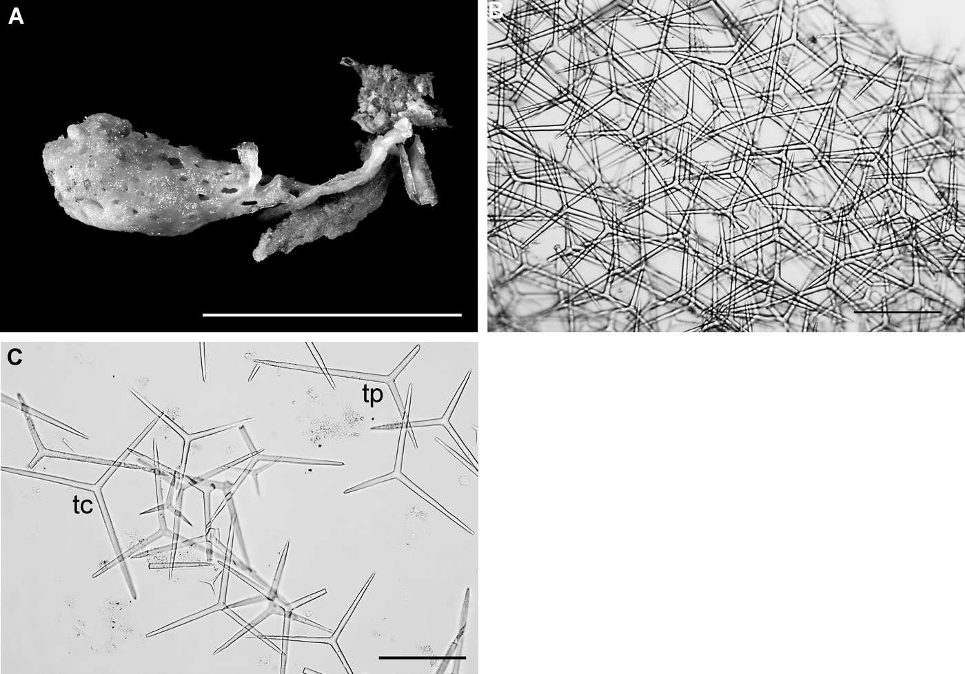
Clathrina cf. blanca: (A) preserved specimen; (B) surface of a asconoid tube; (C) spicules; tc, regular to parasagittal triactine from cormus; tp, parasagittal triactine from peduncle. Scale bars: A, 1 cm; B, C, 100 µm.
Distribution of Clathrina cf. blanca along the coast of Greenland.
Spicule measurements of Clathrina cf. blanca (Miklucho-Maclay, Reference Miklucho-Maclay1868) from Greenland.
Original description: Guancha blanca Miklucho-Maclay, Reference Miklucho-Maclay1868: 221–240.
SYNONYMS AND CITATIONS
Guancha blanca (Barthel & Tendal, Reference Barthel and Tendal1993; Janussen et al., Reference Janussen, Rapp and Tendal2003; Rapp, Reference Rapp2006).
Leucosolenia macleayi (Burton, Reference Burton1934).
Leucosolenia blanca (Breitfuss, Reference Breitfuss1936).
TYPE LOCALITY
Lanzarote, Canary Islands.
PREVIOUS RECORDS
As Leucosolenia macleayi, from the Norwegian Scientific Expeditions to East Greenland (Burton, Reference Burton1934) and as Clathrina coriacea from Jørgen Brønlund fjord (Tendal, Reference Tendal1970).
MATERIAL EXAMINED
17 specimens. Discofjord, 05.08.1988, 7–9 m (1). Hoels Station 1066, Mackenzie Bay, 02.08.1930, 63–83 m (1). Station 1067, Kap Humboldt, 03.08.1930, 20–30 m (1). Station 1081, Alpfjorden, 08.08.1930, 70 m (1). Just and Andersen Station 52, Jørgen Brønlund fjord, off Kap Rasmussen, 29.09.1966, 160–180 m (1). Kap Farvel Station 40, 60.19.7°N 44.15.7°W, 22.07.1970, 100 m (fragmentary). Station 61, 60.08.5°N 44.17.4°W, 03.08.1970, 50–60 m (1). Station 90, 60.09°N 44.10°W, 03.08.1970, 40–60 m (1). Station 132, 59.55.5°N 44.23°W, 23.08.1970, 400–450 m (2). Station 147, 60.06.2°N 43.12°W, 27.08.1970, 15–20 m (1). Bankeundersøkelser Station 22, 66.68°N54.35°W, 01.08.1975, 82 m (1).
DESCRIPTION
Clathrinidae with a cormus normally composed of a clathroid body of regularly and tightly anastomosing tubes and a peduncle (Figure 6A). Up to 13 mm in height and 5 mm in diameter. One or several true tubes with normal choanoderm form the peduncle. Sometimes the peduncle is very short or absent. In specimens without a proper peduncle the cormus is often narrower at the base than in the apical region. Water-collecting tubes converge at the centre of the sponge, ending in one or several apical oscula. The single tubes are about 0.3–0.7 mm in diameter. Surface is smooth. Colour white, greyish-white or beige when alive, and with almost the same colours in alcohol. Dried specimens are usually light beige. The skeleton of the cormus is composed of regular (equiangular and equiradiate) triactines to which slightly parasagittal spicules may be added (Figure 6C). The spicules are irregularly oriented in several layers in the walls of the tubes (Figure 6B), resulting in a wall thickness of about 70–80 μm. In the peduncle the skeleton consists of only parasagittal triactines, with the unpaired actine basipetally oriented. All the actines are straight, cylindrical and with blunt tips.
OCCURRENCE
The species was found in sectors 1, 4, 5, 11, and 14 (Figure 7). Depth 7–450 m.
GEOGRAPHICAL DISTRIBUTION
The species has been reported to have a very wide distribution in the North Atlantic and the Arctic. From shallow sub-littoral to around 1000 m in the Norwegian Sea. The species has previously been reported from great depths down to 3000 m in the Norwegian–Greenland Sea (Barthel & Tendal, 1994; Janussen et al., Reference Janussen, Rapp and Tendal2003; Klautau et al., 2013). However, recent studies have shown that these records represent a new species of Brattegardia (Rapp & Tendal, manuscript in preparation).
REMARKS
In the reticulate/cushion-shaped form the parasagittal spicules are not so numerous, and the unpaired actine is usually shorter than in the pedunculated form. According to Haeckel (Reference Haeckel1872) the spicule dimensions in material from the type locality (Lanzarote) are 50–70 × 3–4 µm (paired actines) and 80–100 × 3–4 µm (unpaired actine), and the gross cormus morphology and composition of the peduncle are highly variable. The great degree of variation observed over the distribution range of the species and the occurrence in highly different habitats and water masses may indicate that C. blanca represents a species complex. All specimens from Greenland have somewhat bigger spicules and may therefore not represent the true C. blanca. The species is therefore here named Clathrina cf. blanca.
Clathrina camura: (A) preserved specimen. Oscular opening to the left; (B) regular to subregular triactines from the cormus; (C) parasagittal triactines with slightly bent paired actines from the peduncle. Scale bars: A, 1 cm; B, C, 100 µm.
Distribution of Clathrina camura along the coast of Greenland.
Spicule measurements of Clathrina camura (Rapp, Reference Rapp2006) from Greenland.
Original description: Guancha camura Rapp, Reference Rapp2006: 355–356.
SYNONYMS AND CITATIONS
Guancha camura Rapp, Reference Rapp2006.
TYPE LOCALITY
Porsanger, north-eastern Norway.
TYPE MATERIAL
Holotype: TMU-185.
Paratype: TMU-184.
PREVIOUS RECORDS
The species is new to Greenland.
MATERIAL EXAMINED
Six specimens. Lille Hellefisk Banke, 65.22°N 54.02°W, 125 m (1). Kap Farvel Station 93, 60.09°N 44.10°W, 12.09.1970, 140 m (4). Station 115, 59.99°N 49.93°W, 19.08.1970, 200 m (1).
DESCRIPTION
Clathrinidae with a cormus normally composed of a clathroid body of irregularly and loosely anastomosing tubes and a peduncle (Figure 8A). Up to 3 cm in height and 2 cm wide. Consistency soft because of the very thin walls of the tubes (about 20 µm). Surface is smooth. Several true tubes with normal choanoderm form the peduncle. The skeleton of the cormus is composed of regular (equiangular and equiradiate) triactines to which parasagittal spicules are usually added (Figure 8B). In the peduncle the skeleton consists of only parasagittal triactines. The unpaired actine of peduncular parasagittal triactines is always basipetally oriented. The paired actines are bent in a manner making them ‘horn-shaped’, while the unpaired actine is straight (Figure 8C). All actines are cylindrical with slightly blunt tips.
OCCURRENCE
The species was found in sectors 1 and 3 (Figure 9). Depth 125–200 m.
GEOGRAPHICAL DISTRIBUTION
The species is also known from the northern part of Norway at 27–90 m depth (Rapp, Reference Rapp2006) and from Iceland and Jan Mayen down to 890 m depth (personal observations).
REMARKS
The material from Greenland fits very well with the type material from Norway both in respect of gross morphology and in size and shape of the spicules.
Clathrina pellucida: (A) preserved specimen; (B) parasagittal triactines. Scale bars: A, 1 cm; B, 100 µm.
Distribution of Clathrina pellucida along the coast of Greenland.
Spicule measurements of Clathrina pellucida (Rapp, Reference Rapp2006) from Greenland.
Original description: Guancha pellucida Rapp, Reference Rapp2006: 357–360.
SYNONYMS AND CITATIONS
Guancha pellucida (Rapp, Reference Rapp2006; Schander et al., Reference Schander, Rapp, Kongsrud, Bakken, Berge, Cochrane, Oug, Byrkjedal, Cedhagen, Fosshagen, Gebruk, Larsen, Nygren, Obst, Plejel, Stöhr, Todt, Warén, Handler-Jacobsen, Kuening, Levin, Mikkelsen, Petersen, Thorseth and Pedersen2010).
TYPE LOCALITY
Porsangerfjord, northern Norway.
TYPE MATERIAL
Holotype: TMU-179.
Paratype: VM-397.
PREVIOUS RECORDS
The species is new to Greenland. One specimen from the Godthåb area was wrongly identified and published as Leucosolenia coriacea by Lundbeck (Reference Lundbeck1909), and four other specimens from Stormbugt were published as L. coriacea by Brøndsted (Reference Brøndsted1916).
MATERIAL EXAMINED
Seven specimens. Stormbugt, 76.46°N 19.02°W, 21.08.1907, 20–40 m (4). 63.48°N 52.23°W, 10.06.1908, 194 m (1). Godthåb Station 305, between Jameson Land and Cape Leslie, 21.08.1933, 179 m (1). Just and Vibe Station 27, 76.27.9°N 69.19°W, 10.08.1968, 30 m (1).
DESCRIPTION
Clathrinidae composed of a globular and highly compressible clathroid cormus of irregularly but tightly anastomosing tubes, and a very short peduncle (Figure 10A). Up to 2 cm in height and 1.8 cm in diameter. Several anastomosing true tubes with a normal choanoderm form the peduncle. In full-grown specimens water-collecting tubes converge into one large apical osculum. Texture soft and fragile. Surface smooth. Colour greyish-white and highly translucent when alive, and yellowish-white in alcohol. The skeleton of the cormus is composed of 1–3 layers of very slender regular to subregular triactines, to which parasagittal spicules may be added. The walls of the tubes are only 10–15 µm thick. In the peduncle the skeleton consists of parasagittal triactines with a highly variable length of the unpaired actine. The unpaired actine of peduncular parasagittal triactines is always basipetally oriented. All actines are undulated, especially close to the centre of the spicule, cylindrical or slightly conical with blunt tips (Figure 10B).
OCCURRENCE
The species was found in sectors 3, 8, 13 and 15 (Figure 11). Depth 30–194 m.
GEOGRAPHICAL DISTRIBUTION
The species is also found in the northern part of Norway at 100–275 m depth (Rapp, Reference Rapp2006) and at hydrothermal vents at 550–700 m depth north-east of Jan Mayen (Schander et al., Reference Schander, Rapp, Kongsrud, Bakken, Berge, Cochrane, Oug, Byrkjedal, Cedhagen, Fosshagen, Gebruk, Larsen, Nygren, Obst, Plejel, Stöhr, Todt, Warén, Handler-Jacobsen, Kuening, Levin, Mikkelsen, Petersen, Thorseth and Pedersen2010).
REMARKS
Specimens examined here correspond entirely with the material from the Norwegian coast (Rapp, Reference Rapp2006).
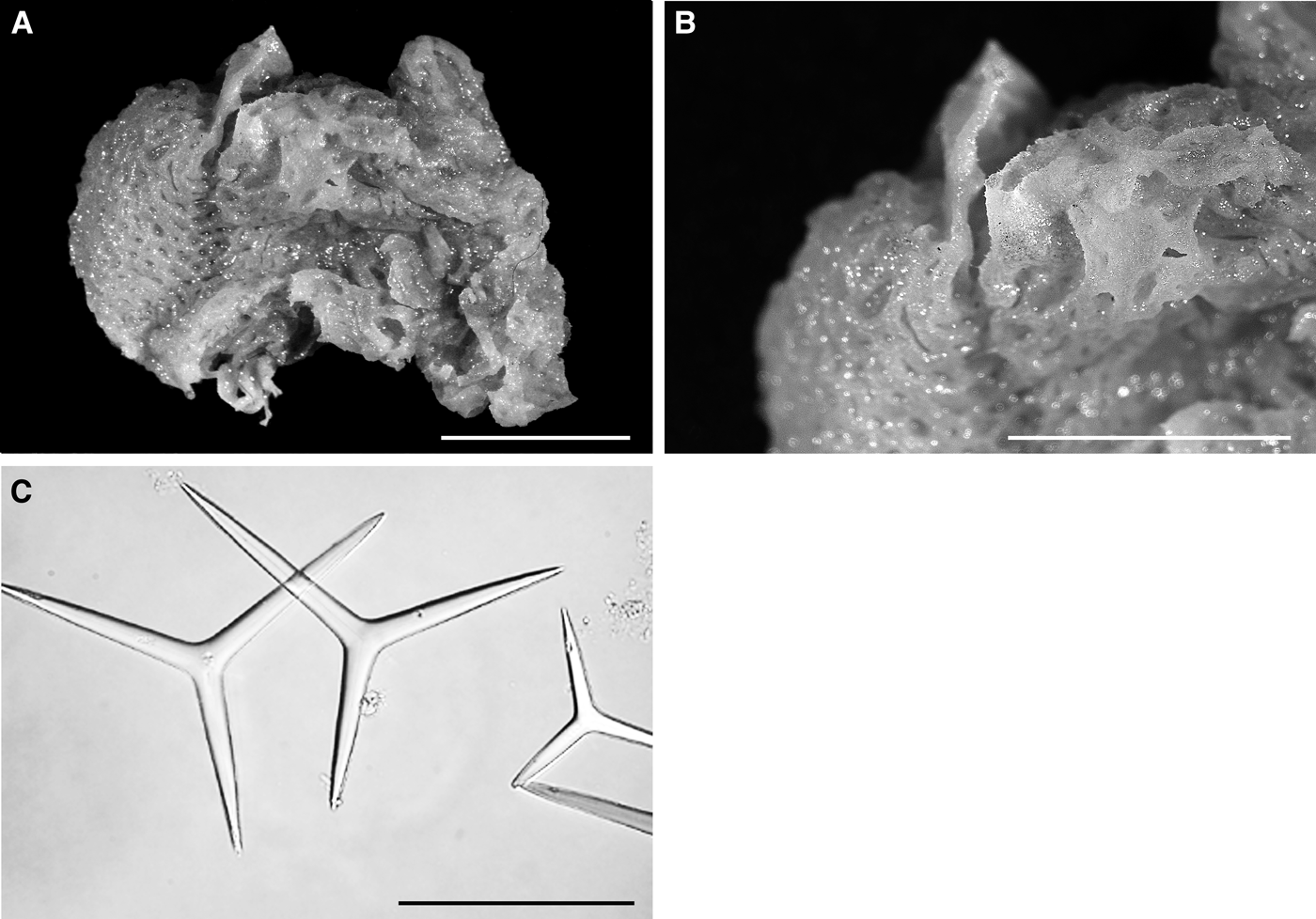
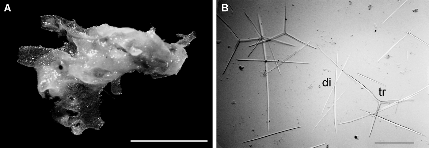
Clathrina tendali sp. nov: (A) preserved holotype (ZMUC CAL-1); (B) oscular region with several osculi; (C) regular triactines. Scale bars: A, 1 cm; B, 0.5 cm; C, 100 µm.
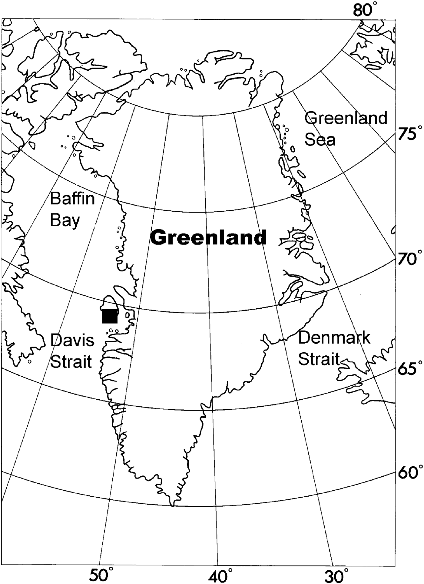
Distribution of Clathrina tendali sp. nov. along the coast of Greenland.
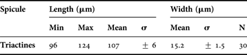
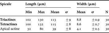
Spicule measurements of Clathrina tendali sp. nov.
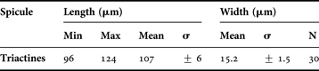
Original description: Nardoa reticulum Schmidt, Reference Schmidt1862. The specimen was identified and published by Schmidt (Reference Schmidt1869).
TYPE LOCALITY
Greenland. No further information is given on the original label.
TYPE MATERIAL
Holotype: ZMUC. CAL-1 (alcohol and slide).
ETYMOLOGY
The species is named after Ole Secher Tendal for his efforts exploring the sponges of the northernmost Atlantic and Arctic Oceans.
PREVIOUS RECORDS
As Nardoa reticulum by Schmidt (Reference Schmidt1869).
MATERIAL EXAMINED
One specimen.
DESCRIPTION
The cormus of this sponge is yellowish-white to light beige in alcohol. The cormus is massive, sub-globular, and composed of densely and regularly to irregularly anastomosing tubes (Figure 12A). Surface is smooth. Texture is soft. Water-collecting tubes converge into several large apical oscula, which are extended by a 5 mm high membrane (Figure 12B). The skeleton is composed of irregularly arranged regular (equiactinal and equiradiate) triactines. The spicules are overlapping but there are not several distinct layers of spicules. The actines are stout, conical, and sharply pointed (Figure 12C).
OCCURRENCE
The exact locality is unknown. All the material treated by Schmidt (Reference Schmidt1869) originates from West Greenland, mainly from the Disco area (sector 5) (Figure 13).
GEOGRAPHICAL DISTRIBUTION
Only known from the type locality.
REMARKS
Ascaltis reticulum (= Nardoa reticulum) (sensu Klautau et al., 2013) possesses a skeleton of triactines, tetractines and diactines. As Clathrina tendali sp. nov. has only triactines; the original identification of this specimen by Schmidt is probably based only on gross outer morphology. Identification and classification of Clathrina with only one category of spicules is always difficult. In the northernmost Atlantic there are only three other species of Clathrina with a skeleton composed entirely of one category of regular triactines, Clathrina cribrata Rapp et al., Reference Rapp, Klautau and Valentine2001, Clathrina coriacea (Montagu, Reference Montagu1818) and Clathrina jorunnae Rapp, Reference Rapp2006. Clathrina tendali sp. nov. is clearly different from C. cribrata and C. coriacea by having conical actines instead of cylindrical actines. In addition C. tendali sp. nov. has considerably longer actines than C. cribrata and C. coriacea (107 µm compared to 69 µm and 87 µm, respectively). Clathrina tendali sp. nov. is more similar to C. jorunnae which also has conical actines. However, the actines of C. tendali sp. nov. are somewhat shorter and considerably thicker than those found in C. jorunnae (107 µm × 15.2 µm compared to 117 µm × 10 µm). The most important differences are actually the shape of the cormus and the anastomosis of the tubes. Clathrina tendali sp. nov. has a massive cormus composed of regularly to irregularly but tightly anastomosed tubes, while the cormus of C. jorunnae is composed of very thin tubes that are very loosely anastomosed, forming a creeping and open meshwork. In addition C. tendali sp. nov. has large and prominent oscular openings, while no oscula were found in C. jorunnae.
Clathrinida with a body differentiated into a cortex and a choanosome whose organization is reminiscent of a clathroid body composed of anastomosed tubes. The cortex contains a specific skeleton and is composed of large triactines and/or tetractines. Choanocyte tubules are often highly ramified and anastomosed. The choanoskeleton is restricted to the walls of the choanocyte tubes, maintaining a distinctly tubular organization (solenoid aquiferous system (Cavalcanti & Klautau, Reference Cavalcanti and Klautau2011)).
TYPE SPECIES
Ascaltis lamarcki Haeckel, Reference Haeckel1872. Leucascidae with a massive cormus composed of ramified and anastomosed tubes covered by a common cortex. The inhalant aquiferous system is represented by spaces delimited by the cortex and the walls of choanosomal tubes. The exhalant aquiferous system is reduced to the osculum or to a secondary atrial cavity without pinacoderm formed by the calyciform growth of the cormus.
KEY TO SPECIES OF ASCALTIS
Only A. lamarcki is known from the area.
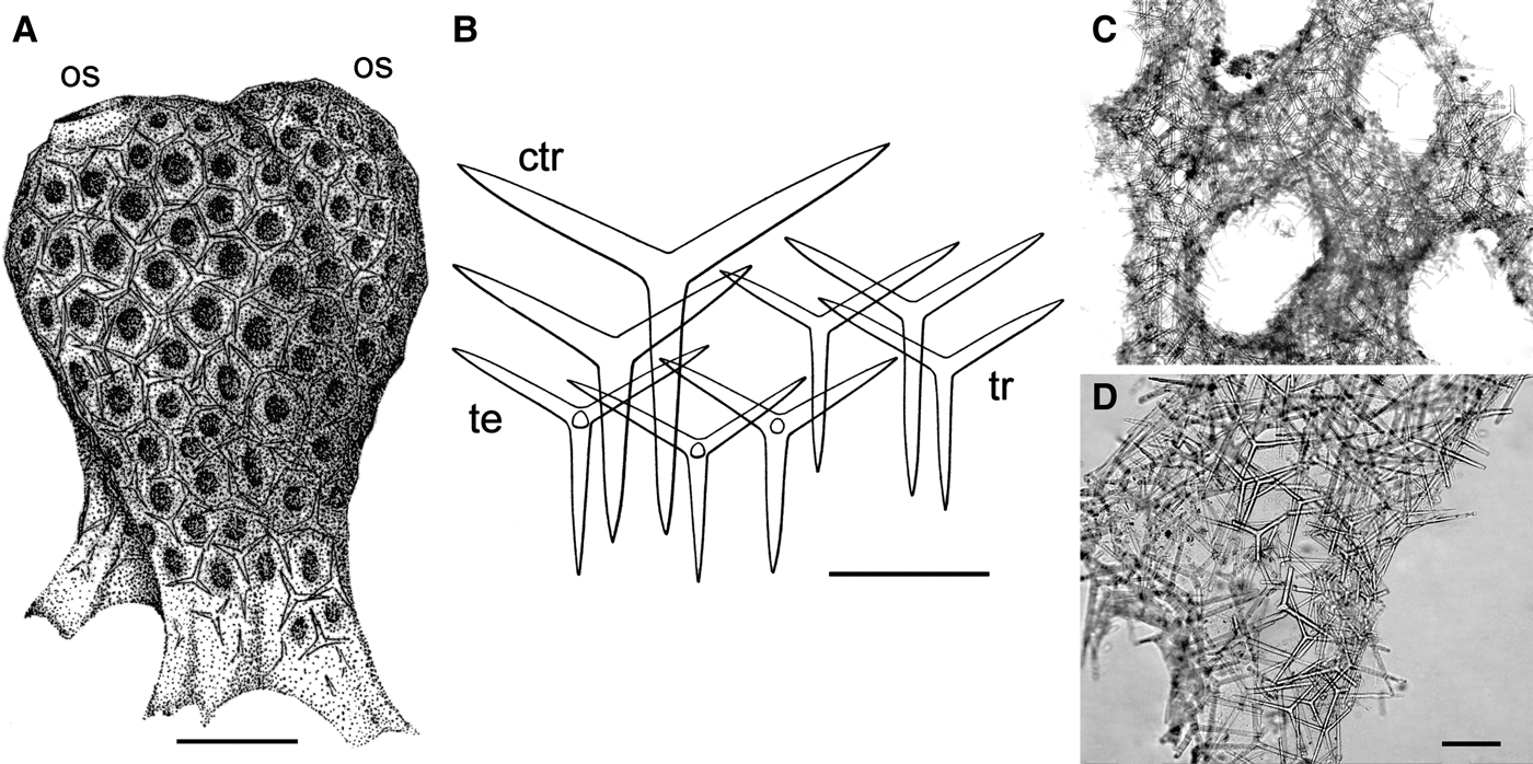
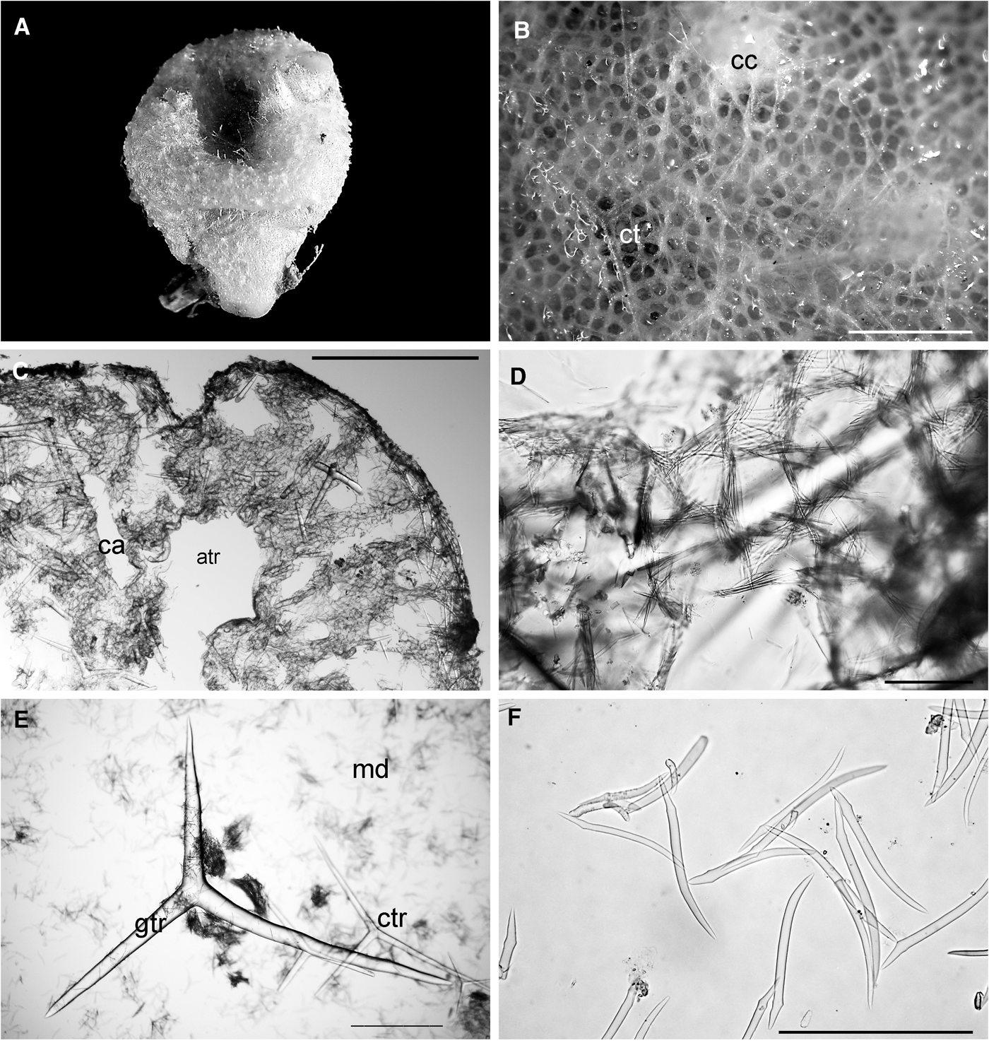
Ascaltis lamarcki: (A) drawing of a specimen from France (from Borojevic, Reference Borojevic1968); os, osculum; (B) spicules; ctr, giant cortical triactine; tr, triactine; te, tetractine (from Borojevic, Reference Borojevic1968); (C & D) photographs of anastomosing asconoid tubes in Haeckel's type slide (MNHM, Paris). Scale bars: A, 1 mm; B, 200 µm.
Distribution of Ascaltis lamarcki along the coast of Greenland.
Original description: Ascaltis lamarcki Haeckel, Reference Haeckel1872: 60–61, plate 9 figure 5, plate 10 figure 4a–d.
SYNONYMS AND CITATIONS
Ascaltis lamarcki (Haeckel, Reference Haeckel1872, Reference Haeckel1874; Lütken, Reference Lütken and Jones1875; Borojevic, Reference Borojevic1968; Rapp, 2004; Rapp et al., Reference Rapp, Janussen and Tendal2011).
Ascetta lamarcki (Arnesen, Reference Arnesen1901).
Leucosolenia lamarcki (Breitfuss, Reference Breitfuss1897, Reference Breitfuss1898b, Reference Breitfussc, Reference Breitfuss1933; Lundbeck, Reference Lundbeck1909; Brøndsted, Reference Brøndsted1914).
TYPE LOCALITY
Greenland. No further information available. Probably from North Shannon.
TYPE MATERIAL
Holotype: fragment, MNHN LBIM C1968.629, Haeckel's slide deposited in the MNHN (examined).
PREVIOUS RECORDS
From North Shannon, East Greenland (Haeckel, Reference Haeckel1874).
MATERIAL EXAMINED
Haeckel's original slide in MNHN.
DESCRIPTION
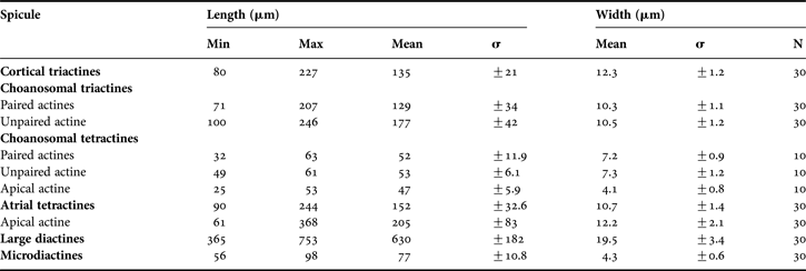
The cormus is massive, globular, and composed of ramified and anastomosed tubes, covered by a thin common cortex. The surface that is perforated by the ostioles that lead into the inhalant aquiferous system, ends in a large central tube covered by choanocytes. Hence the sponge has no true exhalant aquiferous system, but only a large normal tube or cavity that ends in one or several large oscula (Figure 14A). Consistency is soft. The colour is usually greyish-white. Cormus up to 20 mm in diameter, but is usually smaller. The skeleton is composed of two populations of regular triactines and one population of regular tetractines, approximately of the same size as the small triactines (Figure 14B). The large triactines are mainly in the outer part of the cortex, and are visible on the surface (170–270 µm × 15–20 µm) (Figure 14D). Small triactines and tetractines are mainly found in the choanosome (60–120 µm × 4–6 µm) (Figure 14C). The apical actine is of the same length as the basal actines or shorter. All actines are sharply pointed.
OCCURRENCE
Only reported from the type locality. Probably North Shannon in sector 13 (Figure 15).
GEOGRAPHICAL DISTRIBUTION
North Atlantic and the Arctic from littoral to 645 m in the Bay of Biscay (Borojevic & Boury-Esnault, Reference Borojevic, Boury-Esnault, Vacelet and Boury-Esnault1987).
REMARKS
The description is based on Haeckel (Reference Haeckel1872) and the type slide deposited in the MNHN. Haeckel's slide is of very poor quality, the cortical membrane is not visible and the orientation of the embedded pieces makes it very difficult to get photographs of sufficient quality, and especially for examination and illustration of the apical actines of the tetractines. It was not possible to see if the apical actines have spines or not. Spicule measurements are made from the type slide. As not a full set of spicule measurements could be made due to the poor quality of the slide and corrosion of some of the spicules, the spicule size is just presented as minimum–maximum in the text. The type locality for this species is reported to be ‘Greenland’, so it is considered to be a valid constituent of the Greenlandic sponge fauna, even though it must be rare in the coastal waters in the area. ‘Ascaltis lamarcki’ has been reported from most oceans, but the great morphological variation described from worldwide makes it reasonable to believe that it represents a complex of species, each with a more restricted geographical distribution (Rapp et al., Reference Rapp, Janussen and Tendal2011).
TYPE SPECIES
Leucascus simplex Dendy, Reference Dendy1892. Copiously branched and anastomosed choanocyte tubes covered by a continuous membrane. The exhalant aquiferous system is represented by a well-developed atrium delimited by a membrane with no choanoderm, supported by a specific skeleton of triactines and/or tetractines. The size of the spicules of the choanocyte tubes and of the cortical and atrial membranes is very similar. Apical actine of the tetractines ornamented with spines. Solenoid aquiferous system. From Cavalcanti et al. (Reference Cavalcanti, Rapp and Klautau2013).
KEY TO SPECIES OF LEUCASCUS
Only L. lobatus is known from the area.
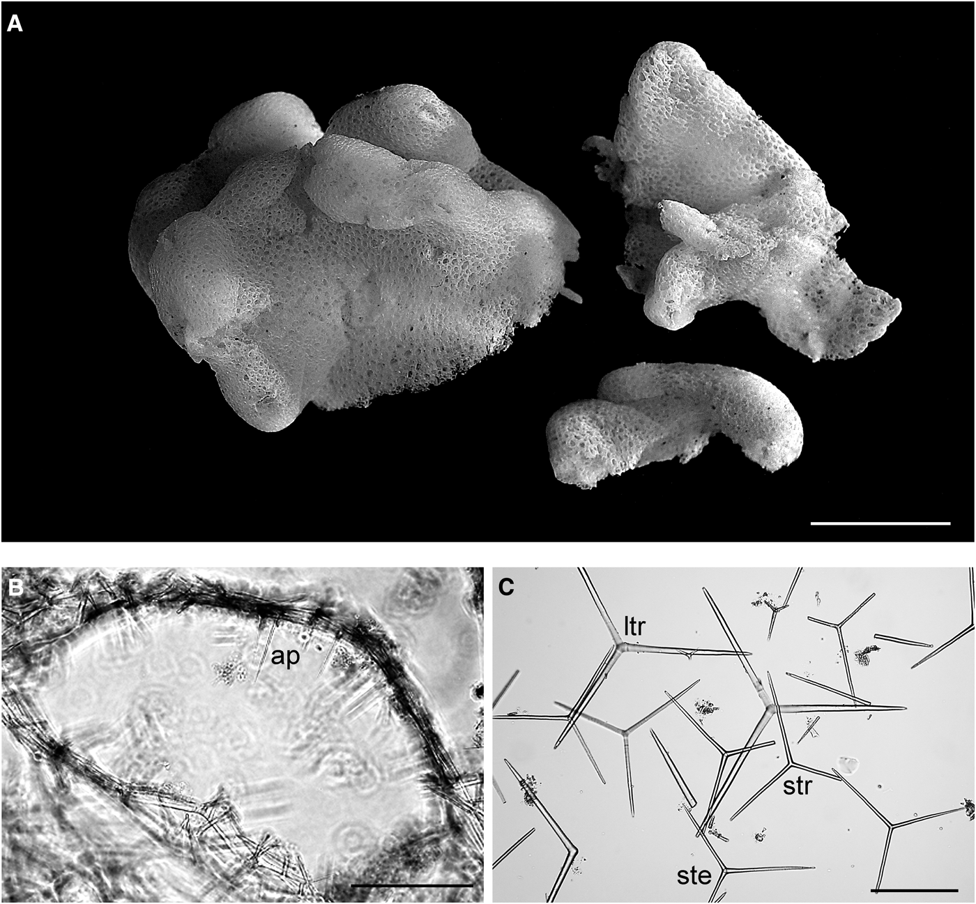
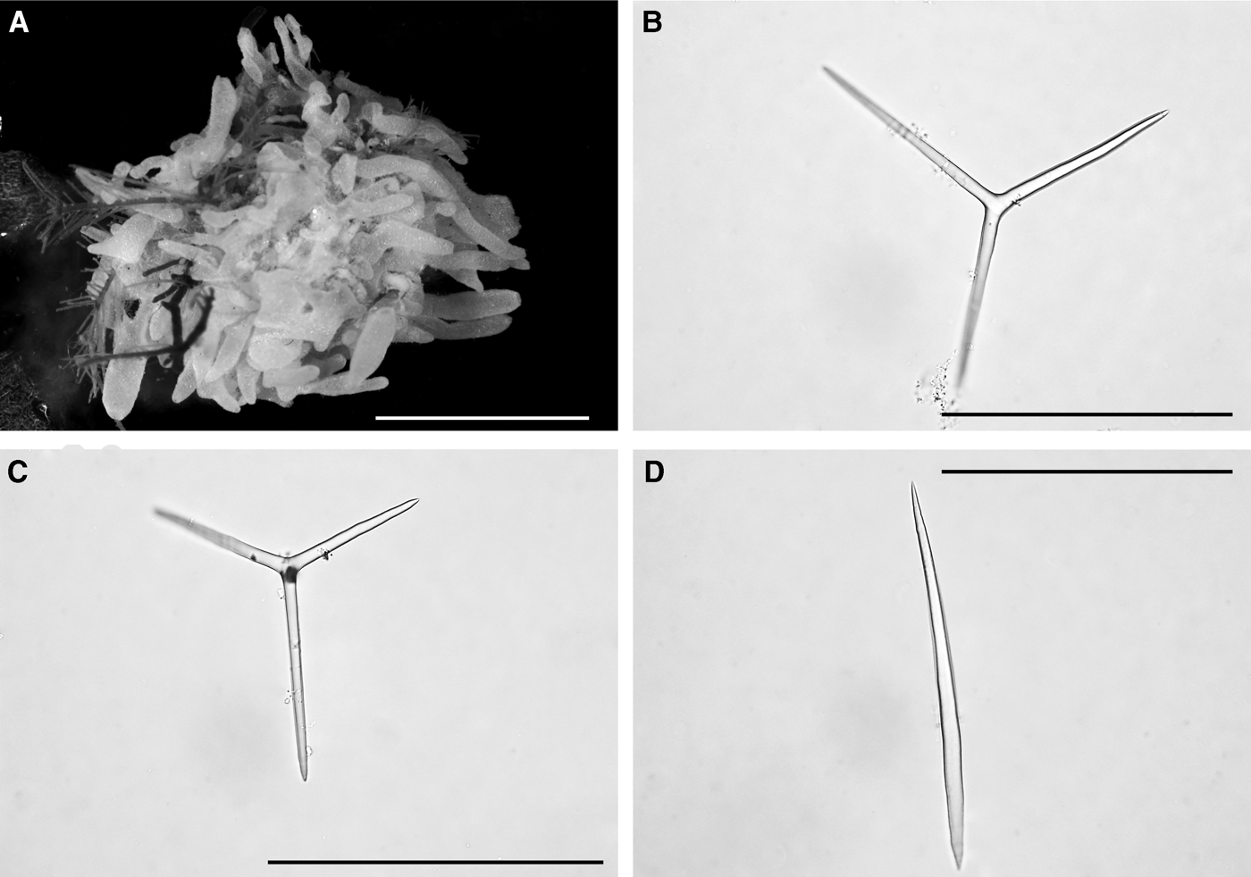
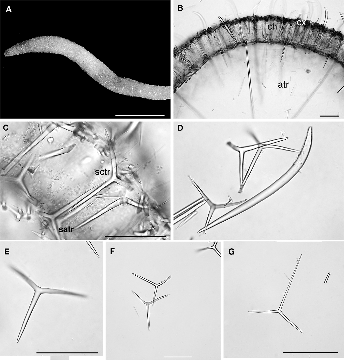
Leucascus lobatus: (A) habitus of preserved specimens (paratypes, ZMUC POR-246); (B) cross-section of an asconoid tube; ap, apical actine of tetractine; (C) spicules; ltr, large triactine; str, small triactine; ste, small tetractine. Scale bars: A, 1 cm; B, C, 100 µm.
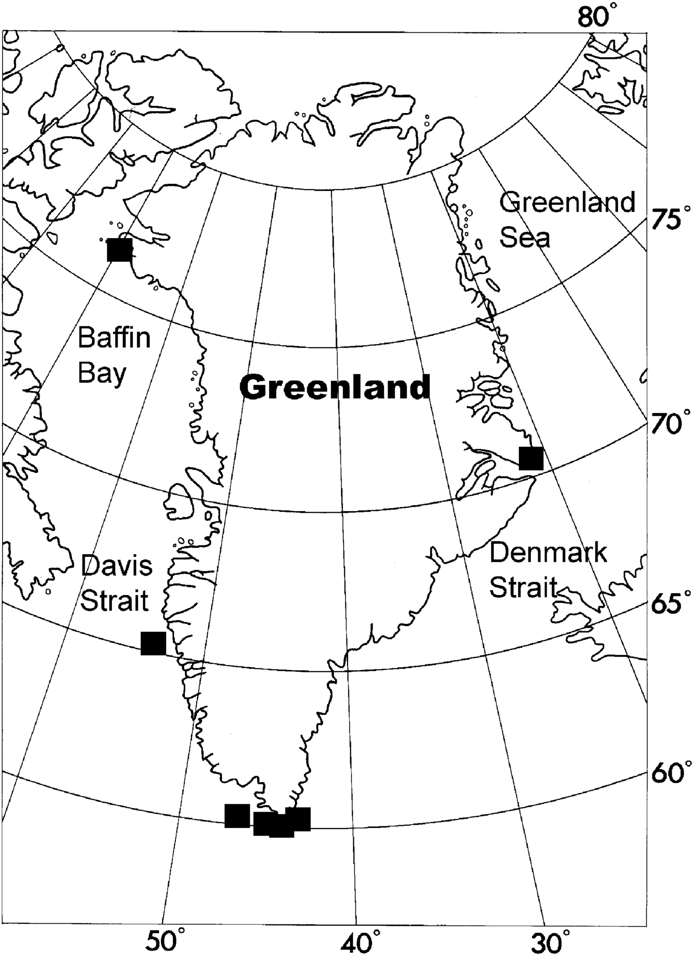
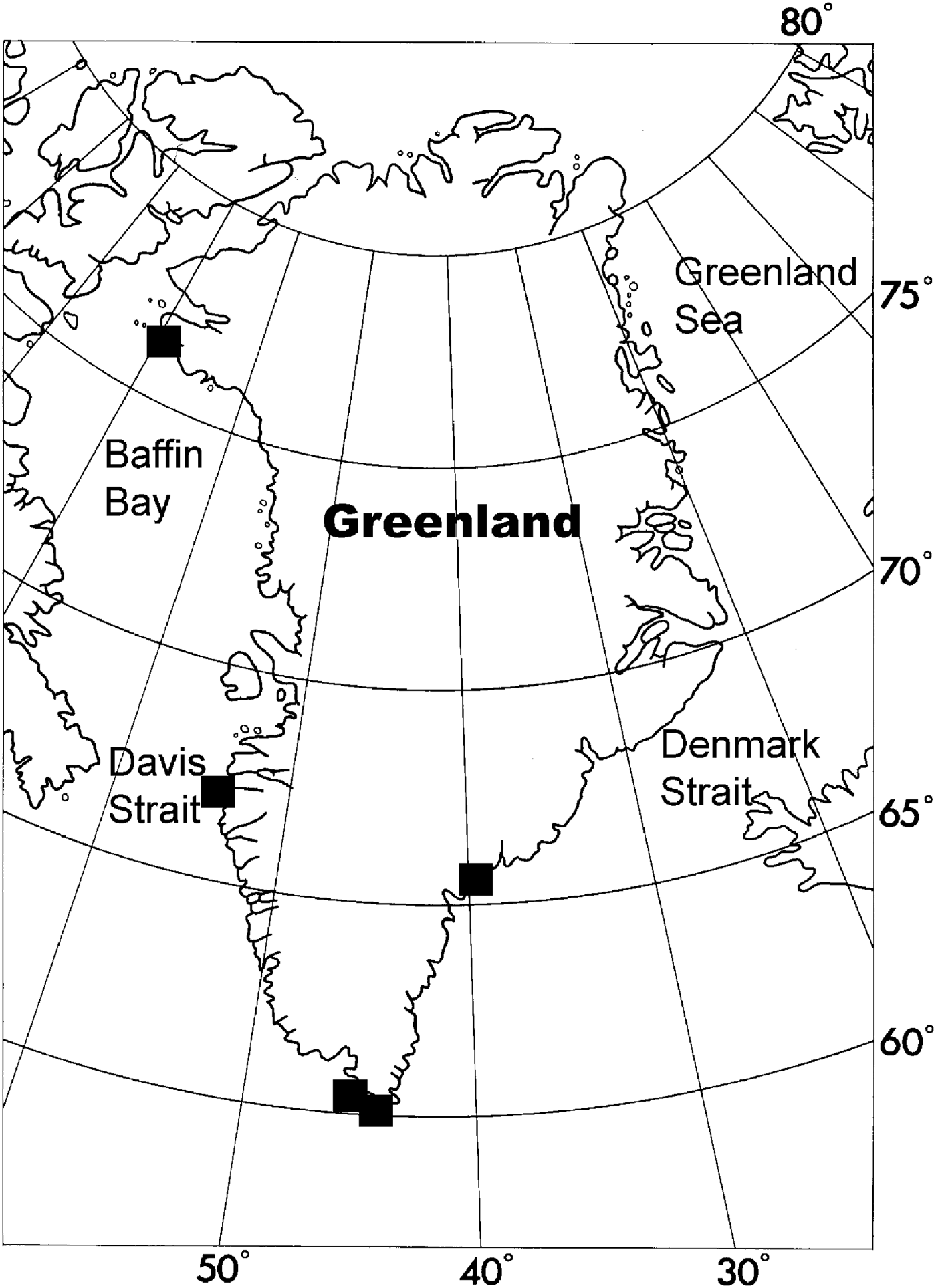
Distribution of Leucascus lobatus along the coast of Greenland.
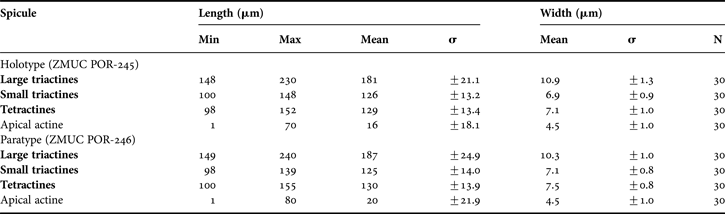
Spicule measurements of Leucascus lobatus Rapp, 2004 from Greenland.
Original description: Leucascus lobatus Rapp, Reference Rapp2004a: 1–9, figures 1a, b and 2a–g.
SYNONYMS AND CITATIONS
Leucascus lobatus (Rapp, Reference Rapp2004a, Reference Rappb; Cavalcanti & Klautau, Reference Cavalcanti and Klautau2011; Rapp et al., Reference Rapp, Janussen and Tendal2011, 2013; Cavalcanti et al., Reference Cavalcanti, Rapp and Klautau2013).
TYPE LOCALITY
Kap Farvel, southern Greenland.
TYPE MATERIAL
Holotype: ZMUC. POR 245 (alcohol and slide).
Paratypes: ZMUC. POR 246 (3 specimens, alcohol), Kap Farvel, southern Greenland. ZMUB no. 67217 (2 specimens, alcohol) and ZMUB no. 67218 (slide).
PREVIOUS RECORDS
From Julianehåb Bank, Godthåb Station 188, identified as Leucosolenia primordialis (Haeckel, Reference Haeckel1872) by Brøndsted (Reference Brøndsted1933a), and from seven localities by Rapp (2004).
MATERIAL EXAMINED
25 specimens. 65.06°N 54.19°W, 07.06.1909, 85 m (2). Godthåb Station 188, 60.22°N 47.27°W, 10.10.1928, 120 m (7). The Danish Three-Year Expedition to East Greenland Station 295, 70.40°N 22°W, 14.08.1933, 17–31 m (1). Just and Vibe Station 20, 76.34.5°N 69.24.5°W, 14.08.1968, 35 m (1). Kap Farvel Station 83, 60.13°N 44,26°W, 08.08.1970, 230–250 m (10). Station 85, 59.53.4°N 44.24.2°W, 09.08.1970, 8–12 m (1). Station 102, 60.15°N 44.17°W, 17.08.1970, 250–400 m (1). Station 136, 60.04.5°N 43.02.7°W, 25.08.1970, 240 m (1).
DESCRIPTION
The collection contains several large specimens with a size up to 6 cm. The cormus of this species is white to light beige in alcohol, and is composed of very regularly and tightly anastomosing tubes forming a massive, laterally compressed, and more or less elongated to folded lobate mass. Large oscula (up to 5 mm in diameter) are located along the upper margin of the cormus (Figure 16A). Remnants of a cortical membrane are found in open spaces between tangential cortical spicules. The surface is smooth, pierced by the openings of the elongated incurrent canals or chambers. The walls of the incurrent canals are smooth and have no choanocytes. The body wall is 2–5 mm thick, and surrounds the flattened central atrium, whose surface is pierced by circular to elongated openings of the exhalant cavities. The choanosome is composed of more or less radially arranged choanosomal tubes that open into exhalant canals. One to several layers of scattered large regular triactines, and several layers of smaller regular to sub-regular triactines and tetractines represent a weakly developed cortical skeleton (about 50 µm thick). The large triactines are slightly tripodite. The choanosomal skeleton is composed of irregularly arranged small tri- and tetractines, and scattered large triactines, of the same types as those in the cortex. The apical actine of the tetractines is ornamented with small spines and is directed inwards into the interior of the choanosomal tubes (Figure 16B). The sponge has a well-developed atrium delimited by an atrial skeleton composed of small, regular to sub-regular triactines and tetractines of the same shape and size as the small spicules in the cortex and choanosome. The atrial skeleton is smooth as the apical actine of the atrial tetractines is directed towards the choanosome. The atrial wall is devoid of choanocytes.
OCCURRENCE
The species was found in sectors 1, 3, 8 and 15 (Figure 17). Depth 8–400 m.
GEOGRAPHICAL DISTRIBUTION
No records from outside Greenlandic waters.
REMARKS
For affinity to other related species and a full review of the genus confer Cavalcanti et al. (Reference Cavalcanti, Rapp and Klautau2013). The species is illustrated in more detail in Rapp (2004) and Cavalcanti et al. (Reference Cavalcanti, Rapp and Klautau2013).
Calcarea with diactines and/or sagittal triactines and tetractines, rarely also with regular spicules. In addition to free spicules there may be a non-spicular basal skeleton in which basal spicules are cemented together or completely embedded in enveloping calcareous cement. In their ontogeny the first spicules to be produced are diactines in the settled larva. Choanocytes are apinucleate, and the basal system of the flagellum is adjacent to the apical region of the nucleus. Calcaronea incubate amphiblastula larvae.
Calcaronea with a skeleton composed of exclusively free spicules, without calcified non-spicular reinforcements. The aquiferous system is asconoid, syconoid, sylleibid, or leuconoid. In the latter case, radial organization around a central atrium can generally be detected by a well formed atrial skeleton tangential to the atrial wall, and/or a subatrial skeleton consisting of subatrial tri- or tetractines with the paired actines tangential to the atrial wall and the unpaired actine perpendicular to it. The post-larval development passes (presumably always) through an olynthus stage.
Leucosolenida with a cormus composed of frequently branched, but rarely anastomosed, asconoid tubes, with a continuous choanoderm that lines all the internal cavities of the sponge. There is neither a common cortex covering the cormus, nor a delimited inhalant or exhalant aquiferous system.
TYPE SPECIES
Spongia botryoides Ellis & Solander, 1786 (by original designation). Leucosoleniidae in which the skeleton can consist of diactines, triactines and/or tetractines. There is no reinforced external layer on the tubes.
KEY TO SPECIES OF LEUCOSOLENIA
1. Unpaired actine of tri- and tetractines usually shorter than paired actine ……… 2
— Unpaired actine longer than paired actines ……… L. complicata
2. Triactines T-shaped with almost straight paired actines ……… L. corallorrhiza
— Triactines with bent paired actines ……… L. variabilis
Leucosolenia complicata: (A) preserved specimen; (B) sagittal triactine; (C) sagittal tetractine; (D) diactine; Scale bars: A, 1 cm; B–D, 100 µm.
Distribution of Leucosolenia complicata along the coast of Greenland.
Spicule measurements of Leucosolenia complicata (Montagu, Reference Montagu1818) from Greenland.
Original description: Spongia complicata Montagu, Reference Montagu1818: 97, pl. ix, figures 2 & 3.
SYNONYMS AND CITATIONS
Ascandra complicata (Haeckel, Reference Haeckel1872; Arnesen, Reference Arnesen1901).
Leucosolenia complicata (Haeckel, Reference Haeckel1870; Levinsen, Reference Levinsen1893).
Ascandra fabricii (Breitfuss, Reference Breitfuss1898c; Lundbeck, Reference Lundbeck1909; Brøndsted, Reference Brøndsted1914).
Ascortis fabricii (Haeckel, Reference Haeckel1872).
Leucosolenia fabricii Schmidt, Reference Schmidt1869: 91.
Leucosolenia fabricii (Schmidt, Reference Schmidt1870).
TYPE LOCALITY
British Isles (Montagu, Reference Montagu1818).
PREVIOUS RECORDS
As Ascandra fabricii from Tasiusak by Lundbeck (Reference Lundbeck1909) and Brøndsted (Reference Brøndsted1914).
MATERIAL EXAMINED
Seven specimens. Ryder Expedition, Tasiusak, 1892 (1). Littoral Station 61, 67.48°N 53.47°W, 1.5 m (1). Just and Vibe Station 27, 76.27.9°N 69.19°W, 10.08.1968, 30 m (1). Kap Farvel Station 22, 60.09.4°N 44.14.9°W, 17.07.1970, 100–120 m (1). Station 40, 60.33°N 44.26°W, 22.07.1970, 100 m (1). Bankeundersøkelser Station 5296, 66.55°N 54.34°W, 22.08.1976, 48 m (1). Station 52.98, 67.05°N 54.67°W, 23.08.1976, 7 m (1).
DESCRIPTION
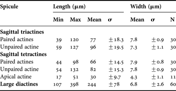
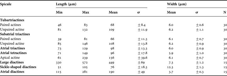
Sponge composed of a basal reticulation of ascon tubes, from which arise erect oscular tubes bearing lateral diverticula (Figure 18A). The surface is minutely hispid and texture is soft. Colour whitish-grey in life and ethanol. The skeleton comprises sagittal triactines and tetractines, and diactines (Figure 18B–D). In the sagittal triactines the unpaired actine is longer and more slender than the paired actines. The tetractines are of approximately the same size and shape as the triactines, and have a short, sharp and bent apical actine projecting into the atrial cavity. Except the apical actine, all the actines are slightly blunt. The diactines are of highly variable length and thickness. There are indications that there are two different populations of diactines with lengths of 100–180 µm and 220–400 µm, respectively, but statistically they are not significantly different. The diactines are with one lanceolate end and one slightly blunted.
OCCURRENCE
The species was found in sectors 1, 4, 8 and 15 (Figure 19). Depth 1.5–120 m.
GEOGRAPHICAL DISTRIBUTION
The species is known from most parts of the northern Atlantic and the Arctic, mainly in the shallow sub-littoral.
REMARKS
Leucosolenia complicata is a very variable species that bears many similarities with L. fabricii Schmidt, Reference Schmidt1869. They have traditionally been distinguished by L. fabricii having more slender spicules than L. complicata (about 6 µm compared to about 12 µm), and more bent paired actines of the sagittal tri- and tetractines. However, when looking at L. complicata over its distribution range, these characters are variable, and L. fabricii falls within this intraspecific variation. The species should therefore be considered synonymous, L. fabricii being a junior synonym of L. complicata.
Leucosolenia corallorrhiza: (A) habitus of a preserved specimen; (B) spicules; tr, sagittal triactine; te, sagittal tetractine; di, diactine. Scale bars: A, 0.5 cm; B, 100 µm.
Distribution of Leucosolenia corallorrhiza along the coast of Greenland.
Spicule measurements of Leucosolenia corallorrhiza (Haeckel, Reference Haeckel1872) from Greenland.
Original description: Ascortis corallorrhiza Haeckel, Reference Haeckel1872: 73–74, plate 11 figure 4, plate 12 figure 4a–i.
SYNONYMS AND CITATIONS
Ascortis corallorrhiza (Haeckel, Reference Haeckel1872; Lütken, Reference Lütken and Jones1875).
Ascandra corallorrhiza (Breitfuss, Reference Breitfuss1898c, Reference Breitfuss1933).
TYPE LOCALITY
Haeckel based his description on one specimen from Greenland and one from Norway, without designating a type specimen.
PREVIOUS RECORDS
Only the one by Haeckel (Reference Haeckel1872) without further indications on locality.
MATERIAL EXAMINED
Two specimens. Greenland, no further information on original label (1). Kangamiut Station 5, 66.15°N 56.12°W, 04.06.1976, 160–200 m (1).
DESCRIPTION
The sponge irregular to cushion-shaped, composed of a reticulation of ascon tubes. Some of the tubes are closed in the end, while others bear an osculum (Figure 20A). Colour greyish-white in ethanol. Tubes are 0.5–1 mm in diameter. The spicules are densely and irregularly to well arranged (with their unpaired actine directed towards the base in the large tubes). The skeleton comprises T-shaped sagittal triactines, tetractines of the same shape and size as the triactines, and one population of diactines (Figure 20B). In the T-shaped tri- and tetractines the unpaired actine is always shorter than the paired ones. All actines are stout and sharply pointed. The smooth apical actine of the tetractines is pointed into the atrium.
OCCURRENCE
The species was found only in sector 4 (Figure 21). Depth 160–200 m.
GEOGRAPHICAL DISTRIBUTION
Also known from Norway (Haeckel, Reference Haeckel1872; Arnesen, Reference Arnesen1901).
REMARKS
The species bears many similarities with the shallow water species Leucosolenia botryoides (Ellis & Solander, 1786) when it comes to size and general shape of the spicules. However, the actines are undulated in L. botryoides while they are straight in L. corallorrhiza. The proportion of tetractines is highly variable between the two examined specimens, a variation also observed by Haeckel (Reference Haeckel1872). However, this difference may be due to variable distribution of the spicule types in the sponge, where tetractines appear to be more abundant in the stolon/basal part than in the oscular tubes.
Leucosolenia variabilis: (A) habitus of a preserved specimen; (B) spicules; tr, di, diactine. Scale bars: A, 0.5 cm; B, 100 µm.
Distribution of Leucosolenia variabilis along the coast of Greenland.
Spicule measurements of Leucosolenia variabilis (Haeckel, Reference Haeckel1872) from Greenland.
Original description: Ascandra variabilis Haeckel, Reference Haeckel1872: 106–112, plate 16 figure 4a–l, plate 18.
SYNONYMS AND CITATIONS
Ascandra variabilis (Breitfuss, Reference Breitfuss1898c; Lundbeck, Reference Lundbeck1909; Brøndsted, Reference Brøndsted1914).
Leucosolenia variabilis (Breitfuss, Reference Breitfuss1933).
TYPE LOCALITY
Gisø on the Norwegian west coast.
PREVIOUS RECORDS
The records of Lundbeck (Reference Lundbeck1909) and Brøndsted (Reference Brøndsted1914) could not be verified.
MATERIAL EXAMINED
Nine specimens. Egedesminde, 1892 (1). Kap Farvel Station 82, 60.07°N 44.17°W, 08.08.1970, 50 m (2). Station 148, 60.07°N 43.20°W, 28.08.1970, 50 m (2). Bankeundersøkelser Station 5297, 67.00°N 54.67°W, 22.08.1976, 64–70 m (1). Station 5298, 67.05°N 54.67°W, 23.08.1976, 7 m (3, fragmentary).
DESCRIPTION
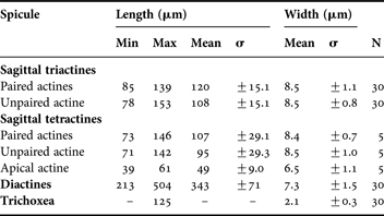
Sponge composed typically of a basal reticulation of ascon tubes, from which arise erect tubular vents bearing lateral diverticula. Surface minutely hispid. Texture soft. Colour in life and in ethanol greyish-white. Skeleton of sagittal triactines, sagittal tetractines with their short apical actine projecting into the atrium, one population of diactines and trichoxea. The unpaired actine of tri- and tetractines is always shorter than the paired ones. Normal lanceolate diactines are of highly variable size, and it is not possible to divide them into well-defined size groups. The second population is composed of slender trichoxea of approximately the same length as the normal diactines. Except for the trichoxea all the spicules are of about the same strength.
OCCURRENCE
The species was found in sectors 1, 4 and 5 (Figure 23). Depth 50–70 m.
REMARKS
The species is widely distributed in the north-east Atlantic in the shallow sub-littoral.
REMARKS
Length of the trichoxea could not be measured with certainty because they are very easily broken, and the maximum length given here should be regarded as an indication only. Sagittal tetractines proved to be very rare, and therefore only a handful of spicules could be measured.
Leucosolenida with a central atrial tube and perpendicular regularly arranged radial tubes lined by choanoderm. The distal cones of the radial tubes, which may be furnished with tufts of diactines, are clearly noticeable on the sponge surface. They are never covered by a cortex supported by tangential triactines and/or tetractines. The proximal skeleton of the radial tubes is composed of a row of subatrial triactines and/or tetractines which are usually followed by only a few or several rows of triactines and/or tetractines. Pseudosagittal spicules are absent. A tangential layer of triactines and/or tetractines supports the atrial wall.
TYPE SPECIES
Sycon humboldti Risso, 1826 (by subsequent designation; Dendy, Reference Dendy1892). Sycettidae with radial tubes partially or fully coalescent; distal cones are furnished with tufts of diactines. The inhalant canals are generally well defined between the radial tubes, and are often closed at the distal end by a membrane that is perforated by an ostium, devoid of a skeleton. There is no continuous cortex covering the distal ends of the radial tubes. Skeleton of the atrium and of the tubes composed of triactines and/or tetractines.
KEY TO SPECIES OF SYCON
1. Surface hispid due to diactines ornamenting the distal cones ……… 2
— Surface smooth without diactines ornamenting the distal cones ……… S. karajakense
2. The short choanocyte chambers are fused in their entire length from the atrium to the edge of the distal cones ……… S. abyssale
— Radial chambers fused only at their base, close to the atrial wall ……… S. ciliatum
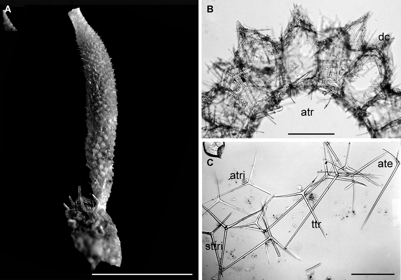
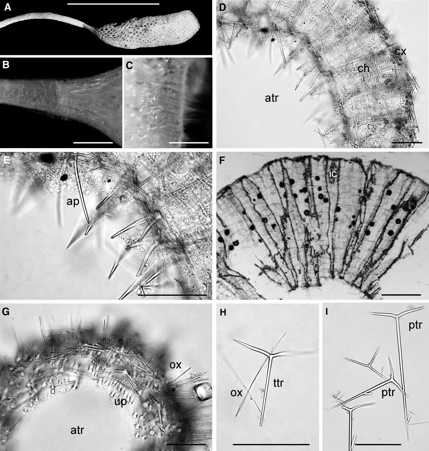
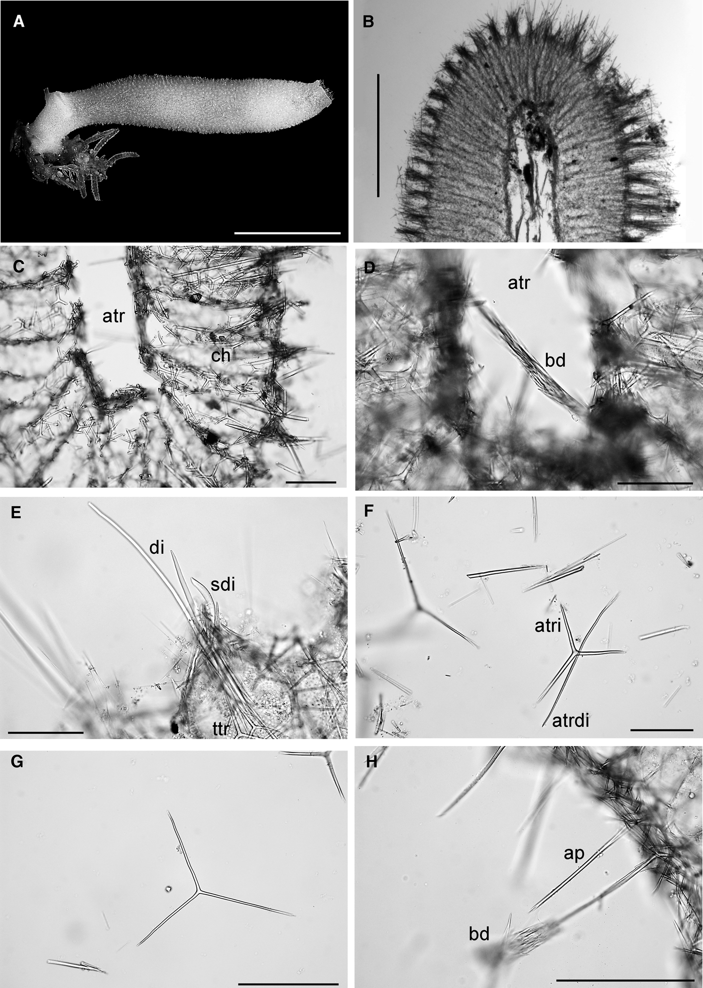
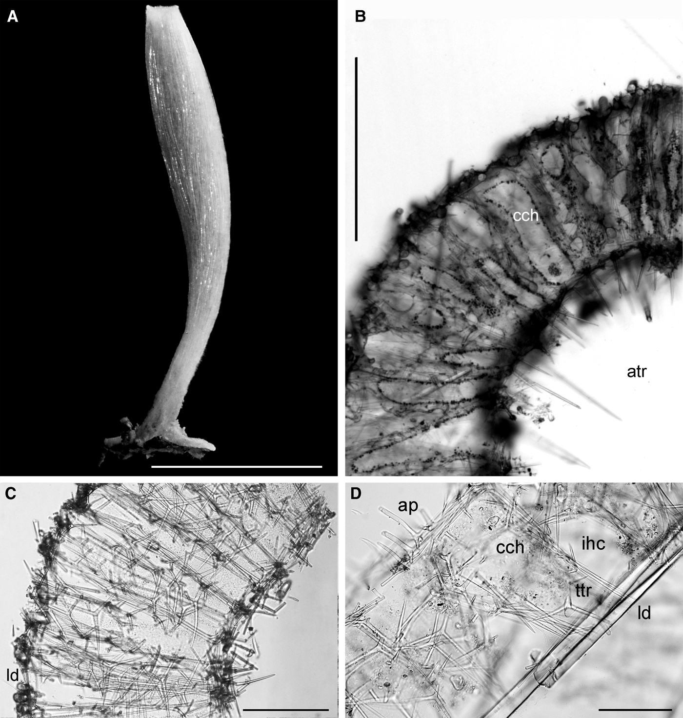
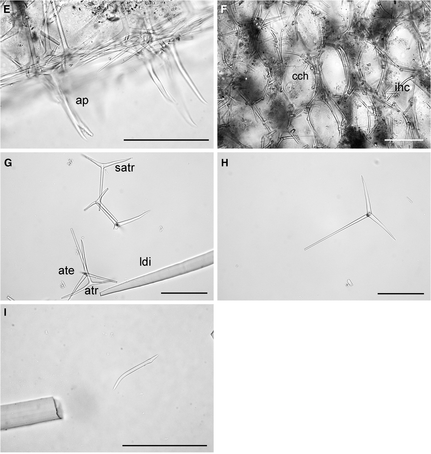
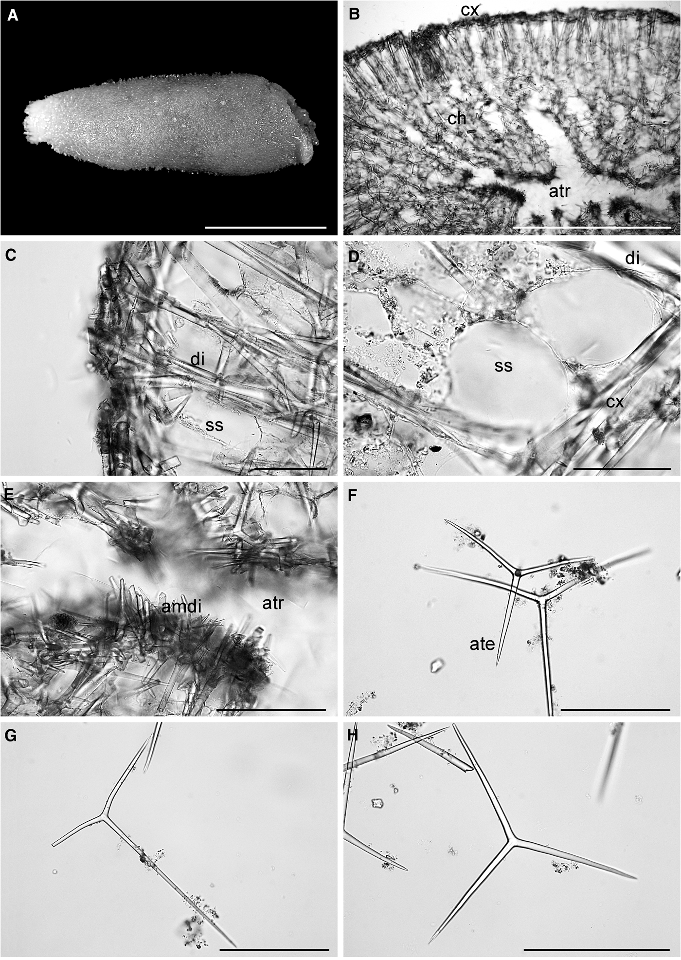
Sycon abyssale: (A) habitus of a preserved specimen; (B) section; dc, distal cone. atr, atrium; (C) spicules; ate, atrial sagittal tetractine; atri, atrial sagittal triactine; ttr, tubar sagittal triactine; sttri, subregular to sagittal triactine from the choanosome and the distal cones. Scale bars: A, 1 cm; B, C, 100 µm.
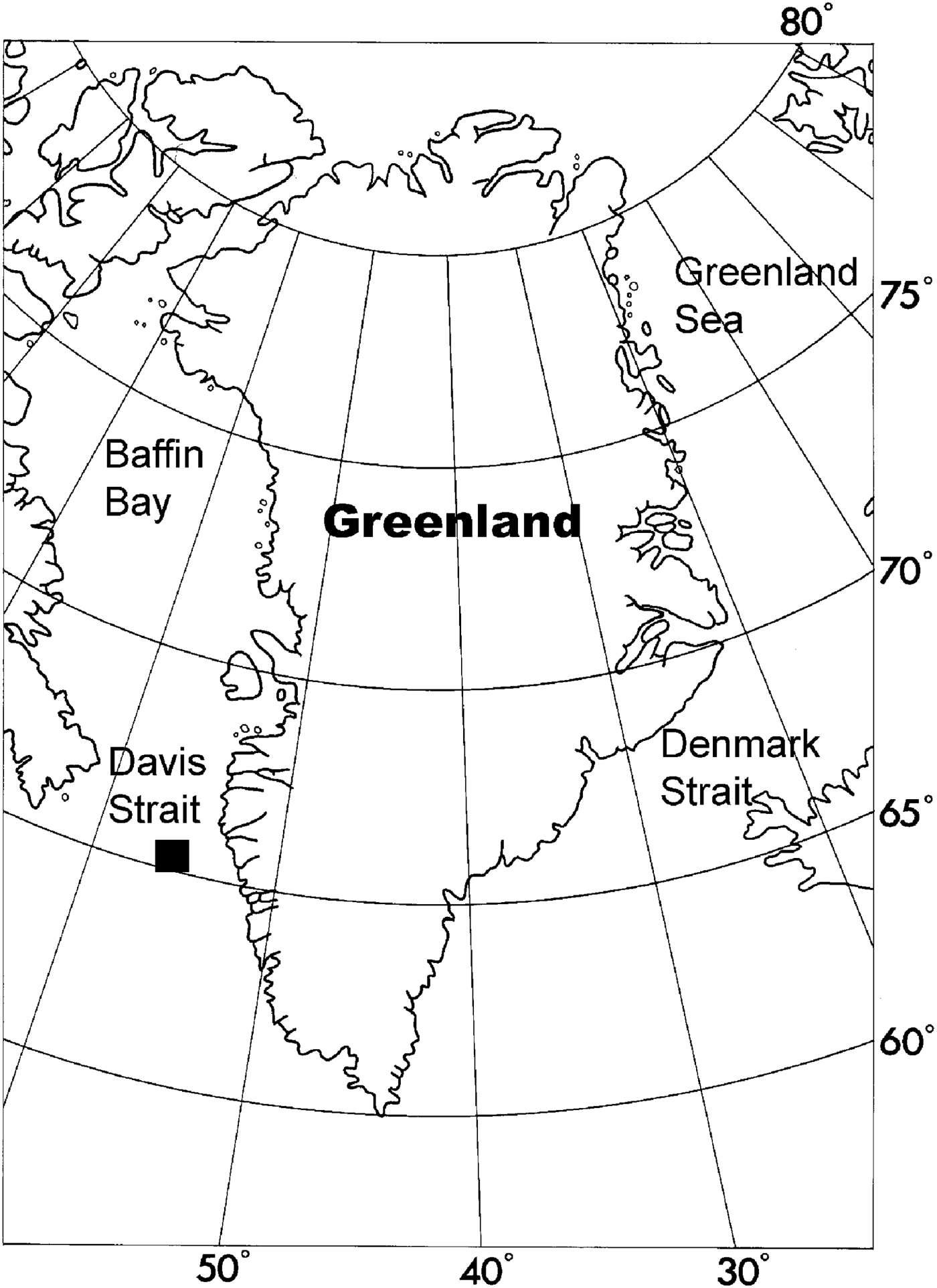
Distribution of Sycon abyssale along the coast of Greenland.
Spicule measurements of Sycon abyssale Borojevic & Graat-Kleeton, Reference Borojevic and Graat-Kleeton1965 from Greenland.
Original description: Sycon abyssale Borojevic & Graat-Kleeton, Reference Borojevic and Graat-Kleeton1965: 81–85, figure 1.
SYNONYMS AND CITATIONS
Sycon abyssale (Tendal, Reference Tendal1989; Barthel & Tendal, Reference Barthel and Tendal1993; Janussen et al., Reference Janussen, Rapp and Tendal2003; Rapp & Tendal, Reference Rapp and Tendal2006; Schander et al., Reference Schander, Rapp, Kongsrud, Bakken, Berge, Cochrane, Oug, Byrkjedal, Cedhagen, Fosshagen, Gebruk, Larsen, Nygren, Obst, Plejel, Stöhr, Todt, Warén, Handler-Jacobsen, Kuening, Levin, Mikkelsen, Petersen, Thorseth and Pedersen2010).
TYPE LOCALITY
North Atlantic at 62°00′N33°00′°W, 28.02.1964, 3800 m depth (holotype).
PREVIOUS RECORDS
The species is new to Greenland.
MATERIAL EXAMINED
One specimen. Ingolf Station 35, 65.16°N 55.05°W, 18.07.1895, 682 m (1).
DESCRIPTION
Sponge slender and tube-shaped with a peduncle (Figure 24A). Surface prominently papillous and slightly hispid due to diactines ornamenting the distal cones of the short radial chambers (Figure 24B). The single apical osculum is ornamented with a thin membrane with sagittal triactines and tetractines. The atrial skeleton comprises sagittal triactines and tetractines of the same size and shape. The apical actine of the tetractines is projected into the atrium. The subatrial skeleton is composed of sagittal triactines with their unpaired actine pointed towards the distal cones. The tubar skeleton consists of subregular and sagittal triactines (Figure 24B). Sharp and only slightly bent diactines are ornamenting the distal cones. In the peduncle the organization of the skeleton is slightly different as there are no choanocyte chambers. The skeleton comprises sagittal triactines and tetractines (Figure 24C) similar to those found in the atrial skeleton, arranged in parallel with their unpaired actine pointing towards the base of the sponge. Some tangential diactines are embedded in the surface layer of the peduncle.
OCCURRENCE
The species was found in sector 3 (Figure 25). Depth 682 m.
GEOGRAPHICAL DISTRIBUTION
The species is found in most parts of the abyssal North Atlantic down to 3800 m depth (Borojevic & Graat-Kleeton, Reference Borojevic and Graat-Kleeton1965; Tendal, Reference Tendal1989; Barthel & Tendal, Reference Barthel and Tendal1993; Rapp & Tendal, Reference Rapp and Tendal2006; Rapp & Tendal, manuscript in preparation). It has also been found on hydrothermal vents off Jan Mayen (Schander et al., Reference Schander, Rapp, Kongsrud, Bakken, Berge, Cochrane, Oug, Byrkjedal, Cedhagen, Fosshagen, Gebruk, Larsen, Nygren, Obst, Plejel, Stöhr, Todt, Warén, Handler-Jacobsen, Kuening, Levin, Mikkelsen, Petersen, Thorseth and Pedersen2010), along the Norwegian shelf and in coastal and fjord areas in more shallow water (personal observations).
REMARK
This record represents the shallowest published record of Sycon abyssale.
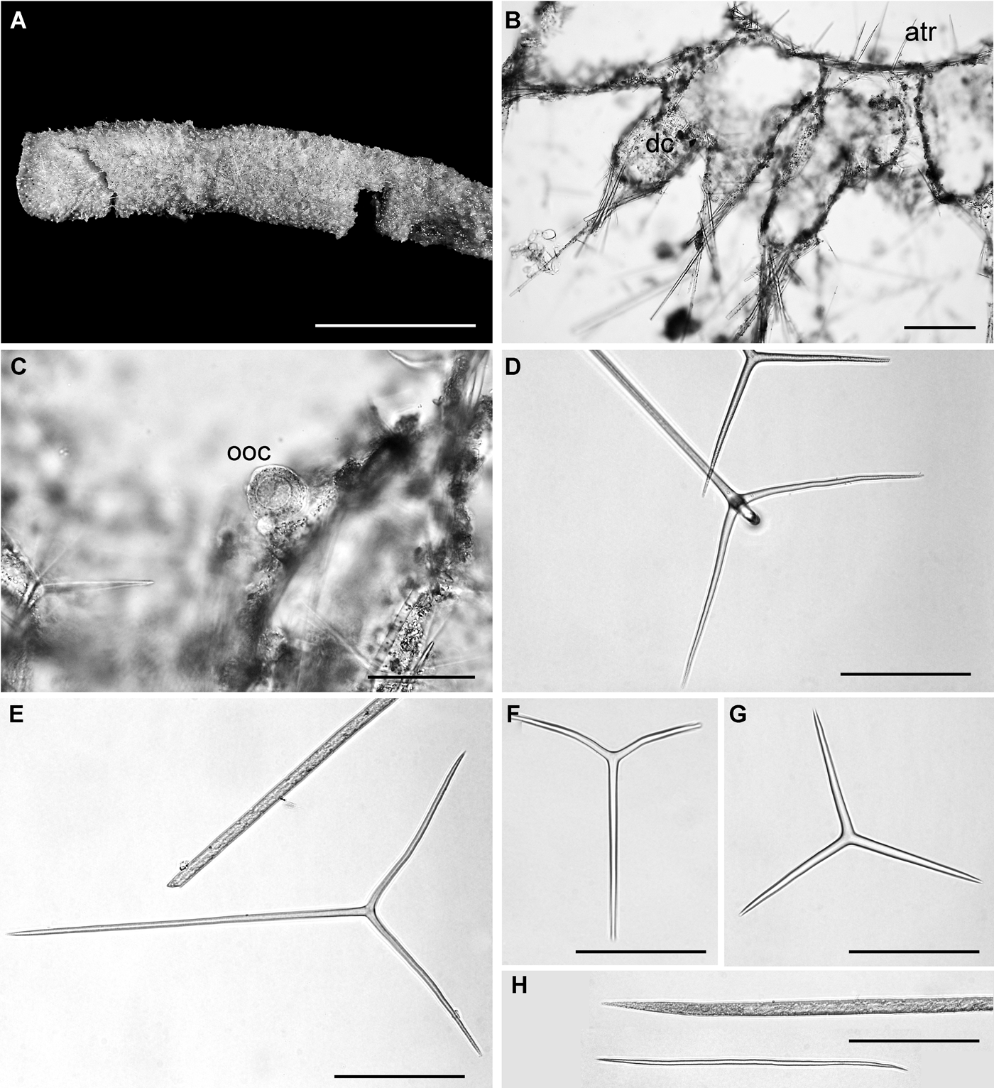
Sycon ciliatum: (A) habitus of a preserved specimen; (B) cross-section; dc, distal cone of radial chamber; atr, atrium; (C) section of a choanocyte chamber with a mature oocyte (ooc); (D) atrial tetractine; (E) subatrial trictine; (F) tubar triactine; (G) atrial triactine. The same type is also found in the outermost part of the choanocyte chambers; (H) diactines from the distal tufts ornamenting the choanocyte chambers. Scale bars: A, 1 cm; B–H, 100 µm.
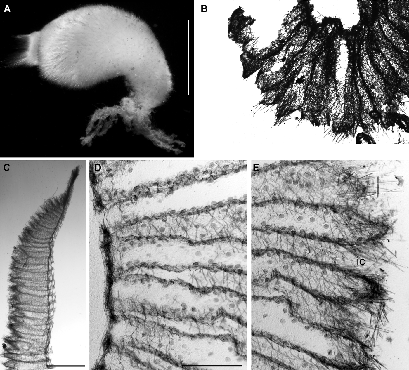
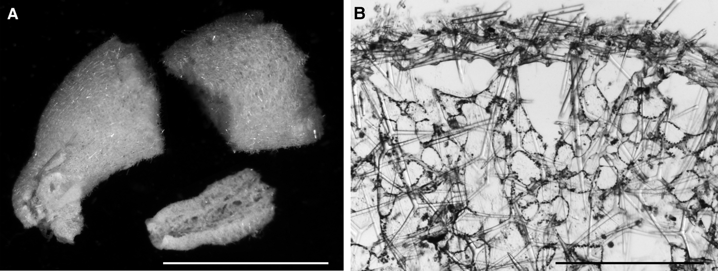
Sycon ciliatum: (A) preserved specimen from the Norwegian west coast; (B) cross-section. Photograph of slide from Haeckel's type collection (MNHN). Specimen was from the Norwegian west coast; (C) longitudinal section of (A); (D) cross-section of (A) from atrial wall to mid-height of the choanocyte chambers. Note that the chambers are free all the way down to the atrial wall; (E) cross-section of (A). Outer part of the choanocyte chambers. Note the wide incurrent canals (ic). Numerous larvae can be observed in D and E (material collected 10 August 2007). Scale bars A, 1 cm; B, 1 mm; C, 2 mm; D, E, 500 μm.
Distribution of Sycon ciliatum along the coast of Greenland.
Spicule measurements of Sycon ciliatum (Fabricius, Reference Fabricius1780) from Greenland.
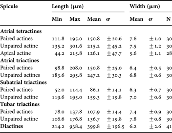
Spicule measurements of Sycon ciliatum (Fabricius, Reference Fabricius1780) from the Norwegian west coast (collected on kelp at Station 6050701 (Tornes, Reference Tornes2008), Toftøy at 60°27.5′N04°56.14′E, 3–25 m depth).
Original description: Spongia ciliata Fabricius, Reference Fabricius1780: 448.
SYNONYMS AND CITATIONS
Sycandra ciliata (Haeckel, Reference Haeckel1872; Arnesen, Reference Arnesen1901).
Sycon ciliatum (Breitfuss, Reference Breitfuss1898b, Reference Breitfussc; Lundbeck, Reference Lundbeck1909; Brøndsted, Reference Brøndsted1914; Tendal, Reference Tendal1970; Fortunato et al., Reference Fortunato, Adamski, Bergum, Guder, Jordal, Leininger, Zwafink, Rapp and Adamska2012).
TYPE LOCALITY
Greenland (Fabricius Reference Fabricius1780). Probably from the Prøven or Disco areas. The type material of Fabricius is lost (O.S. Tendal, personal communication).
PREVIOUS RECORDS
Several previous records, but they cannot be verified, partly because the material has not been found in the collection, or the spicules in the specimens were dissolved (material of Tendal (Reference Tendal1970)).
MATERIAL EXAMINED
One specimen from Ingolf Station 34, 65.17°N 54.17°W, 18.07.1895, 104 m. Haeckel's original slide of Sycandra ciliata from Bergen, Norway (Figure 27B). Terneøya, Norwegian west coast at 60°59.48′N 04°34.45′E, 10.08.2007. 0.5–3 m depth (coll. M. Tornes) (15 specimens). Toftøy, Norwegian west coast 60°27.5′N 04°56.14′E, 06.05.2007, 3–25 m depth (coll. M. Tornes) (1 specimen).
DESCRIPTION
Sponge tubular and minutely papillate and hispid. Osculum apical, with or without a fringe (Figures 26A, 27A). Texture soft to moderately firm. Colour in alcohol is beige. The choanocyte chambers are free in most or all of their length, and if fused only in the basal part close to the atrial wall (Figures 26B, 27D). The atrial skeleton comprises subregular to sagittal tetractines with their apical actine pointing into the atrium and triactines of the same shape and size (Figure 26D, G). The subatrial skeleton comprises sagittal triactines with their unpaired actines lying adjacent to the atrial skeleton, and the unpaired actine pointing towards the exterior (Figure 26E). The skeleton of the choanosome or choanocyte chambers comprises highly variable sagittal to subregular triactines with their unpaired actine pointing towards the distal cones (Figure 26F). The distal cones are ornamented with long diactines extending into the centre of the distal cones (Figure 26H).
OCCURRENCE
The species was only found in sector 3 (Figure 28). Fabricius' type material was probably from sectors 5 or 6. Depth 106 m.
GEOGRAPHICAL DISTRIBUTION
Sycandra ciliatum has been reported world-wide, but it is very likely that the distribution is more restricted (see Remarks below).
REMARKS
Cosmopolitanism is very rare among sponges (Klautau et al., Reference Klautau, Russo, Lazoski, Boury-Esnault, Thorpe and Solé-Cava1999), and most species previously regarded as cosmopolitan have after more thorough investigations by means of a combination of morphological and genetic methods, resulted in the division of morphologically closely but genetically more distantly related species with a more restricted geographical distribution (Solé-Cava et al., Reference Solé-Cava, Klautau, Boury-Esnault, Borojevic and Thorpe1991; Klautau et al., Reference Klautau, Russo, Lazoski, Boury-Esnault, Thorpe and Solé-Cava1999; Wörheide et al., Reference Wörheide, Hooper and Degnan2003). The original description by Fabricius (Reference Fabricius1780) is very short and general, and it basically only describes a white, flexible, tomentose and tubular sponge with an oscular fringe, a description that fits to a range of calcarean taxa. According to Haeckel (Reference Haeckel1872) Sycon ciliatum is a species mainly inhabiting the shallow sub-littoral of the north-east Atlantic and the Arctic. Morphological studies of material from an area ranging from France, the British Isles, the Norwegian and Swedish coasts (personal observations) as well as the material presented here from Greenland, supports Haeckel's statement. The sponge may be slender or chubby and robust, but the organization of the skeleton is remarkably constant. The only exception is that the apical actines of the atrial tetractines are quite variable in length, both within and between populations. Molecular analyses based on the ITS I-5.8S-ITS II gene fragment shows very low variation within this distribution range (M. Adamska, unpublished results). Haeckel based his description on material collected from the Bergen area on the Norwegian west coast. While the studied material from Greenland is a compressed and quite thin-walled tube, specimens from the Norwegian coast are generally more robust and thick-walled. The species is also much more common in boreal waters than in the Arctic, indicating that Greenland is really on the distribution limit for this species.
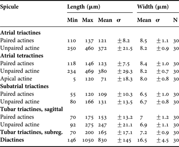
The typical Sycon ciliatum is therefore the one found in shallow waters along the coasts of Western Europe, where it is very common on kelp (Laminaria) in shallow waters (Tornes, Reference Tornes2008). The spicules from specimens from western Norway are slightly smaller (shorter and more slender actines (Table 13)) than in the specimen from Greenland (Table 12). The only difference that should be kept in mind is therefore the robustness of the specimens and the more substantial thickness of the body wall (Figure 27). Sycon ciliatum has recently been established as a model species for studying developmental signalling in sponges (Dr Maja Adamska, Sars International Centre for Marine Molecular Biology, Bergen, Norway). Based on Haeckel's (Haeckel, Reference Haeckel1872) conception of S. ciliatum as well as our own studies we consider the Norwegian west coast to be a core area within the distribution range of the sponge and it is therefore particularly well suited for comprehensive genomic studies.
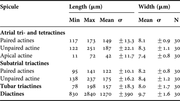
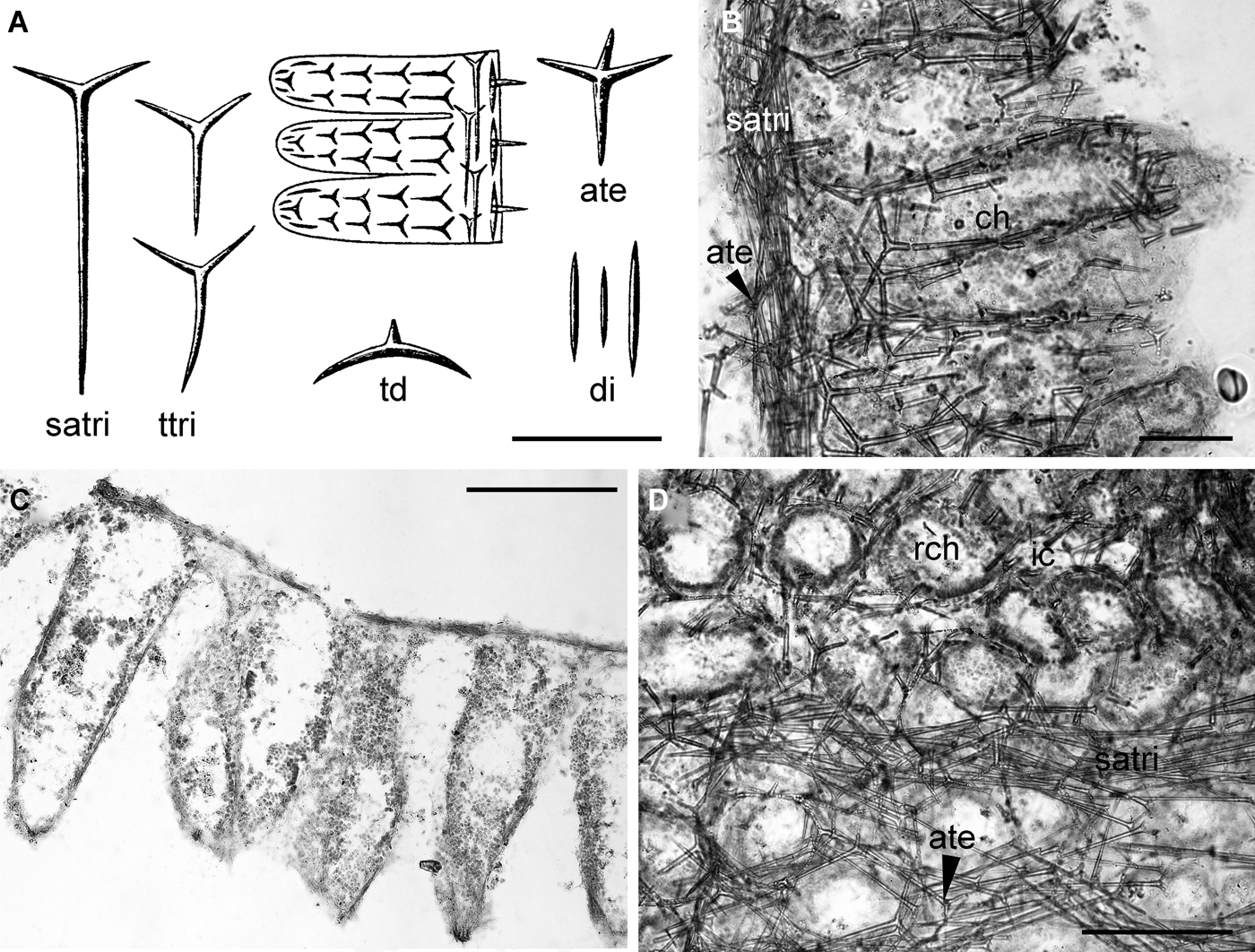
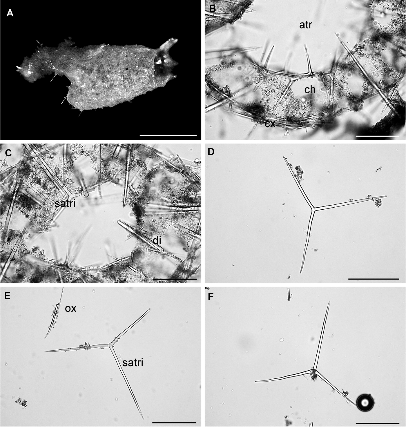
Sycon karajakense: (A) drawing of spicules and longitudinal section of Sycon karajakense (from Breitfuss, Reference Breitfuss1897); satri, subatrial sagittal triactine; ttri, tubar sagittal triactine; td, triactine from the distal part of the choanocyte chambers; ate, atrial tetractine; di, diactines from the distal part of the choanocyte chambers; (B) longitudinal section of the type (ZMB 2705); ate, atrial tetractine; satri, subatial sagittal triactines; ch, choanocyte chamber; (C) cross-section showing the shape of the choanocyte chambers; (D) section made in parallel to the surface; ch, choanocyte chamber; ic, incurrent space between the chambers. Scale bars: A, B, 100 µm; C, D, 200 µm.
Distribution of Sycon karajakense along the coast of Greenland.
Original description: Sycon karajakense Breitfuss, Reference Breitfuss1897: 207–208, figure 1a–g.
SYNONYMS AND CITATIONS
Sycon karajakense (Breitfuss, Reference Breitfuss1898b, Reference Breitfuss1933; Brøndsted, Reference Brøndsted1914, Reference Brøndsted1933b).
TYPE LOCALITY
Lille Karajakfjord (Breitfuss, Reference Breitfuss1897).
TYPE MATERIAL
ZMB Por 2705, Lille Karajakfjord (Breitfuss, Reference Breitfuss1897).
PREVIOUS RECORDS
Only known from Lille Karajakfjord, western Greenland.
MATERIAL EXAMINED
Breitfuss' slides of the holotype (ZMB NO 2705).
DESCRIPTION
Sponge solitary, tube-shaped and with a single naked osculum. The surface is smooth and the consistency is soft. Colour white. The radial chambers are free in most of their length, only fused close to the base (Figure 29A, C). The atrial skeleton comprises scattered and tangentially arranged subregular tetractines with their apical actine pointed into the atrium (actines ~63 µm × 7 µm, apical actine slightly stronger) (Figure 29A, B). The subatrial skeleton is composed of a dense layer of tangential sagittal triactines with their very long unpaired actine pointed towards the base of the sponge (unpaired actine 250–300 µm × 4–5 µm, paired actines 40–50 µm × 4–5 µm) (Figure 29A, B). The tubar skeleton comprises rows of sagittal triactines with their unpaired actine radially arranged. When approaching the distal cones the unpaired actine gradually becomes shorter, and directly beneath the surface the triactines are subregular (Figure 29A). No measurements of these spicules are presented in the original description, but from the figures and the type slide it may be estimated that the actines are about 40–50 µm in length. These triactines seem to be slightly more slender and more sharply pointed than the subatrial triactines. In the end of the distal cones there are some very short diactines. Unlike most other species of Sycon, these diactines never cross the surface of the distal cones (Figure 29A).
OCCURRENCE
Only found in sector 5 (Figure 30). Littoral.
GEOGRAPHICAL DISTRIBUTION
Only known from western Greenland.
REMARKS
Species of Sycon with a smooth surface are very rare, and the arrangement of the skeleton resembles more a Sycetta than a Sycon. However, the type slide is in quite bad condition, and there is considerable etching of the spicules, especially in the choanocyte chambers. The apical actines of the atrial tetractines and the paired actines of the subatrial triactines are also severely damaged. Therefore reliable spicule measurements could not be made and those presented in the description are from Breitfuss (Reference Breitfuss1897). Taken into account that the type material was collected in the littoral and that only a handful of Calcarea specimens from the littoral zone have been available for examination for me and previous authors, S. karajakense may be more abundant than indicated here. The description here is based on the original description (Breitfuss, Reference Breitfuss1897) and examination of slides of the type (ZMB 2705). The remaining parts of the type specimen are just small crumbs on the bottom of the jar (Carsten Lüter (ZMB), personal communication).
Leucosolenida in which there is always a cortex, supported by a skeleton of tangential spicules that can be diactines, triactines, tetractines, or any combination of these. The aquiferous system is either syconoid with radial and elongate choanocytes chambers, or sylleibid or leuconoid with elongate or spherical, scattered choanocytes chambers. The inhalant and exhalant aquiferous systems are always fully developed. The choanoskeleton is always articulate, tubular in syconoid species, and contains a few to several rows of triactines and/or tetractines, or is, in leuconoid species, arranged without apparent order. In the latter case, the choanoskeleton always preserves traces of the radial organization, particularly at the level of the subatrial triactines and/or tetractines. The atrial skeleton consisting of tangential triactines and/or tetractines is well developed.
TYPE SPECIES
Spongia compressa Fabricius, Reference Fabricius1780 (by original designation). Grantiidae with a syconoid organization. The cortex is composed of tangential triactines and/or tetractines, occasionally with small perpendicular diactines. Longitudinal diactines, if present, are not found exclusively in the cortex, but cross obliquely, at least through a part of the choanosome and protrude from the external surface.
KEY TO SPECIES OF GRANTIA
1. Surface hispid or villose due to diactines projecting through the cortex ……… 2
— Surface smooth, sponge with a distinct peduncle ……… G. phillipsi
2. One type of diactines is ornamenting the distal ends of the choanocyte chambers, to which straight trichoxea may be added ……… 3
— Two types of diactines are ornamenting the distal ends of the choanocyte chambers ……… 4
3. Diactines long and sharp making the surface strongly villose ……… G. arctica
— Diactines bent in the distal end which is rounded with spines ……… G. clavigera
4. Large diactines extend through the entire body wall ……… G. capillosa
— Large diactines extend through only a minor part of the body wall ……… G. mirabilis
Grantia arctica: (A) habitus of a preserved specimen; (B) oscular fringe; (C) cortex with tufts of diactines projecting from the end of the choanocyte chambers; (D) opening of inhalant chamber protected by distorted cortical triactines (ih); (E) tubar and cortical skeleton; (F) atrial skeleton; ap, apical actine of an atrial tetractine. Scale bars: A, B, 1 cm; C–F, 100 µm.
Grantia arctica continued; (G) ttri, tubartriactine; tt, triactine from tufts; (H) small distorted cortical triactine; (I) subatrial sagittal triactine and diactine; (J) atrial tetractine. Scale bars: G–J, 100 µm.
Distribution of Grantia arctica along the coast of Greenland.
Spicule measurements of Grantia arctica (Haeckel, Reference Haeckel1872) from Greenland.
Original description: Sycandra arctica Haeckel, Reference Haeckel1872: 353, plate IV figure 1, plate IX figure 15.
SYNONYMS AND CITATIONS
Sycon protectum Lambe, Reference Lambe1896: 204–205, plate 3 figure 6a–g.
Grantia arctica (Marenzeller, Reference Marenzeller1886; Breitfuss, Reference Breitfuss1897; Lundbeck, Reference Lundbeck1909; Brøndsted, Reference Brøndsted1914; Tendal, Reference Tendal1983).
Sycandra arctica (Lütken, Reference Lütken and Jones1875; Fristedt, Reference Fristedt1887).
Sycon arcticum (Vanhöffen, Reference Vanhöffen1897; Brøndsted, Reference Brøndsted1916, Reference Brøndsted1933a; Burton, Reference Burton1934, 1963; Koltun, Reference Koltun1964).
Sycon protectum (Lambe, Reference Lambe1900b; Brøndsted, Reference Brøndsted1914, Reference Brøndsted1933b).
Sycon raphanus (Schmidt, Reference Schmidt1869, Reference Schmidt1870; Breitfuss, Reference Breitfuss1897, 1898, Reference Breitfuss1933; Brøndsted, Reference Brøndsted1933a, Reference Brøndstedb; Koltun, Reference Koltun1964).
TYPE LOCALITY
Haeckel did not designate a type specimen for his species, but he stated that his material originated from Greenland and Spitsbergen.
PREVIOUS RECORDS
Upernavik (Lambe, Reference Lambe1900c), Karajakfjord, Lille Karajakfjord, Egedesminde, Godhavn, Hurry Inlet, Prøven, Scoresby Sound, Tasiusak (Brøndsted, Reference Brøndsted1914, Reference Brøndsted1933a), Kangerdlugssuaq (Brøndsted, Reference Brøndsted1933b), East Greenland (Ob Stations 29 and 33 (Koltun, Reference Koltun1964)), between Renskær and Måtten near Danmarkshavn, Danmarkshavn, Kap Bismarck (Brøndsted, Reference Brøndsted1916) and undefined Greenland (Lundbeck, Reference Lundbeck1909).
MATERIAL EXAMINED
134 specimens. Greenland, no other information on the original label (4). Sukkertoppen, no other information on the original label (2). Egedesminde, 50 m (1). Umanak, no other information on the original label (1). Godhavn, 1861 (3). Greenland, Prøven, 1866 (2). 60.75°N 47.5°W, 1873 (1). Egedesminde, 1877 (1). Frederikshåb, Skorgård, 17.08.1886, 50 m (1). Upernavik, 06.11.1886, 58 m (1). Egedesminde, 1890, 50 m (1). Godhavn, 04.07.1892 (1). Egedesminde, 11.11.1892 (50). Ingolf Station 34, 65.17°N 54.17°W, 18.07.1895, 104 m (2). Tasiusak, 65.35°N 22.08.1900, 60–100 m (1). Angmagsalik, 07.08.1902 (6). Sound between Renskorg and Maroussia, 76.42°N 18.32°W, 19.07.1908, 50–100 m (1). Northern Strømfjord, 06.07.1911, 44–64 m (1). Northern Strømfjord, 24.07.1911 (1). Godthåb Station 188, 16.06.1928, 25 m (1). Kangerdlugssuak, 11.06.1932, 70 m (1). 7′ Thule Expedition Station 7, Kangerdlugssuak, 20.07.1933 (1). Amdrups Havn, Scoresby Sound, 28.07.1933, 22–26 m (2). Godhavn, 16.10.1959 (1). Godthåb, Queqertat, 26.07.1961, 50–60 m (1). East Greenland Expedition, Tasiusak, 01.06.1899, 50–60 m (1). Henry Land, 69.38°N 23.47°W, 21.07.1900, 40 m (1). Just and Vibe Station 20, 76.34.5°N 69.24.5°W, 14.08.1968, 35 m (10). Station 26, 76.51°N 69.89°W, 09.08.1968, 34 m (4). Station 27, 76.27.9°N 69.19°W, 10.08.1968, 30 m (2). Station 63, Murchison Sound, 77.36.1°N 70.40°W, 24.08.1968, 50 m (1). Kap Farvel Station 11, 60.09.4°N 44.14.9°W, 13.07.1970, 60–70 m (3). Station 14, 60.07°N 44.19°W, 15.07.1970, 15–20 m (1). Station 22, 60.09.4°N 44.14.9°W, 17.07.1970, 100–120 m (1). Station 59, 60.08.6°N 44.20.6°W, 03.08.1970, 40 m (1). Station 60, 60.08.5°N 44.17.4°W, 03.08.1970, 40 m (2). Station 82, 60.07.2°N 44.17.2°W, 08.08.1970, 50 m (5). Station 93, 60.09°N 44.10°W, 12.09.1970, 140 m (4). Station 95, 59.54.7°N 44.25.5°W, 13.08.1970, 80–100 m (1). Station 142, 60.03.8°N 43.09.4°W, 26.08.1970, 120 m (2). Bankeundersøkelser Station 5294, 21.08.1976 (1). Station 5299, 67.08°N 54.12°W, 23.08.1976, 80–95 m (3). Station 5306, 66.02°N 54.28°W, 25.08.1976, 176 m (3).
In addition a fragment of Lambe's type specimen of Sycon protectum (deposited in ZMUC) was examined.
DESCRIPTION
Sponge large and egg-shaped, attached with a broadly rounded base. The entire sponge is strongly villose due to numerous tufts of very long diactines covering the surface entirely (Figure 31aA). The single apical osculum is surrounded by a very long and delicate fringe of up to 1 cm long diactines (Figure 31aB). The entire sponge measures up to 5 cm in height and 3 cm in width. The colour in life is grey or yellowish-grey, and light grey, yellowish or brown in ethanol. The texture is firm. The wall of the sponge is very thick, up to 1 cm including the long diactines heavily ornamenting the distal end of the radial chambers (Figure 31aC, F). No true distal cones, but each radial chamber is ornamented by a tuft of diactines protruding through a thin and even cortex. The radial chambers are prismatic, more or less six-sided, and the incurrent canals between them are four-sided. The atrial wall is even and only slightly echinated (Figure 31aF). The cortical skeleton comprises very small and irregular triactines, where the unpaired actine of most spicules are bent at the mid-length at right angles to the plane in which the unpaired actines lie, and they are arranged around the openings of the incurrent canals in a way so that the bent unpaired actines are directed towards the centre of the opening (Figure 31aD). From each choanocyte chamber a thick and long bundle of large diactines extend from the middle of the choanosome and up to 5 mm outside the cortex. Small and slender trichoxea are irregularly arranged in the cortex, making a delicate fur between the tufts of large diactines. The tubar skeleton is composed of several rows of T-shaped sagittal triactines (Figure 31aE) where the paired actines often are of unequal length. The atrial skeleton comprises a thick and dense layer of sagittal tetractines with their short, stout and cone-shaped apical actine directed towards the atrium. Some apical actines protrude into the atrium while others are embedded in the atrial wall (Figure 31aF). The atrial wall is solid and easy to tear off and separate from the choanosomal layer. Some very rare sagittal triactines of the same size and shape as the tetractines are included in the atrial skeleton. The openings of the choanocyte chambers into the atrium are circular and about 110–140 µm in diameter.
OCCURRENCE
The species was found in sectors 1–6, 8 and 14–17 (Figure 32). Depth 22–176 m.
GEOGRAPHICAL DISTRIBUTION
The species has mainly been reported from the northernmost Atlantic and the Arctic, but there are also records from the Philippines and the Caribbean (Poléjaeff, 1883). However, the last two records probably represent other species than Grantia arctica.
REMARKS
Specimens of G. arctica have obviously been subject to a lot of misunderstanding and misidentification in Greenlandic waters, and probably also elsewhere. Schmidt was the first to face the problem with material from Greenland. He identified several specimens as Sycandra raphanus Schmidt, Reference Schmidt1862, a species originally described from the Adriatic, but noted that the specimens were quite different from the specimens he had seen from the Adriatic, mainly by the lack of a proper peduncle in the Greenlandic material, differences in the shape of the radial tubes, and skeleton composition. Haeckel (Reference Haeckel1870) made the first attempt to describe the species, and used the name Sycon arcticum. However, the names used in Haeckel's ‘Prodromus’ have not been in common use since, and fall into the category nomina oblita as defined by the International Code for Zoological Nomenclature (ICZN, 1999). In his monograph he made an excellent description of the species, although he in his typical manner divided the species into several ‘varieties’ (Haeckel, Reference Haeckel1872). The presence of a cortex makes it clear that following modern standards the species belongs to the genus Grantia. A few years later Lambe described Sycon protectum from eastern Canada (Lambe, Reference Lambe1896). His description fits completely with the one of Haeckel, and re-examination of a fragment of Lambe's type material revealed that they are synonymous, S. protectum being a junior synonym of G. arctica. Re-examination of most of the material previously published from the area under the names Sycon arcticum, Sycon protectum, Sycon raphanus, and some of the Grantia capillosa, all belong to the same Grantia arctica.
Grantia capillosa: (A) habitus of a preserved specimen; (B) cross-section; cx, cortex; cch, choanocyte chamber; atr, atrium; (C) spicules; atri, atrial triactine; satri, subatrial triactine; ttri, tubartriactine; di, large diactine. Scale bars: A, 1 cm; B, C, 100 µm.
Distribution of Grantia capillosa along the coast of Greenland.
Spicule measurements of Grantia capillosa (Schmidt, Reference Schmidt1862) from Greenland.
Original description: Ute capillosa Schmidt, Reference Schmidt1862: 17, plate 1 figure 6.
SYNONYMS AND CITATIONS
Grantia capillosa (Breitfuss, Reference Breitfuss1897, Reference Breitfuss1898c, Reference Breitfuss1933; Lundbeck, Reference Lundbeck1909; Brøndsted, Reference Brøndsted1914; Burton, Reference Burton1934; Tendal, Reference Tendal1983).
Sycandra capillosa (Haeckel, Reference Haeckel1872).
TYPE LOCALITY
Sebenico, Adriatic Sea.
PREVIOUS RECORDS
Angmagssalik and Lille Karajakfjord (Brøndsted Reference Brøndsted1914).
MATERIAL EXAMINED
Five specimens. Ingolf Station 94, 64.56°N 36.19°W, 26.06.1896, 384 m (2). East Greenland Expedition, Angmagssalik Fjord, 1902, 63–83 m (1). Just and Vibe Station 18, Bylot Sound, 76.32.5°N 69.23°W, 14.08.1968, 70 m (1). Slope off south-east Greenland, 62.42.1°N 40.37.1°W, 09.10.1973, 604–869 m (1).
DESCRIPTION
Sponge tubular, compressed and with one apical osculum with a well-developed fringe. Surface even but hispid due to long diactines protruding through the cortex (Figure 33A). Texture hard but compressible. Colour grey in life and in ethanol. Aquiferous system syconoid (Figure 33B). Size up to 3 cm in height and 1 cm in width. The wall is approximately 1 mm thick. The atrial skeleton comprises sagittal triactines with their paired actines lying in parallel in the atrial wall, the unpaired actine directed towards the cortex, and the apical actine projected into the atrium (Figure 33B). The apical actine is straight and slightly more slender than the basal ones. The unpaired actine of several atrial tetractines are arranged together in bundles between the excurrent openings in the atrial wall (Figure 33B). Sagittal triactines of the same size and shape as the tetractines are also included in the atrial wall, together with small bundles of slender microdiactines. The subatrial and tubar skeletons are composed of sagittal triactines and tetractines of the same size and shape. Subatrial spicules are aligned with their paired actines adjacent to the atrial skeleton, and like the tubar spicules their unpaired actine is pointed towards the cortex. The paired actines of subatrial and tubar tri- and tetractines are sometimes undulated. The slender and sharply pointed apical actine is pointed towards the atrium or protruding into the choanocyte chambers. The cortical skeleton comprises 2–3 layers of subregular and slender triactines. The distal ends of the choanocyte chambers are ornamented with small diactines in addition to very large diactines protruding through the surface, crossing through the entire body wall and end up close to the atrial skeleton. All the spicules (Figure 33C) are slightly conical.
OCCURRENCE
The species was found in sectors 8, 17 and 18 (Figure 34). Depth 20–869 m.
GEOGRAPHICAL DISTRIBUTION
The species has previously been reported from the Mediterranean, north-east Atlantic and the Arctic from shallow sub-littoral to 340 m depth.
REMARKS
Grantia capillosa bears many similarities with G. arctica. They are mainly distinguished by the shape of the cortical triactines being highly irregular in G. arctica and subregular in G. capillosa. In addition the apical actines of the atrial tetractines are shorter in G. arctica than in G. capillosa. When the large diactines extend through the entire body wall in G. capillosa, they extend only halfway into the body wall in G. arctica. Mature oocytes up to 35 µm in diameter were observed in material of G. capillosa collected in June.
Grantia clavigera: drawing of spicules (from Haeckel, Reference Haeckel1872). di, ornamented diactines from the distal end of the choanocyte chambers; rtri, triactines from the distal end of the radially arranged choanocyte chambers; chtri, triactines from choanocyte chambers; satri, subatrial triactines; ap, apical actine of atrial tetractines. Scale bar: ~100 µm.
Distribution of Grantia clavigera along the coast of Greenland.
Original description: Sycinula clavigera Schmidt, Reference Schmidt1869: 92–93.
SYNONYMS AND CITATIONS
Grantia clavigera (Breitfuss, Reference Breitfuss1933; Brøndsted, Reference Brøndsted1933b).
Sycinula clavigera (Schmidt, Reference Schmidt1870).
TYPE LOCALITY
Western Greenland, probably from the Disco area.
PREVIOUS RECORDS
Only from the type locality.
MATERIAL EXAMINED
No material examined.
DESCRIPTION
Sponge a long, slender and thin-walled cylinder. The aquiferous system is syconoid. Surface is even but regularly hispid due to the diactines ornamenting the ends of the very short choanocyte chambers. The atrial skeleton comprises sagittal tetractines with their long and lanceolate apical actine projecting into the atrium. The tubar skeleton is composed of sagittal triactines. Cortical triactines are subregular. The diactines ornamenting the ends of the choanocyte chambers are deeply buried into the choanosome with their sharp and slender end, and the bent and almost globular end extends from the cortex. The globular end is heavily ornamented with spines.
OCCURRENCE
The type locality is probably in sector 5 (Figure 36). Depth unknown.
GEOGRAPHICAL DISTRIBUTION
Further distribution unknown.
REMARKS
Grantia compressa was originally described from Greenland by Fabricius (Reference Fabricius1780) as a ‘simple compressed and flask shaped sponge’ from the intertidal. No material of G. compressa from Greenland has been available for examination. Fabricius' original description is very short and incomplete and as the type material is lost (O.S. Tendal, personal communication) it is not clear how to define the true Grantia compressa before the type locality has been resampled. One hundred years after Fabricius, Oscar Schmidt (Reference Schmidt1869) described Grantia clavigera from Greenland, a species characterized by the ornamented diactines. Since then several varieties have been described and subsequently put in synonymy with G. compressa: G. clavigera, G. foliacea, pennigera, rhophalodes and polymorpha. I believe that at least some of these are good species, and may be distinguished by the different shape and ornamentation of the diactines (among these the very distinct G. clavigera). No material of G. clavigera has been available for examination, so the description is based on Schmidt (Reference Schmidt1869, Reference Schmidt1870) and partly on Haeckel (Reference Haeckel1872). Schmidt's original specimen was not complete, so nothing can be said about the oscular region. No measurements of the spicules are presented by Schmidt. The measurements given by Haeckel represent the entire ‘compressa’-complex, and are therefore of limited value here. There is great need for a future full revision of this complex of closely related north-east Atlantic and Arctic species of Grantia.
Grantia mirabilis: (A) drawing of habitus (from Fristedt, Reference Fristedt1887); (B) spicules (from Fabricius, 1887); mdi, microdiactines from distal part of choanocyte chambers; otri, triactines from the oscular region; chtri, choanosomal triactines; ate, atrial diactines; rtri, triactines from distal part of the radially arranged choanocyte chambers; di, diactines from the distal part of the choanocyte chambers (shortest) and the collar surrounding the osculum. Scale bars: A, 1 cm; B, 100 µm.
Distribution of Grantia mirabilis along the coast of Greenland.
Original description: Ascandra mirabilis Fristedt, Reference Fristedt1887: 104–108, plate 22 figures 3–13, plate 26 figures 1 & 2.
SYNONYMS AND CITATIONS
Ascandra mirabilis (Breitfuss, Reference Breitfuss1898b).
Grantia mirabilis (Lundbeck, Reference Lundbeck1909; Brøndsted, Reference Brøndsted1914; Breitfuss, Reference Breitfuss1933; Burton, Reference Burton1934).
TYPE LOCALITY
East Greenland at 65°40′°N 35°32′W at 50–80 m depth, the ‘Vega’ Expedition Station 578 S.
PREVIOUS RECORDS
No records could be verified.
MATERIAL EXAMINED
No material examined.
DESCRIPTION
The species is forming an oval tube, up to 15 mm in length and 8 mm in breadth. The surface is rough due to large diactines protruding from the choanocyte chambers. The atrial surface is smooth. The single apical osculum is surrounded by a well-developed fringe. The fringe extends only about 1 mm outside the true osculum, but about 5 mm into the atrium of the sponge (Figure 37A). Along the edge of the oscular opening the long fringe of diactines is surrounded by a rim of triactines. The skeleton consists of a wide range of sagittal triactines, one type of tetractines and two types of diactines (Figure 37B). The paired actines of the tri- and tetractines are about 150 µm, and the unpaired actine is normally slightly longer. The atrial skeleton consists mainly of sagittal tetractines but triactines are also added. The apical actines are pointed towards the atrium, but hardly protrude through the atrial wall. The choanocyte chambers are composed of sagittal triactines where the paired actines are of unequal length. The cortical skeleton comprises triactines of variable shape and angle between the paired actines. Large diactines are ornamenting the distal ends of the choanocyte chambers (1–5 mm in length). Very short diactines are scattered in the cortical skeleton (about 70 µm in length).
OCCURRENCE
The species is only known from sector 17 (Figure 38). Depth 50–80 m.
GEOGRAPHICAL DISTRIBUTION
Only known from East Greenland.
REMARKS
After the description of G. mirabilis based on material from East Greenland (Fristedt, Reference Fristedt1887) the species has been recorded only once in Greenlandic waters. Burton (Reference Burton1934) reported G. mirabilis from the Forsblad Fjord, East Greenland. Re-examination of his specimen (ZMO) revealed that the specimen was not properly fixed, and all the spicules were dissolved. The remaining soft parts indicated that it belongs to Grantia, but the full identity could not be verified. The type material could not be found in the Fristedt collection in SMNH. The description is therefore based on the original description (Fristedt, Reference Fristedt1887).
Grantia phillipsi: (A) habitus of a preserved specimen; (B) transition zone between peduncle and main body; (C) oscular rim with very small tricoxea; (D) cross-section of the body; atr, atrium; ch, choanosome; cx, cortex; (E) atrial wall; ap, apical actine of atrial tetractine projecting into the atrium; (F) cross-section of the sponge wall. Note that the incurrent canals (ic) extend all the way down to the atrial wall and the numerous larvae in the choanocyte chambers; (G) cross-section of the peduncle. Note the many radially arranged trichoxea (ox) and the longitudinally arranged unpaired actines of peduncular triactines (up); atr, atrium; (H) spicules; ttr, tubarsagittal triactine; ox, trichoxea; (I) spicules; ptr, sagittal triactines from the peduncle. Scale bars: A, 1 cm; B, F, 1 mm; C, 500 µm; D, E, G, H, 100 µm.
Distribution of Grantia phillipsi along the coast of Greenland.
Spicule measurements of Grantia phillipsi (Lambe, 1900) from the Denmark Strait (Icelandic zone).
Original description: Grantia Phillipsii Lambe, Reference Lambe1900c: 30–31, plate IV, figure 9a–i.
SYNONYMS AND CITATIONS
Grantia asconoides (Tendal, Reference Tendal1970).
Grantia phillipsii (Lambe, Reference Lambe1900b; Breitfuss, Reference Breitfuss1933).
Sycetta asconoides (Koltun, Reference Koltun1964)?
TYPE LOCALITY
Davis Strait, Cape Aston, 10.08.1891 at about 120 m depth.
PREVIOUS RECORDS
As Sycetta asconoides Breitfuss, Reference Breitfuss1896 from Jørgen Brønlund fjord by Tendal (Reference Tendal1970).
MATERIAL EXAMINED
Nine specimens. Ingolf Station 32, 66.35°N 5638°W, 11.07.1895, 600 m (1). Just and Andersen Station 52, Jørgen Brønlund fjord, 29.07.1966, 160–180 m (1). Kap Farvel Station 92, 60.09.5°N 44.09.5°W, 12.08.1970, 140 m (1). Station 103, 60.08.6°N 44.10.7°W, 17.08.1970, 250 m (1). BIOICE Station 2673, 63.83°N 16.27°W, 15.07.1994, 222 m (5).
DESCRIPTION
Sponge solitary, stipitate, consisting of a well-developed peduncle supporting an elongated, spindle-shaped head that terminates in one apical osculum surrounded by a very short oscular rim (Figure 39A–C). The height is up to 2 cm, the head and peduncle making up about the same length. The width of the head is up to 5 mm and the peduncle about 1 mm. The aquiferous system is syconoid (Figure 39D, F). The surface is perforated by incurrent canals that are evenly distributed. The incurrent canals extend all the way towards the atrial wall (Figure 39F). The wall is about 200 µm thick, and surrounds a quite wide atrium. The atrium becomes narrower towards the base of the head, but is prolonged in the hollow peduncle (Figure 39G). The consistency is soft, and colour is light yellowish-grey in life and greyish-white in ethanol. The skeleton comprises atrial tetractines, tubar triactines, cortical triactines, and very slender trichoxea echinating the surface (Figure 39H, I). Radially arranged slender trichoxea are present in the entire sponge, and are especially abundant in the peduncle (Figure 39G). In addition there are longitudinal and parallel large sagittal triactines in the peduncle (Figure 39B, I). All spicules are slightly conical and sharply pointed except some blunt apical actines of the atrial tetractines. The atrial tetractines are tripodic, regular to slightly sagittal, and with a long slightly bent apical actine projecting into the atrium. The apical actine is a bit stronger than the remaining actines. The tubar triactines are sagittal and T-shaped with their long unpaired actine pointed towards the cortex. The cortical skeleton is composed of one to two layers of tangential sagittal and regular triactines. Trichoxea are radially arranged in the cortex, making the surface slightly microhispid. Some of these slightly bent oxea are also found in the choanosome. The skeleton of the peduncle is composed of several layers of longitudinally arranged sagittal and very robust triactines with long unpaired actine and very short paired actines. The unpaired actine is pointed towards the base of the sponge. In addition there are numerous radially arranged trichoxea.
OCCURRENCE
The species was found in sectors 1, 4 and 11 (Figure 40). Depth 160–600 m.
GEOGRAPHICAL DISTRIBUTION
The species was found on several localities around Iceland during the BIOICE-investigations and in the northernmost part of the Norwegian coast (personal observations). If the records of Sycetta asconoides are included, the distribution extends from eastern and northern Greenland to Iceland, Spitsbergen, Kola and Franz Josef Land down to at least 608 m.
REMARKS
Grantia phillipsi bears strong similarities with Sycetta asconoides Breitfuss, Reference Breitfuss1896, originally described from Spitsbergen, and previously reported from Greenland (Koltun, Reference Koltun1964; Tendal, Reference Tendal1970). The only clear difference is the presence of slender trichoxa in G. phillipsi, especially in the peduncle. However, the trichoxea are always present also in the main body. From Breitfuss' description of Sycetta asconoides it is obvious that he did not examine the composition of spicules in the peduncle, and he might have missed the trichoxea of the head. Presence of trichoxea in S. asconoides would indicate that the species are synonymous, G. phillipsi being a junior synonym of S. asconoides. However, until the type specimen of S. asconoides has been examined the name G. phillipsi is kept. Search for the type has been done in the Breitfuss collections in Berlin and St Petersburg, and even though the material of the other species described in the same publication (Breitfuss, Reference Breitfuss1896) exist, the type of S. asconoides appears to be missing. The name Sycetta is to be used for species of the Sycettidae with a central atrial tube decorated with short completely separate choanocyte tubes, and with no defined inhalant aquiferous system. However, in G. phillipsi and S. asconoides the sides of the radially arranged choanocyte chambers are not free as in the true Sycetta but fused in their entire length, resulting in well-defined incurrent canals. According to Dendy & Row (Reference Dendy and Row1913) who examined the type of S. asconoides, Breitfuss also overlooked the presence of cortical triactines. Like Breitfuss also Dendy & Row (Reference Dendy and Row1913) missed examining the peduncle as they only examined Breitfuss' slides. Based on the cortex and organization of the aquiferous system, the species, here named phillipsi, should therefore be regarded as a Grantia. Re-examination of the specimen reported by Tendal (Reference Tendal1970) as S. asconoides proved to have trichoxea both in the head and peduncle as well as cortical triactines, and it is therefore here included as G. phillipsi. All the examined material from Greenland was in quite bad condition, so the description presented here is supplemented with material from the BIOICE collection Station 2673, collected from the north-eastern part of the Denmark Strait (between Greenland and Iceland).
TYPE SPECIES
Ute utriculus Schmidt, Reference Schmidt1869: 93 (by subsequent designation; Dendy & Row, Reference Dendy and Row1913: 749). Grantiidae with a flattened body. The atrial cavity with a complex network of tissue tracts, supported by parallel diactines.
KEY TO SPECIES OF SYCANDRA
The genus is monospecific.
Sycandra utriculus: (A) habitus of a preserved specimen; (B) cross-section through the middle part of a specimen. Note the meshwork of diactine bundles in the atrium; (C) cross-section of the wall; atr, atrium; ch, choanocyte chamber; (D) cross-section with a bundle of diactines (bd) in the atrium (atr); (E) cross-section of the outer part of the wall including the distal end of choanocyte chambers; di, bundle of long diactines; sdi, sickle-shaped diactines ornamenting the end of the choanocyte chamber; ttr, tubartriactines with their unpaired actine supporting the bundle of diactines; (F) spicules; atri, atrial subregular triactine; adi, diactines from the atrium; (G) tubar sagittal triactine; (H) apical actine of atrial tetractine (ap) supporting atrial bundle of diactines (bd). Scale bars: A, 1 cm; B, 1 mm; C–H, 100 µm.
Distribution of Sycandra utriculus along the coast of Greenland.
Spicule measurements of Sycandra utriculus (Schmidt, Reference Schmidt1869) from Greenland.
Original description: Ute utriculus Schmidt, Reference Schmidt1869: 93.
SYNONYMS AND CITATIONS
Grantia utriculus (Breitfuss, Reference Breitfuss1897, Reference Breitfuss1898b, c; Lundbeck, Reference Lundbeck1909; Brøndsted, Reference Brøndsted1914, Reference Brøndsted1933b; Tendal, Reference Tendal1983).
Sycandra utriculus (Haeckel, Reference Haeckel1872; Fristedt, Reference Fristedt1887).
Ute utriculus (Schmidt, Reference Schmidt1870).
TYPE LOCALITY
Greenland, probably the Disco area.
PREVIOUS RECORDS
Tasiusak and Lille Karajakfjord (Brøndsted, Reference Brøndsted1914) and undefined Greenland (Lundbeck, Reference Lundbeck1909).
MATERIAL EXAMINED
49 specimens. Greenland, no other information on original label (3). Disco Bugt, 50–60 m (1). Littoral Station 75, 67.56°N 53.37°W, 0.5–3 m (2). Greenland, 1856 (1). Egedesminde, 1862, no other information on the original label (1). Greenland, 1866, no other information on the original label (6). Fylla Expedition, Godhavn, 27.07.1886 (3). Egedesminde, 1892, no other information on original label (1). Godhavn, 04.07.1892 (1). Holsteinsborg, 1892, no other information on the original label (1). Egedesminde, 11.11.1892 (1). East Greenland Expedition, Tasiusak, 01.06.1899, 50–60 m (3). Ryder Expedition, Tasiusak, 25.08.1899, 30–40 m (1). Just and Vibe Station 20, 76.58°N 69.41°W, 14.08.1968, 35 m (1). Kap Farvel Station 80, 60.04.9°N 44.15.2°W, 07.08.1970, 1 m (12). Station 82, 60.07.2°N 44.17.2°W, 08–08-1970, 50 m (2). Kangamiut Station 5, 66.15°N 56.12°W, 04.06.1976, 160–200 m (1). Bankeundersøkelser Station 5295, 22.08.1976 (1). Station 5299, 67.08°N 54.12°W, 23.08.1976, 80–95 m (1). Station 5306, 66.02°N 54.28°W, 25.08.1976, 176 m (3). Station 5312, 68.00°N 54.37°W, 28.08.1976, 50–60 m (3).
DESCRIPTION
Sponge tubular, sometimes oval and laterally compressed. Surface even and finely hispid or plush-like. Colour beige in life and in ethanol. Size normally up to 10 cm. Texture leather-like, but compressible. Normally one apical osculum, but irregular specimens with several oscular openings have been reported from elsewhere (Figure 41A). Aquiferous system is syconoid. The choanocyte chambers are fused in their entire length (Figure 41B). The atrial skeleton comprises subregular tetractines with their apical actine projecting into the atrium (Figure 41C). The apical actine is long, nearly straight and thicker than the basal actines (Figure 41H). The subatrial triactines are sagittal and have a very wide angle between the paired actines. The longer unpaired actine is slightly thicker than the paired ones. In the tubar triactines the paired actines are forming a ‘U’ and the unpaired actine is straight. Long diactines are ornamenting the choanocyte chambers, and small, stout and sickle-shaped diactines are found in the distal end of the chambers (Figure 41E). Very slender trichoxea are found scattered in the surface layer. In the atrium there are irregular tissue tracts supported by bundles of diactines (Figure 41B, D). These bundles are at their base often supported by the apical actine of an atrial tetractine (Figure 41H).
OCCURRENCE
The species was found in sectors 1, 4, 5, 8 and 17 (Figure 42). Depth 0.5–200 m.
GEOGRAPHICAL DISTRIBUTION
The species has been reported from the entire north-east Atlantic and the Arctic, from shallow sub-littoral down to 885 m at Jan Mayen (personal observation). See also Remarks below.
REMARKS
One of the specimens examined here was previously identified as Ute utriculus by Schmidt, and it is likely that this is the type material of Sycandra utriculus. Sycandra utriculus sometimes grow to a considerable size, especially when growing on the kelp Laminaria hyperborea (Gunnerus) Foslie at moderately exposed localities, and may reach a length of at least 40 cm and a width of about 5 cm (British Isles (Ackers et al., Reference Ackers, Moss, Picton and Stone1985), Faroe Islands, personal observations). Even in such big specimens the wall is only about 3 mm thick. However, in these shallow water sponges the atrial bundles of diactines are very rare or absent, and the sponge is more similar to sponges within the Grantia ‘compressa’ complex with pointed diactines ornamenting the choanocyte chambers (G. pennigera Haeckel, Reference Haeckel1872 and G. foliacea Breitfuss (1898)).
TYPE SPECIES
Ute glabra Schmidt, 1864. Grantiidae with syconoid organization. The cortex is supported by giant longitudinal diactines, and the choanoskeleton is articulated, composed of several rows of triactines with occasional tetractines. There are no radial fascicles of diactines.
KEY TO SPECIES
Only one species known from the area.
Ute gladiata: (A) habitus of a preserved specimen; (B) stained cross-sections showing the slightly irregular choanocyte chambers; cch, choanocyte chambers; atr, atrium; (C) cross-section of the wall; ld, longitudinally arranged large diactines of the cortex; (D) longitudinal section of the wall; ld, longitudinally arranged large cortical diactines; ttr, tubar sagittal triactines supporting the cortical skeleton; ihc, inhalant canal; cch, choanocyte chamber; ap, apical actine of the atrial tetractines. Scale bars: A, 1 cm; B, 0.5 mm; C, D, 100 µm.
Ute gladiata continued: (E) section of the atrial wall; ap, apical actine of atrial tetractines; (F) longitudinal section of the wall parallel to the cortex; cch, choanocyte chamber; ihc, inhalant canal; (G) spicules; satr, subatrial triactine; atr, atrial triactine; ate, atrial tetractine; ldi, large diactine; (H) subatrial tetractine; (I) microdiactine. Scale bars: E–I, 100 µm.
Distribution of Ute gladiata along the coast of Greenland.
Spicule measurements of Ute gladiata Borojevic, Reference Borojevic1966 from Greenland.
Original description: Ute gladiata Borojevic, Reference Borojevic1966: 713–722, figures 4 & 5.
SYNONYMS AND CITATIONS
Ute ensata (Bowerbank, Reference Bowerbank1858; Burton, Reference Burton1959).
Ute glabra (Breitfuss, Reference Breitfuss1897; Vanhöffen, Reference Vanhöffen1897; Brøndsted, Reference Brøndsted1914, Reference Brøndsted1933b).
TYPE LOCALITY
Roscoff.
PREVIOUS RECORDS
From Lille Karajakfjord as Ute glabra (Breitfuss, Reference Breitfuss1897; Vanhöffen, Reference Vanhöffen1897; Brøndsted, Reference Brøndsted1914).
MATERIAL EXAMINED
Nine specimens. Ingolf Station 35, 65.16°N 55.05°W, 18.07.1895, 682 m (9).
DESCRIPTION
Grantiidae with syconoid organization. The sponge is long and slender, with a distinct peduncle. The cortex is supported by a dense layer of giant longitudinal diactines (Figure 43aA). The choanoskeleton is articulated, composed of several rows of triactines with occasional tetractines (Figure 43aC). There are no radial fascicles of diactines. Colour bright white or light grey. Surface is smooth, only slightly microhispid due to scattered microdiactines and the unpaired actine of tubar tri- and tetractines protruding through the dense layer of cortical diactines (Figure 43aD). The tubar skeleton comprises sagittal tri- and tetractines of the same size and shape (Figure 43bH). Tetractines with their short apical actine pointing towards the centre of the sponge (Figure 43aB). Subatrial sagittal tri- and tetractines are alligned with their paired actines adjacent to the atrial wall and with their unpaired actine pointing towards the cortex, extending through about two-thirds of the body wall. The atrial skeleton comprises several layers of sagittal tri- and tetractines with a very long unpaired actine. The tetractines have a very long and robust apical actine extending into the atrium (Figure 43bE). The apical actine is thickest in its middle part.
OCCURRENCE
The species was found in sectors 3 and 5 (Figure 44). Depth 682 m.
GEOGRAPHICAL DISTRIBUTION
The species is widely distributed in the north-east Atlantic and the Arctic, from the shallow sub-littoral down to about 1000 m.
REMARKS
The record from Lille Karajak Fjord is included here as the species can hardly be misidentified. The species is represented in the ‘Ingolf’ material at several localities in the Icelandic zone of the Denmark Strait (personal observations), so the species is likely to be present also in the south-eastern part of Greenland. Traditionally the name Ute glabra (Schmidt, 1864) has been used for this species. However, Borojevic (Reference Borojevic1966) described U. gladiata to substitute the Mediterranean species U. glabra in the North Atlantic and the Arctic. The two species may be differentiated based on the presence of cortical microdiactines in U. gladiata, while these are absent in U. glabra.
TYPE SPECIES
Sycinula egedii Schmidt, Reference Schmidt1869 (by subsequent designation; Dendy & Row, Reference Dendy and Row1913). Grantiidae with sylleibid or leuconoid organization. Longitudinal large diactines, if present, are not restricted to the cortex, but lie obliquely across the external part of the sponge wall and protrude from the surface of the sponge.
KEY TO SPECIES OF LEUCANDRA
1. Surface smooth or slightly hispid due to radially arranged diactines ……… 2
— Surface even with regularly scattered conules ……… L. penicillata
2. Surface smooth ……… L. egedii
— Surface irregularly and only slightly hispid ……… 3
3. Subcortical spaces supported by the radially arranged diactines, atrial wall smooth ……… L. polejaevi
— No subcortical spaces, atrial wall strongly hispid ……… L. valida
Leucandra egedii: spicules (drawing from Haeckel, Reference Haeckel1872); ate, atrial tetractine; ap, apical actine of atrial tetractine; chtr, choanosomal and cortical triactines. Scale bar: 100 µm.
Distribution of Leucandra egedii along the coast of Greenland.
Original description: Sycinula egedii Schmidt, Reference Schmidt1869: 92.
SYNONYMS AND CITATIONS
Leucandra egedii (Haeckel, Reference Haeckel1872; Lütken, Reference Lütken and Jones1875; Hansen, Reference Hansen1885; Breitfuss, Reference Breitfuss1897, Reference Breitfuss1898c, 1911; Arnesen, Reference Arnesen1901; Lundbeck, Reference Lundbeck1909; Brøndsted, Reference Brøndsted1914, Reference Brøndsted1933a, b; Palerud et al., Reference Palerud, Gulliksen, Brattegard, Sneli, Vader, Prestrud, Strøm and Goldman2004).
Leuconia egedii (Tendal, Reference Tendal1983).
Sycinula egedii (Schmidt, Reference Schmidt1870).
TYPE LOCALITY
Greenland, probably the Disco area or Prøven.
PREVIOUS RECORDS
See Remarks.
MATERIAL EXAMINED
No material examined.
DESCRIPTION
Sponge vase-shaped, smooth and with one single apical and naked osculum or with a very short fringe. The texture is tough, and hardly compressible. The outer surface is smooth. The aquiferous system is leuconoid with numerous globular choanocyte chambers irregularly scattered throughout the choanosome. The colour in ethanol is yellowish to light brown. Up to 2 cm in height and 1 cm in width. The wall is very thick, resulting in a narrow atrium. The skeleton is highly diverse, comprising several groups of triactines, tetractines and diactines (Figure 45). The atrial skeleton is composed of tangentially arranged sagittal tetractines with their apical actine pointing into the atrium (paired actines ~200 µm × 20 µm, unpaired actine 300 µm × 20 µm). The apical actine is short (~50 µm × 20 µm). The basal actines are of approximately equal length. The choanosomal skeleton comprises irregularly arranged subregular triactines (200–300 µm × 15–20 µm). The cortical slightly sagittal triactines (very similar to the choanosomal ones) are arranged in several layers tangential to the surface (paired actines 200 µm × 15–20 µm, unpaired actine 300 µm × 15–20 µm). Large radially arranged diactines are embedded in the cortex, but never extend through it (500–800 µm × 20–30 µm) (Schmidt, Reference Schmidt1869; Haeckel, Reference Haeckel1872).
OCCURRENCE
The original material is probably from sector 5 (Figure 46).
GEOGRAPHICAL DISTRIBUTION
The species has been reported from most of the Atlantic Arctic down to 2195 m.
REMARKS
Except from the subcortical spaces and the diactines slightly projecting through the cortex, Leucandra polejaevi and Leucandra egedii are very similar. The most detailed descriptions of L. egedii (Schmidt, Reference Schmidt1869; Haeckel, Reference Haeckel1872) do not mention anything about subcortical spaces even though they describe in detail the subcortical zone consisting of large diactines. The collection in ZMUC contains many specimens previously identified and published as Leucandra egedii; however, re-examination revealed that all specimens have subcortical spaces, and the cortex appeared smooth only because the outer end of the diactines were broken close to the cortex. These specimens are therefore identified as L. polejaevi. No material of the true L. egedii has been available for examination, so the description here is based on the descriptions of Schmidt (Reference Schmidt1869, Reference Schmidt1870) and Haeckel (Reference Haeckel1872).
Leucandra penicillata: (A) habitus of a preserved specimen; (B) surface with conules; (C) cross-section of wall; atr, atrium; ch, choanosome; cx, cortex; cco, cortical conule of diactines. Note the circular choanocyte chambers; (D) section of a cortical conule (cco); str, subcortical triactine supporting the cortical conule; (E) section of the atrial wall; ap, apical actine of atrial tetractines projecting into the atrium; satr, subatrial triactine; (F) spicules; chtr, choanosomal sagittal triactine; chte, choanosomal sagittal tetractine; (G) ate, atrial tetractine; atr, atrial triactine. Scale bars: A, 1 cm; B, 0.5 cm; C–G, 100 µm.
Distribution of Leucandra penicillata along the coast of Greenland.
Spicule measurements of Leucandra penicillata (Schmidt, Reference Schmidt1869) from Greenland.
Original description: Sycinula penicillata Schmidt, Reference Schmidt1869: 91–92.
SYNONYMS AND CITATIONS
Leucandra ananas (Breitfuss, Reference Breitfuss1897, Reference Breitfuss1898c; Brøndsted, Reference Brøndsted1914, Reference Brøndsted1916, Reference Brøndsted1933a, Reference Brøndstedb).
Sycinula penicillata (Schmidt, Reference Schmidt1870).
TYPE LOCALITY
West Greenland, probably the Disco area.
PREVIOUS RECORDS
Godhavn and Lille Karajakfjord (Brøndsted, Reference Brøndsted1914) and Kangerdlugssuak (Brøndsted, Reference Brøndsted1933b).
MATERIAL EXAMINED
37 specimens. Greenland, 1856, no more information on the original label (1). Prøven, 1866, no more information on original label (2). Sydøstbugten, Disco, 1892, 10–50 m (1). Godhavn, 04.07.1892 (1). Egedesminde, 11.11.1892 (12). Northern Strømfjord, 25.06.1911, 325–330 m (1). Just and Vibe Station 15, 76.33.7°N 69.09.3°W, 13.08.1968, 160–170 m (1). Station 26, 76.51°N 69.89°W, 09.08.1968, 34 m (2). Kap Farvel Station 14, 60.13°N 44.31°W, 15.07.1970, 70–90 m (2). Station 20, 76.34.5°N 69.24.5°W, 14.08.1968, 35 m (4). Station 21, 60.09.4°N 44.14.9°W, 17.07.1970, 70–80 m (1). Station 93, 60.09°N 44.10°W, 12.09.1970, 140 m (3). Station 142, 60.06°N 43.16°W, 26.08.1970, 100 m (1). Bankeundersøkelser Station 5299, 67.08°N 54.12°W, 23.08.1976, 80–95 m (2). Station 5306, 66.02°N 54.28°W, 25.08.1976, 176 m (3).
DESCRIPTION
Sponge ovoid or sac-shaped (Figure 47A). The surface is smooth with large conules (Figure 47B). One apical osculum without fringe. Consistency hard. Colour in ethanol white to light grey. Size up to 5 cm in height and 2.5 cm in width. Aquiferous system leuconoid with globular choanocyte chambers (Figure 47C). The wall is about 500–600 µm thick. The species comprises a very complex and diverse skeleton (Figure 47F, G). All spicules are cylindrical. The atrial skeleton is composed of sagittal tetractines with approximately equal length of the actines. The apical actine is cylindrical, sharply pointed or slightly blunt. The apical actine is projected into the atrial cavity (Figure 47E). Subatrial T-shaped sagittal triactines are aligned with their paired actines adjacent to the atrial skeleton and with their long unpaired actine centrifugally oriented (Figure 47E). Similar tetractines with a very short and blunt apical actine are arranged in the same way as the subatrial triactines. The choanosomal skeleton comprises irregularly arranged sagittal to subregular triactines with slightly undulating paired actines (Figure 47C). Some of the choanosomal spicules have a very short, cone-shaped and bent apical actine. The subcortical and cortical sagittal triactines are of the same shape and size. The subcortical triactines are oriented with their unpaired actine towards the cortex, and are usually included in the large conules of diactines radially projecting from the cortex (Figure 47D). In the cortex the triactines are arranged in a dense layer. The diactines from the conules extend about halfway through the choanosome (Figure 47C).
OCCURRENCE
The species was found in sectors 1, 4–6, 8 and 16 (Figure 48). Depth 10–330 m.
GEOGRAPHICAL DISTRIBUTION
See Remarks below.
REMARKS
Spongia ananas Montagu, Reference Montagu1818 (from Devon, the British Isles) and Sycinula penicillata Schmidt, Reference Schmidt1869 (from Greenland) have since Haeckel (Reference Haeckel1872) been regarded as synonymous, even though there are some very important differences between them. First and foremost the description by Montagu (Reference Montagu1818) is very incomplete, but he describes well the main difference between the two species, namely the distribution of radially arranged large diactines extending through the cortex. These are evenly distributed in ananas, while they are arranged in distinct bundles in penicillata. In the material examined here the arrangement of the diactines in bundles is consistent, and I therefore re-establish the species Leucandra penicillata (Schmidt, Reference Schmidt1869). The published records from the north-east Atlantic and the Arctic have to be re-evaluated before anything can be said about the distribution of the two species. One of the specimens examined here is probably Schmidt's original material.
Leucandra polejaevi: (A) habitus of a preserved specimen; (B) cross-section of the wall; atr, atrium; ch, choanosome; cx, cortex; (C) section of the cortical zone with radially arranged diactines (di) and subcortical spaces (ss); (D) subcortical spaces (ss) arranged between the bundles of diactines (di) and the cortex (cx); (E) cross-section of the atrial wall; atr, atrium; amdi, atrial microdiactine; (F) ate, atrial tetractine; (G) choanosomal slender triactine; (H) cortical triactine. Scale bars: A, 1 cm; B, 0.5 cm; C–H, 100 µm.
Leucandra polejaevi continued: (A) habitus of the preserved holotype of Leucandra polejaevi (deposited in ZIL); (B) cross-section of the body wall. Note the large subcortical spaces and the small rounded choanocyte chambers. Scale bars: A, 5 mm; B, 500 µm.
Distribution of Leucandra polejaevi along the coast of Greenland.
Spicule measurements of Leucandra polejaevi (Breitfuss, Reference Breitfuss1896) from Greenland.
Original description: Pericharax polejaevi Breitfuss, Reference Breitfuss1896: 431–432.
SYNONYMS AND CITATIONS
Pericharax polejaevi (Breitfuss, Reference Breitfuss1898a, Reference Breitfuss1933; Arnesen, Reference Arnesen1901).
Leuconia polejaevi (Van Soest, Reference Van Soest, Costello, Emblow and White2001).
TYPE LOCALITY
Eastern Spitsbergen, east of the Thymen Strait at about 80 m depth. The type specimen is deposited in the Breitfuss collection in ZIL. The type was examined (and illustrated in Figure 49b).
PREVIOUS RECORDS
From Kangerdlugssuaq by Brøndsted (Reference Brøndsted1933b). Many records have been published under the name L. egedii (Schmidt, Reference Schmidt1869).
MATERIAL EXAMINED
10 specimens. Ikertok Fjord, 66.45°N 53.20°W, 60 m (1). Godhavn (2). Prøven, 1866 (1). Egedesminde, 1890, 50 m (3). Disco, 04.07.1892 (1). The Three-Year Expedition to East Greenland, Moskusoksefjorden, 3.08.1932 (1). Kangerdlugssuak, 11.08.1932, 50 m (1).
DESCRIPTION
Sponge vase-shaped, smooth, with one single apical naked osculum or with a very short fringe. The texture is tough, and hardly compressible. The outer surface is irregularly hispid due to large diactines protruding through the surface (Figure 49aA). Approaching the osculum the surface becomes more hispid than at the base. The aquiferous system is leuconoid with numerous globular choanocyte chambers (about 100 µm in diameter) irregularly scattered throughout the choanosome (Figure 49aB, bB). The colour in ethanol is yellowish to light brown. Up to 2 cm in height and 1.3 cm in width. The wall is very thick, and measures up to 5 mm. The atrium is very narrow and the atrial wall is slightly folded (Figure 49aB). The skeleton is highly diverse, comprising several groups of triactines, tetractines and diactines. The atrial skeleton is composed of tangentially arranged small and sagittal tetractines with their apical actine pointing into the atrium (Figure 49aF). The apical actine is short, stout and slightly bent in the outer part. The basal actines are of approximately equal length. The choanosomal skeleton comprises two populations of irregularly arranged triactines. The first are sagittal and T-shaped with very slender actines (Figure 49aG). The other population of triactines is close to parasagittal with more robust actines than the T-shaped triactines. The cortical small and slightly sagittal triactines are arranged in several layers parallel to the surface (Figure 49aH). Triactines similar to those of the choanosome are sometimes found also in the cortex. Large radially arranged diactines are embedded in the cortex, projecting with about one-fifth of their length off the cortex. They are arranged in small bundles and make up a very distinct subcortical layer, all ending in a distance of about 400 µm beneath the cortex (Figure 49aC). Between the cortex and the bundles of diactines there are quite large subcortical spaces (without choanocytes (Figure 49bB)), measuring about 200 µm in diameter. The subcortical spaces are not supported by a specialized skeleton, except the radial bundles of diactines (Figure 49aC–D, bB). A few small diactines are found, mainly in the cortex, and some in the choanosome and the atrial wall (Figure 49aF).
OCCURRENCE
The species was found in sectors 4, 5, 6, 14 and 16 (Figure 50). Depth 17–60 m.
GEOGRAPHICAL DISTRIBUTION
Arctic.
REMARKS
The species was originally described as Pericharax polejaevi by Breitfuss (Reference Breitfuss1896). He placed the polejaevi in the genus Pericharax based on the presence of subcortical spaces. However, Pericharax belongs to the subclass Calcinea, characterized by possessing mainly regular spicules, while in polejaevi they are mainly sagittal. The arrangement of the skeleton and aquiferous system resembles a typical Leucandra. Van Soest (Reference Van Soest, Costello, Emblow and White2001) allocated the polejaevi in the genus Leuconia, and following the most recent revision the name should be Leucandra (Borojevic et al., Reference Borojevic, Boury-Esnault, Manuel, Vacelet, Hooper and van Soest2002b). As noted by Rapp (Reference Rapp2006) presence of subcortical spaces is not unique for Pericharax within the Calcarea (Rapp et al., Reference Rapp, Janussen and Tendal2011, Reference Rapp, Göcke, Tendal and Janussen2013; Klautau et al., 2013), and may also be found in some Leucandra. Except for the subcortical spaces and diactines slightly projecting through the cortex, L. polejaevi is almost identical with Leucandra egedii (Schmidt, Reference Schmidt1869). The most detailed descriptions of L. egedii (Schmidt, Reference Schmidt1869; Haeckel, Reference Haeckel1872) do not mention anything about subcortical spaces even though they describe in detail the subcortical zone of large diactines. The collection at the ZMUC contains many specimens previously identified and published as Leucandra egedii; however, re-examination revealed that all specimens have subcortical spaces, and the cortex appeared smooth only because the outer end of the diactines were broken close to the cortex. The type specimen of P. polejaevi (deposited in the ZIL) has been examined, and the subcortical spaces are evident (Figure 49bA).
Leucandra valida: (A) habitus of a preserved specimen; (B) cross-section of the atrial wall; ap, apical actine of atrial tetractines; (C & D) rough sections close to the oscule; di, diactines; ox, trichoxea of the oscular fringe; (E) atrial skeleton with microdiactines (md) and apical actines of atrial tetractines; (F) spicules; ctr, cortical triactine; (G) sagittal triactine of the choanosome. Scale bars: A, 1 cm; B, E–G, 100 µm, C, D, 500 µm.
Distribution of Leucandra valida along the coast of Greenland.
Spicule measurements of Leucandra valida Lambe, 1900 from Greenland.
Original description: Leucandra valida Lambe, Reference Lambe1900c: 32–34, plate IV, figure 10a–e and plate V, figure 11a–e.
SYNONYMS AND CITATIONS
Leucandra valida (Lambe, Reference Lambe1900b, Reference Lambec).
Leucandra cylindrica (Rapp, Reference Rapp2004b).
TYPE LOCALITY
Davis Strait, Exeter Harbour at 18 m depth.
TYPE MATERIAL
Holotype: according to Lambe (Reference Lambe1900b) the type specimen is in the museum of the University College, Dundee, Scotland, and his second specimen in the museum of the Geological Survey of Canada. A small piece of the type is deposited in the ZMUC.
PREVIOUS RECORD
The species is new to Greenland.
MATERIAL EXAMINED
Six specimens. Egedesminde, 11.11.1892, no more information on the original label (6).
Comparative material: Leucandra cylindrica (Fristedt, Reference Fristedt1887). SMNH-Type-1676. Pitlekai, Russia. Sand and stones, depth 12 fathoms. Vega Expedition 1878–1880 Station 1015 (dry) (examined).
DESCRIPTION
The sponge forms a cylindrical, arcuated or straight tube, attached to the substrate by a rounded base. Most specimens have a short oscular rim of straight and slender trichoxeas. The surface is slightly roughened or smooth (Figure 51A). The wall is thick, and the entire sponge has a tough consistency. Colour in life light grey and yellowish-grey in ethanol. Examined specimens up to 2 cm in height and 1 cm in width. The aquiferous system is leuconoid with globular to slightly elongated choanocyte chambers. The skeleton comprises atrial tetractines and microdiactines, diactines, triactines and tetractines, and cortical triactines (Figure 51C–G). The atrial skeleton is composed of tangential subregular tetractines with a very long apical actine projecting into the atrium (Figure 51B). All actines are sharply pointed. In between the tetractines there are numerous microdiactines (Figure 51E). The microdiactines have an inflation with small spines near one of the ends. The choanosomal skeleton comprises regular tetractines with an apical actine of approximately the same length, and bluntly pointed as the other actines, in addition to sharply pointed sagittal and regular triactines. The radially arranged diactines extend to a variable extent through the cortex (Figure 51C, D). Trichoxea are found scattered throughout the choanosome or in bundles that can also extend through the cortex (Figure 51C, D). The cortex is composed of tangential subregular to sagittal and sharply pointed triactines. All actines are cylindrical.
OCCURRENCE
The species was only recorded in sector 5 at unknown depth (Figure 52).
GEOGRAPHICAL DISTRIBUTION
The only previously published record of the species is from the type locality in the Davis Strait.
REMARKS
Leucandra valida has not been recorded since it was described by Lambe in 1900. Lambe (Reference Lambe1900c) did not mention atrial microdiactines in his original description, however, after examination of a fragment of his type specimen (ZMUC) these spicules were also found in the type. It appears that the calcareous sponge faunas of southern Greenland and the north-east coasts of Canada are quite similar with many species in common. Leucandra valida resembles Leucandra cylindrica (Fristedt, Reference Fristedt1887), originally described from Pitlekai in the Beaufort Sea (collected in 1879 during the ‘Vega’ Expedition). However, examination of the type of L. cylindrica (SMNH) revealed that the spicules in L. cylindrica are almost two times bigger than in L. valida.
Leucosolenida with syconoid, sylleibid or leuconoid organization. The thin wall surrounding the large atrial cavity is supported by tangential atrial and cortical skeletons, and essentially an inarticulated choanoskeleton consisting of unpaired actines of the subatrial triactines and/or tetractines, and occasionally with small radial diactines. The proximal part of the large radial diactines that protrude from the external surface, or the tangential triactines scattered irregularly in the cortex, may also form the choanoderm. Large cortical tetractines or subcortical pseudosagittal triactines are not present.
TYPE SPECIES
Ebnerella schulzei Breitfuss, Reference Breitfuss1896 (by original designation). Jenkinidae with a simple tubular body and syconoid organization. The choanoskeleton is reduced to the unpaired actines of the subatrial triactines, and occasionally contains the proximal part of radial diactines.
KEY TO SPECIES OF BREITFUSSIA
Only B. schulzei is known from the northern hemisphere.
Breitfussia schulzei: (A) habitus of a preserved specimen; (B) cross-section of the wall; cx, cortex; ch, choanosome; atr, atrium; (C) cross-section of the wall; di, radially arranged diactine; satri, subatrial triactine; (D) cortical triactine; (E) satri, subatrial triactine. ox, trichoxea; (F) atrial tetractine. Scale bars: A, 1 mm; B–F, 100 µm.
Distribution of Breitfussia schulzei along the coast of Greenland.
Spicule measurements of Breitfussia schulzei (Breitfuss, Reference Breitfuss1896) from Greenland.
Original description: Ebnerella schulzei Breitfuss, Reference Breitfuss1896: 429–430.
SYNONYMS AND CITATIONS
Acramorpha schulzei (Tendal, Reference Tendal1970).
Breitfussia schulzei (Borojevic et al., Reference Borojevic, Boury-Esnault and Vacelet2000, Reference Borojevic, Boury-Esnault, Manuel, Vacelet, Hooper and van Soest2002b).
Ebnerella schulzei (Breitfuss, Reference Breitfuss1898a; Lundbeck, Reference Lundbeck1909; Brøndsted, Reference Brøndsted1914).
TYPE LOCALITY
Spitsbergen, Barentsland, at about 80 m depth.
PREVIOUS RECORDS
From Forsblad Fjord (Brøndsted, Reference Brøndsted1914) and Jørgen Brønlund Fjord (Tendal, Reference Tendal1970).
MATERIAL EXAMINED
Four specimens. East Greenland Expedition, Forsblad fjord, 1900, 100–170 m (1). Just and Andersen Station 69, Jørgen Brønlund fjord, 82.14°N 30.08°W, 03.08.1966, 40 m (1). Just and Schiøtte Station 17, Peary Land, 82.7°N 29.58°W, 14.08.1983, 50 m (1). Station 23, Peary Land, 82.14°N 30.05°W, 17.08.1983, 38 m (1).
DESCRIPTION
Sponge elongated to ellipsoid, up to approximately 2 cm in height and 3 mm wide. Surface finely hispid due to numerous slender diactines protruding through the cortex. One apical osculum with well developed fringe of long and slender diactines (Figure 53A). Colour greyish-brown in life and in alcohol. Aquiferous system syconoid. The wall of the tube is 300–400 µm thick (Figure 53B). The cortical skeleton is composed of subregular to sagittal triactines arranged in parallel to the surface (Figure 53B). In addition there are radially arranged diactines (Figure 53B, C). Trichoxea are found in the cortex, while the larger diactines extend through the entire body wall and end up adjacent to the atrial wall or extend into the atrium. The choanosomal skeleton is composed solely of the long unpaired actines of subatrial sagittal triactines. Atrial skeleton composed of subregular tetractines with their apical actine pointing into the atrium.
OCCURRENCE
The species was found in sectors 11 and 14 (Figure 54). Depth 38–170 m.
GEOGRAPHICAL DISTRIBUTION
Arctic. Spitsbergen (Breitfuss, Reference Breitfuss1896, Reference Breitfuss1898a), Franz Josefs Land and eastern Barents Sea (Koltun, Reference Koltun1964).
REMARKS
The material examined here differs slightly from the description of Breitfuss (Reference Breitfuss1896) by the fact that the large diactines extend into the atrium instead of ending up adjacent to the atrial skeleton, and that the spicules in general have a wider angle between the paired actines than figured by Breitfuss. However, the organization of the skeleton is the same as figured by Breitfuss. The trichoxea are easily broken, and no proper measurements could be made. The type specimen deposited in the ZIL was examined. The radial chambers in the type are a bit more elongated than in the Greenlandic material but the spicules and organization of the skeleton are similar. The two other known species, B. vitiosa (Brøndsted, 1931) and B. chartacea (Jenkin, 1908), are known only from the Antarctic.
Leucosolenida with syconoid or leuconoid organization. The choanoskeleton is composed of a proximal layer of subatrial triactines and a distinct distal layer of pseudosagittal triactines and/or pseudosagittal tetractines, often separated by an intermediate layer that is supported by several rows of triactines and/or tetractines. The atrial skeleton is well developed.
TYPE SPECIES
Sycetta (Sycettusa) staurida Haeckel, Reference Haeckel1872 (by monotypy). Heteropiidae with syconoid organization. Atrial and cortical skeletons are formed by tangential triactines and/or tetractines. The choanoskeleton is inarticulated, and is composed of unpaired actines of the subatrial triactines, and of centripetal rays of the pseudosagittal subcortical triactines.
KEY TO SPECIES OF SYCETTUSA
1. Surface smooth and without diactines ……… S. glacialis
— Surface hispid due to radially arranged diactines ……… S. thompsoni
Sycettusa glacialis: (A) habitus of a large preserved specimen; (B) cross-section of the wall; cx, cortex; ch, choanosome; sctr, subcortical pseudosagittal triactine (arrow); atr, atrium; (C) section of the wall; ap, apical actine of atrial tetractine; satr, subatrial sagittal triactine; (D) cortical triactine; (E) subcortical pseudosagittal triactine; p, paired actines; up, unpaired actine; (F) atrial tetractine. Scale bars: A, 1 cm; B–F, 100 µm.
Distribution of Sycettusa glacialis along the coast of Greenland.
Spicule measurements of Sycettusa glacialis (Haeckel, Reference Haeckel1872) from Greenland.
Original description: Sycaltis glacialis Haeckel, Reference Haeckel1872, plate 45 figures 4–7.
SYNONYMS AND CITATIONS
Amphoriscus glacialis (Breitfuss, Reference Breitfuss1897, Reference Breitfuss1898a, b, c).
Grantessa glacialis (Brøndsted, Reference Brøndsted1933a).
Sycaltis glacialis (Lütken, Reference Lütken and Jones1875).
Sycettusa glacialis (Lundbeck, Reference Lundbeck1909; Brøndsted, Reference Brøndsted1914; Breitfuss, Reference Breitfuss1933; Burton, Reference Burton1959; Tendal, Reference Tendal1970).
TYPE LOCALITY
Spitsbergen or East Greenland (Haeckel, Reference Haeckel1872).
PREVIOUS RECORDS
North Shannon (Haeckel, Reference Haeckel1872; Brøndsted, Reference Brøndsted1914), Davis Strait (Brøndsted, Reference Brøndsted1933a) and Jørgen Brønlund Fjord (Tendal, Reference Tendal1970).
MATERIAL EXAMINED
Ten specimens. Ingolf Station 34, 65.17°N 54.17°W, 18.07.1895, 104 m (2). Three Year Expedition to East Greenland, Kempes Fjord, 14.07.1932, 70–74 m (1). Jørgen Brønlund Fjord, 21.07.1966, 30 m (1). Kap Farvel Station 40, 60.33°N 44.26°W, 22.07.70, 100 m (3). Station 63, 60.08.5°N 44.18.5°W, 03.08.1970, 30–40 m (1). Grønlands Geologiske Undersøkelser, UTM 5324.4990, 28.08.1975, 100 m (2).
DESCRIPTION
Sponge tubular with a very smooth surface. Wall about 400 µm thick. Diameter of the tube is up to 5 mm (Figure 55A). The texture is soft, and the colour is greyish-white to light brown in life and in ethanol. Aquiferous system syconoid with large oval choanocyte chambers delimited by the longest unpaired actine of the subcortical triactines and the unpaired actine of the subatrial triactines (Figure 55B, C). All spicules are cylindrical and sharply pointed. The atrial tetractines are subregular with a slightly bent and blunt apical actine projecting into the atrium (Figure 55C, F). The subatrial triactines are sagittal, some even pseudosagittal with their paired actines or unpaired and paired actines respectively, adjacent to the atrial skeleton (Figure 55C). The unpaired actine or the longest paired actine crosses the entire body wall and ends up close to or even into the cortical skeleton. The subcortical skeleton comprises pseudosagittal triactines with their longest paired actine directed towards the atrial skeleton, and covers one-half to two-thirds of the body wall (Figure 55B, E). The unpaired actine and the shortest paired actine are adjacent to the cortical skeleton. The cortical triactines are all sagittal (Figure 55D).
OCCURRENCE
The species has been found in sectors 1, 3, 5, 11 and 14 (Figure 56). Depth 30–104 m.
GEOGRAPHICAL DISTRIBUTION
Arctic and north-east Atlantic to at least 1000 m depth (Haeckel, Reference Haeckel1872; Koltun, Reference Koltun1964; personal observations).
Sycettusa thompsoni: (A) habitus of a preserved specimen; (B) cross-section of the wall; cx, cortex; ch, choanosome; atr, atrium; (C) section of the wall; sctr, subcortical pseudosagittal triactine; satr, subatrial triactine; (D) diactine with small spines; (E) cortical tripodic triactine; (F) subcortical pseudosagittal triactines; (G) subatrial triactine. Scale bars: A, 1 cm; B–G, 100 µm.
Distribution of Sycettusa thompsoni along the coast of Greenland.
Spicule measurements of Sycettusa thompsoni (Lambe, 1900) from Greenland.
Original description: Amphoriscus thompsoni Lambe, Reference Lambe1900c: 36–37, plate 3, figure 8a–j.
SYNONYMS AND CITATIONS
Amphoriscus thompsoni (Lambe, Reference Lambe1900b, Reference Lambec).
Grantessa thompsoni (Dendy & Row, Reference Dendy and Row1913; Breitfuss, Reference Breitfuss1933; Burton, Reference Burton1935).
Sycettusa thompsoni (Borojevic et al., Reference Borojevic, Boury-Esnault and Vacelet2000).
TYPE LOCALITY
Strait of Belle Isle, off Norman's Light, 09.04.1892 at about 120 m depth.
PREVIOUS RECORDS
New to Greenland.
MATERIAL EXAMINED
21 specimens. Holsteinsborg, no more information on the original label (1). Davis Strait, 09.07.1884, 188 m (1). Egedesminde, 1892 (6). Egedesminde, 11.11.1892 (1). The Three-Year Expedition to East Greenland Station 310, Kempes Fjord, 10.07.1932, 38–40 m (1). Station 346, Kempes Fjord, 13.07.1932, 59–69 m (2). Just and Vibe Station 47, 76.32°N 69.16°W, 1968, 168 m (2). Kap Farvel Station 21, 60.09.4°N 44.14.9°W, 17.07.1970, 70–80 m (1). Station 40, 60.33°N 44.26°W, 22.07.1970, 100 m (1). Station 62, 60.08.5°N 44.17.4°W, 03.08.1970, 50–60 m (1). Station 82, 60.07.2°N 44.17.2°W, 08.08.1970, 50 m (1). Station 93, 60.09°N 44.10°W, 12.08.1970, 140 m (1). Bankeundersøkelser Station 5299, 67.08°N 54.12°W, 23.08.1976, 80–95 m (1).
DESCRIPTION
Sponge solitary, erect and cylindrical, broadest at mid-height. One apical osculum without oscular fringe. Surface even but echinated by stout projecting diactines (Figure 57A). Texture firm. The sponge wall is very thin, measuring only about 180–200 µm (Figure 57B). The atrium is wide, and the entire sponge measures up to 4 cm in height and 3 mm in width. Aquiferous system syconoid with circular to oval choanocyte chambers. Colour in ethanol greyish-white. The skeleton comprises atrial tri- and tetractines, subatrial triactines, subcortical triactines, and cortical triactines with large diactines and small linear spicules (trichoxea) projecting beyond the cortex. The atrial tetractines are subregular and stout with sharply pointed actines. The apical actine is long, straight and is projected into the atrium (Figure 57B). The atrial triactines are of the same shape and size as the atrial tetractines. Occasionally large diactines are present in the atrium. The subatrial triactines are sagittal and sharply pointed (Figure 57G). The paired actines are adjacent to the atrial skeleton and the straight unpaired actine reaches well across the body wall to the paired actines of the subcortical triactines (Figure 57C). The subcortical triactines are pseudosagittal and sharply pointed (Figure 57F). The unpaired actine and the shortest paired actine lie adjacent to the cortical triactines, and the longest paired actine almost reaches into the paired actines of the subatrial triactines (Figure 57C). The cortical triactines are subregular and slightly tripodic with stout and tapering actines (Figure 57E). The large diactines are straight and tapering to a sharp point proximally, curved in the outer half of their length and terminating abruptly in a minutely spined, more or less blunted and bent extremity (Figure 57D). The diactines are deeply embedded in the wall at right angles to the cortex beyond which one-half or less of their length projects (Figure 57B). In addition there are some very slender and straight trichoxea in between the large diactines.
OCCURRENCE
The species was recorded in sectors 1, 4, 5, 8 and 14 (Figure 58). Depth 38–188 m.
GEOGRAPHICAL DISTRIBUTION
Previously known from north-east Canada (Lambe, Reference Lambe1900a, Reference Lambeb, Reference Lambec) and the Okhotsk Sea (Burton, Reference Burton1935). However, the species has also been found in Icelandic waters down to 912 m depth (personal observations).
REMARKS
The apical actine of the atrial tetractines is considerably longer in my material than figured by Lambe (Reference Lambe1900c), but the remaining skeletal features are the same.
Leuconoid Calcarea with the skeleton either composed exclusively of microdiactines, or in which microdiactines constitute exclusively or predominantly a specific sector of the skeleton, such as choanoskeleton or atrial skeleton. Large or giant spicules are frequently present in the cortical skeleton, from which they can partially or fully invade the choanoderm. In sponges with a reinforced cortex, the inhalant pores can be restricted to a sieve-like ostia-bearing region. Dagger-shaped small tetractines (pugioles) are frequently the sole skeleton of the exhalant aquiferous system. Although the skeleton may be highly reinforced by the presence of dense layers of microdiactines in a specific region, an aspicular calcareous skeleton is not present.
Baerida with a choanoskeleton consisting of giant triactines and/or tetractines in no particular order, and/or of very numerous microdiactines. No traces of radial organization can be seen in the choanoskeleton. The cortical skeleton consists of triactines, giant diactines, and/or numerous microdiactines, and occasionally the basal actines of cortical giant tetractines. The choanoskeleton consists of scattered spicules similar to those observed in the cortex, to which numerous microdiactines can be added, or which can be entirely replaced by microdiactines. The exhalant aquiferous system is formed by ramified canals that have no tangential skeleton, being loosely or densely covered by harpoon-shaped pugioles and/or microdiactines.
TYPE SPECIES
Leuconia stilifera Schmidt, Reference Schmidt1870 (by monotypy). Baeriidae in which the cortex is formed by tangential triactines and microdiactines. The choanoskeleton is composed almost exclusively of irregularly scattered giant tetractines, and numerous microdiactines. Both the cortical and atrial surfaces are covered by a dense layer of microdiactines. While the cortical microdiactines overlay the continuous layer of tangential triactines, they are the sole skeleton of the exhalant aquiferous system.
KEY TO SPECIES
The genus is monospecific.
Leucopsila stilifera: (A) habitus of a preserved specimen; (B) surface. Note the meshwork of microdiactines and the embedded cortical triactines (ct); cc, cortical conule; (C) section of the wall; ca, canal; atr, atrium; (D) reticulation of microdiactines in the choanosome; (E) spicules; gtr, giant triactine; ctr, cortical triactine; md, microdiactines; (F) microdiactines. Scale bars: A, 1 cm; B, E, 500 µm; C, 2 mm; D, F, 100 µm.
Distribution of Leucopsila stilifera along the coast of Greenland.
Spicule measurements of Leucopsila stilifera Schmidt, Reference Schmidt1870 from Greenland.
Original description: Leuconia stilifera Schmidt, Reference Schmidt1870: 73.
SYNONYMS AND CITATIONS
Leucandra stilifera (Lütken, Reference Lütken and Jones1875).
Leuconia stilifera (Schmidt, Reference Schmidt1870; Breitfuss, Reference Breitfuss1898c; Brøndsted, Reference Brøndsted1914).
Leucopsila stilifera (Hozawa, 1919; Breitfuss, Reference Breitfuss1933; Brøndsted, Reference Brøndsted1933a).
Leucopsila stylifera (van Soest et al., Reference Van Soest, Boury-Esnault, Hooper, Rützler and de Voogd2013).
TYPE LOCALITY
Greenland, probably Godhavn or Disco.
PREVIOUS RECORDS
Undefined Greenland (Brøndsted, Reference Brøndsted1914).
MATERIAL EXAMINED
Eight specimens. Greenland, no more information on the original label (4). Greenland, 20.11.1961 (1). Kap Farvel Station 102, 60.15°N 44.17°W, 17.08.1970, 250–400 m (1). Station 115, 59.99°N 43.93°W, 19.08.1970, 200 m (1). Station 117, 59.52.8°N 43.31.5°°W, 20.08.1970, 225 m (1).
DESCRIPTION
Sponge globular or barrel-shaped (Figure 59A). Surface with small conules or projections of the cortex made by one of the actines of the choanosomal tri- and tetractines supporting the cortical skeleton (Figure 59B). Consistency hard and barely compressible. The colour in ethanol is light beige. Globular specimens are up to 4 cm in diameter. Aquiferous system leuconoid with circular choanocyte chambers (Figure 59C). The single osculum is surrounded by a thin membrane. Cortex formed by T-shaped sagittal tangential triactines of very variable size, and lanceolate microdiactines (Figure 59C). The cortical triactines are more or less embedded in a dense layer of microdiactines (Figure 59B). The microdiactines of the cortex are arranged in a fine and regular meshwork. The choanoskeleton is composed almost exclusively of irregularly scattered and merely irregularly arranged giant triactines and tetractines, and numerous microdiactines (Figure 59D). A small number of small and slender sagittal triactines are scattered in the choanosome. The atrial skeleton is composed of a dense layer of microdiactines. While the cortical microdiactines overlay the continuous layer of tangential triactines, they are the sole skeleton of the exhalant aquiferous system. Microdiactines of the cortex, choanosome and atrial wall are all of the same size and shape (Figure 59F). All actines are cylindrical.
OCCURRENCE
The species has been recorded in sectors 1 and 5 (Figure 60). Depth 200–400 m.
GEOGRAPHICAL DISTRIBUTION
Arctic and sub-Arctic waters of the Atlantic and Pacific regions (Borojevic et al., Reference Borojevic, Boury-Esnault, Manuel, Vacelet, Hooper and van Soest2002c).
REMARKS
Some of the specimens examined here were previously examined by Schmidt, and the type material is probably among the specimens labelled ‘Greenland’. The original description does not include giant triactines in the choanosome, but they were probably overlooked by Schmidt. The species was recorded from Iceland by Burton (Reference Burton1959), but re-examination of his specimens revealed that they are not L. stilifera, but an unidentified species of Leucandra.
DIVERSITY AND DISTRIBUTION OF CALCAREOUS SPONGES IN GREENLANDIC WATERS
A total of 28 species of Calcarea are identified here, from which six are new to Greenland and one is new to science. New records for Greenland are: Clathrina camura (Rapp, Reference Rapp2006); Clathrina pellucida (Rapp, Reference Rapp2006); Clathrina arnesenae (Rapp, Reference Rapp2006); Sycon abyssale Borojevic & Graat-Kleeton, Reference Borojevic and Graat-Kleeton1965; Leucandra valida Lambe, 1900; and Sycettusa thompsoni (Lambe, 1900). Clathrina tendali sp. nov. is here being described from western Greenland. A list of recorded species and their distribution is presented in Table 26. The species are arranged according to their distribution from the southern coast, facing the western-, northern-, eastern- and back to the southern coast. The distribution of the species is presented with reference to the division of the coast into 18 sectors as proposed by Tendal & Schiøtte (Reference Tendal and Schiøtte2001) (Figure 1).
Distribution of Calcarea along the coasts of Greenland. Division into sectors follows Figure 1.
The present work includes samples from the entire Greenlandic coast with the samples from Peary Land (sector XI) representing some of the northernmost records of calcareous sponges ever. Greenland is a transition zone between the western and eastern Atlantic boreal/boreoarctic calcareous sponge faunas, housing species from both sides of the North Atlantic combined with a strong Arctic element. There are also species mainly found in the southern boreal/Mediterranean Atlantic regions. This southern element is represented by Grantia capillosa and Ute gladiata, species only occasionally found in the northern boreal and Arctic regions. Leucosolenia complicata, L. variabilis, Sycandra utriculus and Sycon ciliatum are mainly found in the boreal/boreoarctic region, but also in the Arctic. Brattegardia nanseni, Grantia arctica, Grantia phillipsi, Clathrina cf. blanca, Clathrina camura, Clathrina pellucida, Clathrina arnesenae, Leucosolenia corallorrhiza and Sycon abyssale represent the boreoarctic element. Leucandra polejaevi, Leucascus lobatus and Sycettusa glacialis are mainly found in the Arctic region, and only occasionally in the northernmost boreal. Breitfussia schulzei, Leucandra valida, Leucandra penicillata, Leucopsila stilifera and Sycettusa thompsoni are only found in the Arctic region. Leucopsila stilifera and Grantia arctica are also known from the Russian and Canadian coasts of the northernmost Pacific. The remaining species are only known from type locality or from very few records, and their distribution range is therefore difficult to judge.
Within the Greenlandic Calcarea only Sycon karajakense is found solely in the littoral zone, while Leucosolenia complicata and Sycandra utriculus are found from littoral/shallow sub-littoral down to 120 m and 200 m, respectively. Most species are found in the depth range 30–200 m. Clathrina cf. blanca was here found from 7 to 450 m depth, and it is known to occur at bathyal depths in the Norwegian Sea (personal observations). Grantia capillosa and G. phillipsi were found at 20–869 m and 160–600 m, respectively. Ute gladiata was found at one locality at 682 m depth, and one locality in more shallow waters (exact depth not known). Sycon abyssale was found at one locality at 682 m depth, and it is known to be the main constituent of the calcareous sponge fauna in the lower bathyal and the abyssal zones in the Nordic Seas (Borojevic & Graat-Kleeton, Reference Borojevic and Graat-Kleeton1965; Tendal, Reference Tendal1989; Barthel & Tendal, Reference Barthel and Tendal1993; Rapp & Tendal, Reference Rapp and Tendal2006; Rapp & Tendal, unpublished data). A more detailed overview of depth distribution of species is presented in Table 27.
Depth distribution of calcareous sponges in Greenlandic waters. Records are given as number of localities (and specimens) of each species found in the different depth intervals. −, no record; +, previously published record with no more detailed information available; and ?, no information about depth available.
UNVERIFIED RECORDS AND EXCLUDED SPECIES
This work has revealed that quite a substantial part of the specimens of Calcarea previously reported from the coast of Greenland have been erroneously identified, or following current standards, should be allocated to other taxa. These misidentifications have also resulted in that several species are no longer considered as a part of the Greenlandic calcareous sponge fauna: Clathrina cancellata (Verrill, Reference Verrill1874) was reported by Lambe (Reference Lambe1900c) and Brøndsted (Reference Brøndsted1933a) from ‘Greenlandic waters’, but they actually refer to records from the Canadian north-eastern coast. For further information on C. cancellata see the ‘Remarks’ section of Brattegardia nanseni. Clathrina coriacea has been reported by several previous authors. None of the specimens examined here previously identified as coriacea can be allocated to this species. As presented here these specimens should be identified as C. nanseni, Clathrina cf. blanca or Clathrina arnesenae. Clathrina coriacea was recently reported from the Ikka Fjord (Thorbjørn & Petersen, Reference Thorbjørn and Petersen2003), but from the outer shape of the sponge in their pictures it is not C. coriacea. A more plausible identification is Brattegardia nanseni. However, identifications based on pictures only should be treated with caution. Clathrina primordialis reported by Brøndsted (Reference Brøndsted1933a) is now identified as Leucascus lobatus Rapp, 2004. Schmidt (Reference Schmidt1869, Reference Schmidt1870) reported Clathrina reticulum (now Ascaltis reticulum) from Greenland, obviously identified from outer shape only, and re-examination of his specimens revealed that they have no diactines. These specimens are here identified as Clathrina tendali sp. nov. and Brattegardia nanseni. Later reports of C. reticulum only refer to Schmidt's misidentified specimens. Guancha lacunosa was previously reported by Johnston (Reference Johnston1842), but the record could not be verified here. Specimens previously identified as Guancha macleayi should be named Clathrina cf. blanca (also confer Rapp, Reference Rapp2006). Previous records of Leucosolenia fabricii Schmidt, Reference Schmidt1869 should be named Leucosolenia complicata. Sycon arcticum (Haeckel, Reference Haeckel1870) and Sycon protectum (Lambe, Reference Lambe1896) are here considered as synonymous with Grantia arctica. The reports of Sycon raphanus Schmidt, Reference Schmidt1862 should also be considered as G. arctica. Grantia pennigera (Haeckel, Reference Haeckel1872) has been reported from Neuherrnhut (Breitfuss, Reference Breitfuss1897) and Hurry Land (Lundbeck, Reference Lundbeck1909). The record of Breitfuss (Reference Breitfuss1897) was based on material collected by Vanhöffen. The material has not been available for examination. Lundbeck's specimens from Hurry Land have been re-examined. Unfortunately all the spicules were dissolved, probably due to bad fixation.
Grantia compressa was originally described from Greenland by Fabricius (Reference Fabricius1780). However, his description is very incomplete, and as the type material is probably lost it is very difficult to judge what the ‘true’ G. compressa is, and what belong to the many ‘varieties’ described later, many of which are now regarded as separate species (G. clavigera, G. pennigera and G. rhophalodes).
CONCLUDING REMARKS
Greenlandic waters host a rich fauna of calcareous sponges and represent the type locality for a range of species described in the early days of sponge taxonomy. Some of these species have not been encountered since, or have been very poorly defined and are in great need of taxonomic revision.
This study has attempted to revise their taxonomy, and all together 28 species have been included and described. Six species are new to Greenlandic waters, two species have been resurrected and one is new to science. The new records include species originally described from the north-east Atlantic and the Arctic as well as from the Canadian east coast. Material from the shallow sublittoral, deeper than around 500 m and the northernmost coasts is underrepresented in the material available, and therefore the actual diversity of calcareous sponges is expected to be higher than the 28 species described here.
ACKNOWLEDGEMENTS
This study has been heavily dependent upon access to museum collections. I want to express my gratitude to the following people giving access to their respective collections and for their effort trying to find sampling data on a lot of old material: Dag Gammelsæter and Marit E. Christiansen (Zoological Museum, University of Oslo, Norway), Ole S. Tendal and Tom Schiøtte (Zoological Museum, University of Copenhagen, Denmark), Anders Warén (Swedish Museum of Natural History, Stockholm, Sweden), Carsten Lüter (Museum für Naturkunde, Berlin), Olga Sheiko (The Zoological Institute of the Russian Academy of Sciences, St Petersburg, Russia), Isabelle Domart-Coloun (Muséum National d'Histoire Naturelle, Paris) and Gudmundur Gudmundsson (Icelandic Institute and Museum of Natural History, Iceland, the BIOICE collection). Karin Engelstad is thanked for assistance with some of the spicule measurements. Dag Altin and Elena Gerasimova are heartily thanked for the effort they have put into helping with editing and layout of the figures. Dorte Janussen is thanked for making the spicule measurements of the type of Ascaltis lamarcki in MNHN. I also owe a great debt to Ole Tendal for being my host during several visits in Copenhagen, and for helpful discussions and critical reading of an earlier version of the manuscript. Two anonymous referees are thanked for their useful comments on the manuscript.
FINANCIAL SUPPORT
This project was supported by the University of Bergen, the Norwegian Research Council (project number 158923) and the European Community—Access to Research Infrastructure action of the Improving Human Potential Programme, through Copenhagen Biosystematics Centre (COBICE), project numbers 232 and 638.