The enzyme β-glucuronidase (EC 3.2.1.31) is generally an ubiquitous protein in living organisms. In mammalians, is it well recognized in ‘pentose and glucuronate interconversions’, ‘glycosaminoglycan degradation’, and in ‘starch and sucrose metabolism’ (BRENDA). β-Glucuronidase is, like many other hydrolases, mainly situated in the cellular lysosomes. Kiermeyer & Güll (Reference Kiermeier and Güll1966) inferred from previous studies that β-glucuronidase was likely to be indigenous in milk and abundant in mastitic milk owing to invasion of leucocytes. Furthermore, they measured activity in cow milk by a colorimetric method. Hurley (Reference Hurley1987) observed the same β-glucuronidase activity in leucocyte fractions (polymorphonuclear leucocytes and mononuclear) in bovine blood and later showed a fairly constant activity of β-glucuronidase during non-lactating and post-partum periods in non-mastitic mammary secretions Hurley et al. (Reference Hurley, McKee and Cue1987). A limited number of studies have positively correlated β-glucuronidase activity in bovine milk with mastitis. Kitchen (Reference Kitchen1976) found a good correlation to leucocyte counts (Wisconsin Mastitis Test) and electronic cell counts. Perdigon et al. (Reference Perdigon, Medici, Cecilia, Nader de Macias, Haedo, Oliver and Peche de Ruiz Holgado1986) showed a correlation to cell counts (visual) in quarter milk samples and later in bulk milk samples (Perdigon et al. Reference Perdigon, Medici, Oliver and Peche de Ruiz Holgado1988). Nagahata et al. (Reference Nagahata, Saito and Noda1987) introduced the fluorometric quantification of β-glucuronidase activity in milk and demonstrated a significant correlation between milk DNA (California Mastitis Test, CMT) and β-glucuronidase activity. This latter study also observed ∼3–20-times increase in enzyme activity during natural cases of clinical mastitis, estimated on quarter milk. Oliszewski et al. (Reference Oliszewski, Nunez de Kairuz, Gonzales de Elias and Oliver2002) reported a positive correlation among somatic cell counts, CMT and β-glucuronidase in goat milk.
The use of β-glucuronidase activity in milk for the determination of mastitis has gained little attention. There are several possible explanations for this: 1) the colorimetric assay used formerly was tedious, timeconsuming and needed organic solvents for extraction; 2) the activity of the enzyme was low, resulting in longer incubation times to obtain reliable results; 3) the range in activity between healthy milk and (sub)clinically infected is apparently more narrow than for other candidates for mastitis detection (i.e. lactate dehydrogenase, LDH, and N-acetyl-β-d-glucosaminidase, NAGase). That warrants better precision in activity assessment.
The present study intended to investigate the possibilities of optimizing the fluorometric determination of β-glucuronidase activity in milk in order to evaluate its use as a potential marker for degree of mastitis. Furthermore, we investigated whether β-glucuronidase activity might strengthen the possibility of identifying mastitis-causing pathogens alone or in combination with LDH-activity and NAGase-activity.
Materials and Methods
Sample collection
Fresh composite milk samples were collected at random from both Danish Jersey and Danish Holstein cows from one local herd. The milking system was a robotic system, where representative milk samples were taken using a 10-ml tube with the preservative Bronopol® to obtain 100 mg/kg sample. Milk samples were collected daily every morning over an 8-week period and stored at 4°C until analysis on that same day.
Enzymic-fluorometric determination of β-glucuronidase activity
The present analysis was based on the enzyme's ability to recognize and cleave 4-methylumbelliferyl-β-d-glucuronide (4-MUG; Melford Laboratories Ltd., Ipswich, UK) to the fluorescent product 4-methylumbelliferone (4-MU). Being a determination of enzyme activity, the assay was a kinetic measurement of product formation.
Reagents
Substrate was 6 mm-4-MUG in citrate buffer, 0·2 m, pH 5·5 containing 3% Brij 35©. Stop buffer was 0·2 m-sodium carbonate buffer, pH 10·5. Dilution buffer was 67 mm-carbonate buffer, pH 10·5.
Procedure
Dosage and dilution of samples and addition of reagents were performed in a robotic system (Biomek® 2000; Beckman Coulter). A 50-μl milk sample or standard was pipetted into a 96-well plate where 100 μl substrate was then added. The plate was sealed with a tight film and incubated for 100 min at 40°C. Then 120 μl stop buffer was added and mixed and 33 μl of the blend was transferred to a new plate where 167 μl dilution buffer was added (a 6-fold dilution). The plate was read in a fluorometer (FLUOstar Galaxy, BMG Labtechnologies, Offenburg, Germany), excitation λ=355 nm and emission λ=460 nm.
Standard and control material
Standards and controls were based on heat-inactivated milk (4% fat; 75°C, 30 min). A 20 μm–4-MU (Na2-salt) stock solution was prepared and standards of 0; 1·5; 3·0; 5·0; 9·0; 14; and 20 μm were developed. Correspondingly, control material was based on a 10 μm–4-MU stock solution, and 2·0 μm and 10 μm controls were developed. Standards and controls were used in duplicate on every plate.
Precision
Intra (within plate) assay variation was tested using a total of 192 fresh milk samples run in triplicate following the assay procedure described above. For determination of inter (between plate) assay variation, 144 milk samples were analysed in 4 replicates on separate plates; between-day variation was assessed by analysing 84 samples in triplicate on two consecutive days (i.e. two thawing-freeze cycles).
Linearity and recovery
I) Linearity by dilution. A total of 25 fresh milk samples were subjected to dilution with heat-inactivated milk before measuring. Each sample was divided into 4 tubes (replicates) and diluted 2+1 and 1+2 before analyses. The observed activity of β-glucuronidase was compared with the expected activity based on calculations (n=200).
II) Linearity in time (i.e. incubation). Seven milk samples were each replicated four times and incubated for 60, 80, 100, or 120 minutes according to the present procedure. Enzyme activity was recorded for the specific incubation times.
Temperature stability
Four milk samples with initial β-glucuronidase activity of 177, 215, 112, and 217 nmol min−1 l−1 were each divided into 6 samples with 2 replicates each and incubated for 5 min at 21, 65, 70, 75, 80 and 85°C. After incubation the samples were placed at 4°C for 30 min and then analysed according to the present procedure.
Substrate concentration
Various substrate concentrations were tested in order to obtain optimal analytical conditions under the present circumstances. Substrate concentrations of 0; 1; 1·6; 2·4; 3·4; 4·6; 6·0; 7·0; and 8·0 mm were added in the process, corresponding to 0; 0,67; 1·07; 1·60; 2·27; 3·07; 4·0; 4·67; and 5·33 mm in the reaction medium during incubation.
Optimum pH
Eight milk samples were used for determination of the pH optimum in the present analyses. Each sample was tested using substrate buffers of pH 4·6; 4·8; 5·0; 5·2; 5·4; 5·6; 5·8; 6·0; and 6·2, respectively, and repeated in four replicates, i.e. on four plates. The enzyme activity was determined according to the present procedure.
Comparison of β-glucuronidase activity with other potential mastitis indicators and milk constituents
From a commercial herd containing Danish Jersey and Danish Holstein breeds, 825 milk samples were sampled in order to compare the β-glucuronidase activity with other potential mastitis indicators as well as other available cow data. Milk protein, fat, lactose and SCC were determined at a commercial laboratory (Eurofins, Holstebro, Denmark) using a standard MilkoScan 4000 and a Fossomatic cell counter (EN ISO 13366-3; Foss Electric Ltd., Hillerød, Denmark). Mastitis indicators were determined by kinetic fluorometric measurements, i.e. lactate dehydrogenase activity (LDH, EC 1.1.1.27; Larsen, Reference Larsen2005), N-acetyl-β-d-glucosaminidase activity (NAGase, EC 3.2.1.30; Larsen et al. Reference Larsen, Røntved, Ingvartsen, Vels and Bjerring2010) and alkaline phosphatase activity (AP, EC 3.1.3.1; Larsen et al. Reference Larsen, Røntved, Ingvartsen, Vels and Bjerring2010).
Identification of milk pathogens
Milk samples were taken from cows in the experimental dairy herd at the Institute of Organic Farming in Trenthorst, Germany. Aseptic quarter milk samples were collected after pre-milking and cleaning of the teat ends according to DVG Standards (2009). Samples were kept at 4°C until SCC analysis (EN ISO 13366-3; Foss Electric Ltd., Hillerød, Denmark) and bacteriological examination (DVG, 2009). All samples used were from cases of latent or subclinical mastitis. Identification of the pathogens was performed by PCR methods. Bacterial DNA was isolated from cultivated milk samples (Aulrich & Barth, Reference Aulrich and Barth2008) and inactivated. For the identification of the Streptococcus and Staphylococcus species the following methods were used: (1) PCR-RFLP analysis of the 16S-23S ribosomal DNA according to McDonald et al. (Reference McDonald, Fry and Deighton2005) described for the identification of Streptococcus spp. and adapted for identification of Staphylococcus spp. (Aulrich Reference Aulrich2011); (2) Internal transcribed spacer-PCR (ITS-PCR) according to Couto et al. (Reference Couto, Pereira, Miragaia, Sanches and de Lencastre2001). Reference strains from the DSMZ (German Collection of Microorganisms and Cell Cultures, Braunschweig, Germany) used in this study were Staph. chromogenes (DSM 20454), Staph. xylosus (DSM 20266), Staph. haemolyticus (DSM 20263), Staph. simulans (DSM 20322), Staph. capitis subsp. capitis (DSM 20326), Staph. cohnii subsp. cohnii (DSM 20260), Staph. sciuri subsp. sciuri (DSM 20345), Str. dysgalactiae subsp. dysgalactiae (DSM 20662), and Str. uberis (DSM 20569).
The samples were analysed for β-glucuronidase activity (in triplicate) at pH 5·5; 6·0; 6·5; and 7·0 in order to elucidate the optimum activity for identified strains of pathogens. Furthermore, LDH, NAGase, and AP activity were measured.
Results
Standards and controls
All standard curves produced revealed a highly straight line correlated association between standards and signal (n=40; r >0·99 in all instances). Control material was compared between plates (n=40); low control: mean 17·9 (range 14·1–25·4 nmol min−1 l−1), CV% 14·4, bias −10·6%; high controls: mean 108·45 (range 87·6–120·6 nmol min−1 l–1), CV% 7·1, bias +8·5%.
Precision
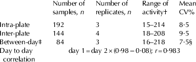
The obtained results concerning intra- and interassay variation and between-day determinations of milk samples are given in Table 1. CV was in all instances below 10% and determinations of the same sample after a freeze-thawing cycle (next day) were highly correlated (r=0·983; n=252; average CV%=7·5).
The precision of the analyses, within plate, between plates and between days

† nmol product l−1 min−1
‡ two thawing-freeze cycles
§ based on means of replicates
Linearity and recovery
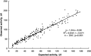
Results relating to dilution of milk samples (recovery of activity) are shown in Fig. 1a. The association between observed and expected (calculated) activities of β-glucuronidase activity were highly significant (r=0·97; P <0·001; n=200). On average we found 15·3% higher activities than expected from calculations of the diluted samples (82 U/l v. 71·1 U/l).
Linearity by dilution of sample. Twenty-five milk samples were each divided into four replicates and diluted 2+1 or 1+2 with heat treated and in-activated milk. The observed activity of β-glucuronidase was compared with the expected (calculated) activity (n=200)

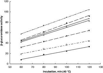
Linearity based on time of incubation. Seven milk samples were each replicated four times and incubated for 60, 80, 100 or 120 min and analysed for β-glucuronidase activity. Lines represent mean of replicates. For details see the text

Linearity as a function of incubation time is shown in Fig. 1b. The seven milk samples exposed to varying incubation time showed a straight-line increase in activity between 60 and 120 min of incubation (r 2 >0·98).
Temperature stability
None of the samples (replicates) exposed to 65, 70, 75, 80, or 85°C for 5 min had detectable enzymic activity.
Substrate concentration
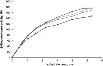
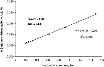
The preliminary investigation of substrate concentration (before the final concentration was chosen) is shown in Fig. 2a. Four milk samples showed increased response to increased substrate concentration up till 5·33 mm in the reaction medium. The average of the four samples was the basis for a Lineweaver-Burk plot and calculations (Fig. 2b) where maximum velocity of the reaction (under the chosen circumstances) was 250 nmol min−1 l−1. The corresponding Michaelis constant (i.e. substrate concentration supporting 50% maximum velocity) was calculated as 2·5 mm of substrate.
Four milk samples were assayed under standard conditions where only substrate concentration was varied (from 0 to 5·33 mm in the reaction medium)

Double reciprocal plot (Lineweaver-Burk plot) of the average of the four milk samples response to increasing substrate concentration. Maximum reaction velocity (Vmax) under the present circumstances was calculated to 250 nmol product min−1 l−1. Corresponding calculations showed that a substrate concentration of 2·5 mm (Km) would support a velocity of Vmax/2. U/l=nmol product min−1 l−1

Reaction pH
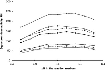
The initial studies of pH optimum for the enzyme under the present conditions are shown in Fig. 3. The activity was apparently optimal at pH 5·4–5·6. At pH 4·6 the activity was approx. 65% of the optimal activity, whereas the activity declined in more alkaline environments than pH 5·6; at pH 6·2 the activity reached 86% of the maximum. Therefore we chose pH 5·5 as standard for the substrate-buffer solution and the incubation medium.
Eight milk samples were assayed under standard conditions where only pH in the reaction medium was varied. U/l=nmol product min−1 l−1

Comparisons among mastitis indicators
The range in β-glucuronidase activity for the 825 milk samples was (5–1789) nmol min−1 l−1 (0·05–0·95 inter quartile 26–270). The association between β-glucuronidase activity and other mastitis indicators are given in Table 2. Activity of β-glucuronidase was significantly positively correlated to LDH, NAGase, and AP activities, days in milk, and milk protein and SCC (P<0·001), and negatively correlated to lactose concentration and milk yield (r=−0·383; n=407 and r=−0·161, n=820, respectively). Apparently, lactation number (1 v. 2 and 1 v. 3+4) did not affect milk β-glucuronidase activity.
The association between milk β-glucuronidase activity and other mastitis indicators (somatic cell count, SCC; N-acetyl-β-d-glucosaminidase, NAGase; lactate dehydrogenase, LDH; alkaline phosphatase, AP). Figures represent Pearsons correlation coefficient (r); figures in parentheses are numbers of observations

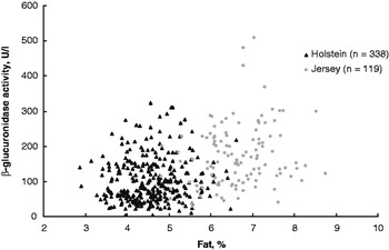
Breed, from an isolated point of view, affected β-glucuronidase activity: Danish Holsteins showed on average 102·2 nmol min−1 l−1 (0·05–0·95 inter quartile 26–227, n=338), Danish Jerseys showed on average 171 (0·05–0·95 inter quartile 53–302, n=119), P<0·001. Similarly, the fat percentage of the milk was positively correlated to the activity (r=0·237; n=768; P<0·001). However, breed and milk fat, are confounded in the present context (simple correlations); Fig. 4 illustrates this situation.
The association between milk fat and β-glucuronidase activity in milk from Danish Holstein and Danish Jersey cows. Animals were from the same herd and fed the same diet. U/l=nmol product min−1 l−1

Identification of pathogens
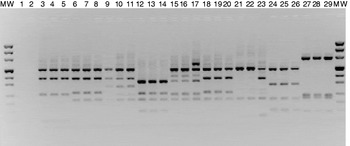
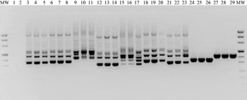
The RFLP patterns after restriction with the enzymes HaeIII and RsaI are shown in Fig. 5. Specific patterns were obtained for all samples of bovine origin and could be identified by comparison with the reference strains. The results were confirmed by the ITS-PCR as shown in Fig. 6.
Restriction digestion of 16S-23S rDNA with HaeIII and RsaI for field and reference strains (RF). Lanes: MW standard DNA size marker; 1,2 negative control; 3,4 field strains; 5 RF Staphylococcus chromogenes; 6,7 field strains; 8 RF Staph. xylosus; 9,10 field strains; 11 RF Staph. haemolyticus; 12,13 field strains; 14 RF Staph. simulans; 15,16 field strains; 17 RF Staph. capitis; 18,19 field strains; 20 RF Staph. cohnii; 21,22 field strains; 23 Staph. sciuri; 24,25 field strains; 26 Streptococcus dysgalactiae; 27,28 field strains; 29 RF Str. uberis

ITS-PCR amplification pattern for field and reference strains (RF). Lanes: MW standard DNA size marker; 1,2 negative control; 3,4 field strains; 5 RF Staphylococcus chromogenes; 6,7 field strains; 8 RF Staph. xylosus; 9,10 field strains; 11 RF Staph. haemolyticus; 12,13 field strains; 14 RF Staph. simulans; 15,16 field strains; 17 RF Staph. capitis; 18,19 field strains; 20 RF Staph. cohnii; 21,22 field strains; 23 Staph. sciuri; 24,25 field strains; 26 Streptococcus dysgalactiae; 27,28 field strains; 29 RF Str. uberis

Measurement of samples identified for pathogens
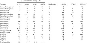
Details for this study are shown in Table 3. Increasing pH in the reaction medium from 5·5 to pH 7·0 in all cases reduced the response (production of product per time unit; P<0·001). None of the milk samples that tested positive for pathogens showed greater activity in reaction media closer to pH 7·0; on the contrary the response declined as the pH increased. This is in fairly good accordance with the preliminary general pH-test of the reaction medium.
Nineteen milk samples from subclinical and latent cases of mastitis (individual glands) were identified for specific pathogens and measured for β-glucuronidase activity at four different pH levels and for N-acetyl-β-d-glucosaminidase (NAGase), lactate dehydrogenase (LDH) and alkaline phosphatase (AP) activity

† Staph=Staphylococcus (genus)
‡ Str.=Streptococcus (genus)
§ U/l=nmol product min−1 l−1
¶ U/l=μmol product min−1 l−1
Discussion
The activity of β-glucuronidase in milk was considerably lower than the activity of NAGase, LDH and AP; i.e. median values of β-glucuronidase activity were 6%, 9%, and 0·03% of that of NAGase, LDH and AP activities, respectively. It is therefore a necessity in our system that we use a stronger concentration of sample in the reaction medium and a longer incubation time to obtain sufficient product in order to assess the activity with a comparable precision. However, with these alterations, the automated analysis of β-glucuronidase activity was comparable to that of other mastitis indicators (i.e. NAGase and LDH) in our laboratory.
The present β-glucuronidase analysis set-up seems reliable with regard to accuracy as tested by linearity, and with regard to precision, which is comparable to other enzymic-fluorometric analysis in milk not pretreated. Our procedure revealed a general pH optimum between 5·4 and 5·6, which is in accordance with observations by Nagahata et al. (1985) who used pH 5·2 in milk samples not pretreated, as well as with the work of Fang et al. (Reference Fang, Vikerpuur and Sandholm1995) where a pH between 5·3 and 6·2 was used for assays for activity in bacterial strains. Both research groups used 4-MUG as a substrate for the enzymic activity as in the present study. Other researchers analysing milk activity (Kiermeier & Güll, Reference Kiermeier and Güll1966; Kitchen, Reference Kitchen1976; Perdigon et al. Reference Perdigon, Medici, Cecilia, Nader de Macias, Haedo, Oliver and Peche de Ruiz Holgado1986; Hurley et al. Reference Hurley, McKee and Cue1987; Perdigon et al. Reference Perdigon, Medici, Oliver and Peche de Ruiz Holgado1988; Oliszewski et al. Reference Oliszewski, Nunez de Kairuz, Gonzales de Elias and Oliver2002) have used considerably lower pH in the reaction (pH 4–5), possibly due to differences in substrate and the reaction process (spectrophotometry).
Heat stability of indigenous milk enzymes may play an important role in relation to dairy processing and utilization of the milk. Our limited test of temperature stability of the β-glucuronidase activity in milk showed that the present enzyme is not heat resistant. Compared with data gathered by Jenness & Patton (Reference Jenness and Patton1959), β-glucuronidase stability seems to be similar to lipase and alkaline phosphatase and is considerably more labile than catalase, xanthine oxidase, peroxidase and acid phosphatase. The β-glucuronidase stability observed in the present study supports the work of Kiermeyer & Doruk (Reference Kiermeyer and Doruk1972) and Griffiths (Reference Griffiths1986).
The association between β-glucuronidase activity in bovine milk and SCC, the most widely used mastitis indicator, was weaker than the SCC association with NAGase and LDH activity. Similarly, the association between β-glucuronidase activity and LDH or NAGase was weaker than associations between LDH and NAGase. This suggests that the use of β-glucuronidase activity alone may not be a reliable method to detect mastitis. However, when combined with other indicators such as indices based on ratios between, for example, β-glucuronidase activity and LDH activity it may prove useful in describing the degree of mastitis established by SCC, CMT or clinical observations. We tested simple combinations between β-glucuronidase and LDH or NAGase activity in reference to SCC. The correlations between combined β-glucuronidase and LDH or NAGase activity to SCC were inferior to LDH or NAGase alone, indicating that β-glucuronidase measurements would not improve diagnosis; however, we did not elaborate further in this area.
The positive correlation of β-glucuronidase activity to days in milk supports that of Chagunda et al. (Reference Chagunda, Larsen, Bjerring and Ingvartsen2006) who compared LDH and NAGase activity to SCC; however, the steadily increasing levels of the mastitis indicators may be a question of management within the herd. Chagunda et al. (Reference Chagunda, Larsen, Bjerring and Ingvartsen2006) further found an effect of parity on NAGase, LDH and SCC levels for cows with mastitis but not for healthy animals. For the present study, no differentiation was made between healthy and clinically mastitic animals with regard to concentration of SCC, LDH and NAGase activity; however, the majority of samples were considered healthy, as determined from SCC, LDH and NAGase measurements.
Several fluorogenic substrates have been used to identify specific enzyme activities in bacterial pathogens and to thereby identify bacteriological species or strains (e.g. Schaufuss et al. Reference Schaufuss, Lämmler and Blobel1986; Iritani & Inzana, Reference Iritani and Inzana1988; Matthews et al. Reference Matthews, Oliver and King1991; Sarhan & Foster, Reference Sarhan and Foster1991). Fang et al. (Reference Fang, Vikerpuur and Sandholm1995) used this technique (also making use of 4-MUG as substrate) and reported that different species of pathogens apparently possess different pH optima for β-glucuronidase activity. Str. uberis and Str. agalactiae revealed optimum activity between pH 5·3 and 6·2, whereas Escherichia coli strains apparently had a pH optimum closer to neutral. The present study was not able to detect marked differences in pH optima between the milk samples infected by various pathogens but rather described a general decreasing activity with increasing pH in the reaction medium. One potential reason for this may be that the enzyme activity from the bacterial cells per se may be ignorable when compared with contributions from the mammary epithelium and the immunological defence mechanism of the cow.
The fluorometric determination of β-glucuronidase activity in milk can be analysed on a large-scale basis similar to that of LDH and NAGase fluorometric determinations. The precision of the assay was comparable to that of the other mastitis indicators; however, incubation of reagents needs to be longer owing to a generally lower activity of the enzyme. Whether β-glucuronidase activity can improve mastitis detection on-farm warrants further investigation.
Jens Clausen and Martin Bjerring are kindly acknowleged for their skilful technical assistance during the course of the work.