Introduction
Superovulatory response and embryo recovery rates in buffalo are lower than in bovines (Baruselli & Cavalho, Reference Baruselli and Carvalho2003). The number of stimulated follicles and embryos collected in buffalo normally corresponds to one-third of that obtained in cattle (Singh et al., Reference Singh, Nanda and Adams2000). Despite efforts to use different hormones/protocols (Misra et al., Reference Misra, Kasiraj, Rao, Ragareddy, Jaiswal and Pant1998; Carvalho, Reference Carvalho2001; Baruselli et al., Reference Baruselli, Carvalho, Cavalcante, Nichi and Zicarelli2003), only 50 to 55% of the animals respond: these buffaloes ovulate two to four structures, producing one or four viable embryos (Manik et al., Reference Manik, Singla, Palta and Chauhan2002). Considering this, in vitro embryo production (IVEP) technology represents the best tool to improve maternal contribution to genetic progress in buffalo. Besides the progress obtained in the percentage of in vitro produced transferable embryos (Gasparrini et al., Reference Gasparrini, Boccia, Marchandise, Di Palo, George, Donnay and Zicarelli2006, Manjunatha et al., Reference Manjunatha, Ravindra, Gupta, Devaraj and Nandi2009), the pregnancy rate achieved by transferring these structures remains poor (Gasparrini, Reference Gasparrini2002; Nandi et al., Reference Nandi, Raghu, Ravindranatha and Chauan2002a).
In vitro embryo production (IVEP) in buffalo is based on the bovine model. Despite some modifications made to improve the process (Gasparrini, Reference Gasparrini2002; Presicce, Reference Fresicce2007), it is generally observed by different research groups that embryo production is only between 15 and 30% (Presicce, Reference Fresicce2007, Liang et al., Reference Liang, Lu, Chen, Zhang, Lu, Zhang, Pang, Huang and Lu2008, Manjunatha et al., Reference Manjunatha, Ravindra, Gupta, Devaraj and Nandi2008). Ultrastructural studies on the oocyte during in vitro maturation in different mammalian species [mouse (Merchant & Chang, Reference Merchant and Chang1971), human (Zamboni & Thomson Reference Zamboni and Thomson1972), cattle (Hyttel et al., Reference Hyttel, Fair, Callesen and Greve1997), camel (Kafi et al., Reference Kafi, Mesbah, Nili and Khalili2005)] have resulted in a better understanding of the biology of the oocyte and, as a consequence, improvements in IVM and IVF. However, systematic studies on ultrastructure of buffalo oocytes during IVM have not been reported. Therefore, the objective of the present study was to describe ultrastructural changes in the nucleus and cytoplasmic organelles of buffalo oocytes during in vitro maturation.
Materials and Methods
Animals
The experiments were conducted between July and August, winter in the Southern hemisphere, in Planaltina–Federal District–Brazil (15°38′ S and 47°43′ W). The average maximum temperature during the period was 29.3°C, while the average minimum temperature was 16.2°C. During the experimental period there was no rainfall and the animals were fed ad libitum with Panicum spp., mineral supplement and received corn silage. The 10 primiparous Murrah females submitted to ovum pick-up (OPU) were aged between 3 and 4 years, the average body weight was 360 kg and the corporal score between 2 and 3 (Moreira et al., Reference Moreira, Risco, Pires, Ambrose, Drost and Delorenzo2000). Before OPU sessions began, the animals were submitted to a complete gynaecological examination, including ultrasonography of the ovaries.
To eliminate the dominant follicle, selected animals were fitted for 8 days with a progesterone implant (CIDR® – Pfizer, São Paulo, Brazil) and estradiol benzoate (Estrogin® – FarmaVet) i.m. on progesterone implant removal. Two days after progesterone implant removal, animals were subjected to three sessions of OPU with an interval of 7 days between sessions. On any given day, OPU from 10 animals was undertaken.
Ovum pick-up (OPU)
Just before OPU procedure, the animals received 3 ml of lidocaine 2% (Anesthetic Pearson® – Eurofarma, São Paulo, Brazil) via epidural route, without any further sedation. The ultrasound equipment used was an Aloka SSD-500 with a micro convex 5.0 MHz probe coupled to a vaginal support equipped with aspiration guide (Watanabe Tecnologia Aplicada). All follicles, from 2 to 8 mm, were punctured with an 18-gauge needle connected to a vacuum system (Watanabe Tecnologia Aplicada) adjusted to –40 mmHg. During aspiration procedure, the aspiration line was constantly washed with LAV medium (100 μl of heparin (Liquemine® – Roche), 500 μl of fetal calf serum (FCS) and q.s.p. 50 ml of PBS). The collected COCs were classified under a stereomicroscope, and only grade I and II (Gupta et al., Reference Gupta, Ravindranatha, Nandi and Sarma2002) structures were used.
Ultrastructural changes during in vitro maturation
All media and supplements used in the experiment were donated by Nutricell® Nutrientes Celulares Ltda (Campinas).
In vitro maturation
A total number of 85 COCs were matured in TCM199 with Earle's salts supplemented with 10% FCS, LH, FSH, l-glutamine, penicillin and streptomycin. Each group of 10 to 15 COCs was matured in 150 μl microdrops of medium, covered with mineral oil (AMRESCO®), previously stabilized in an incubator at 38.5°C, saturated humidity and 5% CO2 in air. The IVM procedure was repeated three times.
Each repetition used an average of 28.3 COCs. Five to six structures were removed from maturation drops at 0, 6, 12, 18 and 24 h of maturation and processed as described below. Approximately five structures were evaluated by transmission electron microscopy at each time point.
Preparation of COCs for transmission electron microscopy (TEM)
The structures were fixed in Karnovsky solution (2% glutaraldehyde, 2% paraformaldehyde, 3% sucrose, in 0.1 M sodium cacodylate buffer, pH 7.2) at 4°C for 24 h. Before the post-fixation performed with osmium tetroxide, the COCs were embedded in 4% agar (Difco®) to facilitate manipulation (Hyttel & Madsen, Reference Hyttel and Madsen1987). The material was then contrasted in block with uranyl acetate 3% and the structure was dehydrated with acetone. Dehydrated COCs were included in Spurr (Polysciences) and semi-thin sections (2 μm) were performed. To allow detection of the nucleus (equatorial region), the serial sections were dyed with toluidine blue and observed under light microscopy. The ultrathin sections (90 nm) were made from COCs with intact nucleus and were contrasted with uranyl acetate and lead citrate to be observed with transmission electron microscopy (Jeol 1011) operated at 80 kV.
Results
The average number of COCs collected by OPU/animal/session was 6.4, and 44% of them were of grades I and II. During successive OPU sessions the average number of collected COCs/animal decreased, from 8.3 in the first session to 3.9 total structures per animal in the last session. In addition, the percentage of grade I and II (Gupta et al., Reference Gupta, Ravindranatha, Nandi and Sarma2002) structures also decreased from 47% to 35%. It was also important to note the remarkable individual variation: one animal produced an average of 11 structures, while another just produced three COCs per OPU section. Animal individuality also affected the percentage of viable structures, with some animals producing 100% usable structures and others just 5.88%.
Ultrastructural changes during in vitro maturation
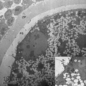
Figure 1 shows an immature COC obtained from antral follicle. The oocyte nucleus with loose chromatin is peripherally located. Observing the structural aspects, such as ooplasm and granulosa cell (GC) nuclei, it is possible to infer that the structure is functionally viable. The GC nucleus presented loose chromatin, showing the high activity in protein synthesis of these cells, which will support the oocyte maturation. It is also important to observe the intimate relation between corona radiata cells and zona pellucida (ZP), including the presence of cytoplasmic projections in ZP. The perivitelline space (PS) is well developed and the presence of a large number of bent oocyte microvilli is also noted. Mitochondria, as well as the Golgi complex, are clustered in the oocyte cortical region. Most ooplasm is occupied by a large number of coalescent lipid vacuoles.
Immature oocyte (TEM). showing lipid droplets (LD), peripherally located mitochondrial clusters (m) and nucleus (N); granulosa cells (GC) intimately related to zona pellucida (ZP), and well developed perivitelline space (PS). Note inset showing a well developed Golgi complex (G).

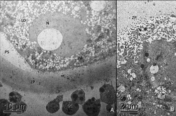
After 6 h of IVM the oocyte nucleus changed its round shape to a more flattened aspect (Fig. 2). Near the nucleus the PS is larger than in the rest of the oocyte, and not filled with ooplasm villi, unlike the rest of the structure. Mitochondria, Golgi complex and lipid vacuoles did not show significant changes during the first 6 h of maturation. Considerable loosening of GC from ZP took place during this time.
(A) Six-hour matured oocyte (TEM), showing a large number of lipid droplets (LD), peripherally located mitochondrial clusters (m) and nucleus (N); granulosa cells (GC) are coming loose from zona pellucida (ZP) and the perivitelline space (PS) is larger near the nucleus, possibly preparing for polar body extrusion. In (B) it is also possible to note a well developed Golgi complex (G), probably involved in cortical granule production and ooplasm vilosities (*) embedded in ZP.

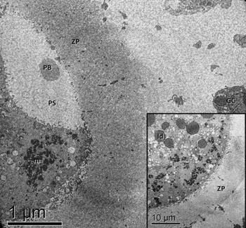
At 12 h of IVM (Fig. 3), the first polar body (PB) was extruded into a large PS, showing that the oocyte had reached metaphase II stage. The GC separated completely from ZP. Morphological features of lipid droplets also changed at this time point, which may indicate chemical changes.
Twelve-hour matured oocyte (TEM), showing an extruded polar body (PB) in a large perivitelline space (PS), peripherally located mitochondrial clusters (m). The granulosa cells (GC) are completely separated from zona pellucida (ZP). Note inset showing the change in the lipid droplets’ (ld) aspect, probably denoting a chemical alteration in lipid molecules.

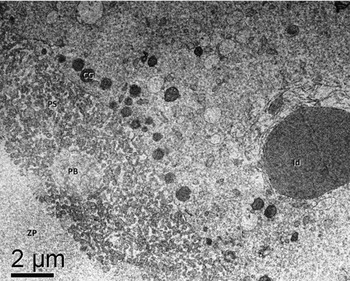
Figure 4 shows the peripheral region of an oocyte matured for 18 h. The lipid droplets maintain a dark appearance and the PB is located in a PS filled with ooplasm villi. The most important change observed at this stage is the presence of cortical granules located in the ooplasm cortical region.
Eighteen-hour matured oocyte (TEM), showing an extruded polar body (PB) in a large perivitelline space (PS) filled with ooplasm villosites, apparently not embedded in zona pellucida (ZP). Note the peripherally located cortical granules (CG) and the large lipid droplet (ld).

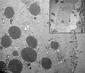
At the end of the IVM period (24 h), it is possible to note (Fig. 5) the large dark lipid droplets and numerous cortical granules on the oocyte periphery.
Twenty-four-hour matured oocyte (TEM), showing large lipid droplets (ld) and cortical granules (arrows). Note inset with extruded polar body (PB) in a large perivitelline space (PS) and an apparently loose zona pellucida (ZP).

Discussion
The low number of COCs collected by OPU is probably the result of some peculiarities inherent to buffaloes, such as the reduced number of antral and preantral follicles, approximately ten times lower than in cattle (Drost, Reference Drost2007; Mondadori et al., Reference Mondadori, Santin, Fidelis, Porfirio and Bao2008). In addition, some hormonal protocols could increase the number of collected structures (Sá Filho et al., Reference Sá Filho, Carvalho, Gimenes, Torres Júnior, Garcia, Tonhati, Gasparrini and Baruselli2005). The number of COCs per ovary (approximately three structures), was slightly higher than previously described in buffalo (Gasparrini, Reference Gasparrini2002; Drost, Reference Drost2007; Manjunatha et al., Reference Manjunatha, Ravindra, Gupta, Devaraj and Nandi2008), the higher number of structures is probably caused by the longer interval between OPU sessions. The percentage of viable structures (44%) was also higher than in one study (Baruselli et al., Reference Baruselli, Gimenes, Carvalho, Sá Filho, Ferraz and Barnabé2007) and lower than in another (Manjunatha et al., Reference Manjunatha, Ravindra, Gupta, Devaraj and Nandi2008). As previously described by Ferraz et al. (Reference Ferraz, Gimenes, Filho, Watanabe, Joaquim, Accorsi, Meirelles and Baruselli2007), in our study, a decrease in the number and in the quality of the oocytes was observed from the first to the last OPU.
Ultrastructural changes during in vitro maturation
Immature COCs showed typical structure previously described for buffalo (Boni et al., Reference Boni, Santella, Dale, Roviello, Di Palo and Barbieri1992; Mondadori et al., Reference Mondadori, Santin, Fidelis, Porfirio and Bao2008), as well as for bovine (Hyttel et al., Reference Hyttel, Xu, Smith and Greve1986; Kacinskis et al., Reference Kacinskis, Lucci, Luque and Bao2005; Nagano et al., Reference Nagano, Katagiri and Takahashi2006), ovine (O'Brien et al., Reference O'Brien, Dwarte, Ryan, Maxwell and Evans2005) and camel (Kafi et al., Reference Kafi, Mesbah, Nili and Khalili2005) oocytes. Confirming previous observations (Boni et al., Reference Boni, Santella, Dale, Roviello, Di Palo and Barbieri1992; Mondadori et al., Reference Mondadori, Santin, Fidelis, Porfirio and Bao2008), the most important difference observed between the species is the larger number of lipid droplets in buffalo ooplasm. The same sort of GC–oocyte junctions previously described for buffalo (Boni et al., Reference Boni, Santella, Dale, Roviello, Di Palo and Barbieri1992; Mondadori et al., Reference Mondadori, Santin, Fidelis, Porfirio and Bao2008) and bovines (Fair & Hyttel et al., Reference Fair, Hyttel and Motta1997) was also observed in some immature oocytes. It is well known that these junctions play an important role during oogenesis (in buffalo after ZP formation – Mondadori et al., Reference Mondadori, Luque, Santin and Bao2007) and IVM in different species (Zhang et al., Reference Zhang, Jiang, Wozniak, Yang and Godke1995; Suzuki et al., Reference Suzuki, Jeong and Yang2000). The PS in most analysed immature oocytes was well developed, denoting that the structures were obtained from large antral follicles (Mondadori et al., Reference Mondadori, Santin, Fidelis, Porfirio and Bao2008), whereas PS in immature bovine oocytes is absent or narrow (Hyttel et al., Reference Hyttel, Xu, Smith and Greve1986). Evaluation of cattle and buffalo immature oocytes allows us to affirm that cortically located mitochondrial clusters and Golgi complex are ultrastructures that are characteristic of these structures in both species. It is also possible to infer that these functional complexes are involved in CG synthesis. This feature could also be used as a marker for oocyte competence because it only appears in oocytes originating from larger antral follicles (Mondadori et al., Reference Mondadori, Santin, Fidelis, Porfirio and Bao2008).
From the start of IVM, as a result of the resumption of meiosis, nucleus morphology changes and PS grows, preparing to receive the polar body. After 6 h of IVM, the GC–oocyte junctions become loose; considering this, these cells probably do not play an important role in oocyte maturation during the rest of the IVM period. Instead, the separation is probably caused by GC hyaluronic acid production induced by gonadotrophins (Chen et al., Reference Chen, Wert, Hendrix, Russel, Cannon and Larsen1990).
In most oocytes (three of five) studied, metaphase II stage was achieved much earlier (12 h of IVM) in contrast to earlier reports (Yadav et al. Reference Yadav, Katiyar, Chauhan and Madan1997; Santos et al., Reference Santos, Dantas, Miranda and Ohashi2002; Nandi et al., Reference Nandi, Ravindranatha, Gupta and Sarma2002b; Gasparrini et al., Reference Gasparrini, Rosa, Attanasio, Boccia, Di Palo, Campanile and Zicarelli2008). From our point of view, as this experiment was not designed to determine oocyte maturation time point, this finding does not have a high biological value, because the number of observed oocytes (five structures) was low and the literature describes great biological variability in female buffalo reproductive patterns.
The most important change observed between 6 and 12 h of IVM is the change in lipid droplets, probably caused by chemical alteration in lipid molecules. It is well known that buffalo oocytes are more sensitive to oxidative damages because of their high lipid content (Boni et al., Reference Boni, Santella, Dale, Roviello, Di Palo and Barbieri1992; Mondadori et al., Reference Mondadori, Santin, Fidelis, Porfirio and Bao2008). Boni et al. (Reference Boni, Santella, Dale, Roviello, Di Palo and Barbieri1992) did not observe the lipid droplet changes, although our observation could explain the increasing proportion of tight morula and blastocyst-stage embryos when cysteamine is used on IVM medium (Gasparrini et al., Reference Gasparrini, Neglia, Di Palo, Campanile and Zicarelli2000), and the confirmation that thiol compounds increase glutathione synthesis in buffalo oocytes (Gasparrini et al., Reference Gasparrini, Boccia, Marchandise, Di Palo, George, Donnay and Zicarelli2006). Finally, it is important to observe that at the end of the IVM period, from 18 to 24 h, the cortical granules are located on the ooplasm periphery, denoting preparation for polyspermy block (Cran & Cheng, Reference Cran and Cheng1985).
It is concluded that: (1) immature oocytes in buffalo have a high lipid content; (2) the PS increases during IVM; (3) Golgi complexes and mitochondrial clusters migrate to the oocyte periphery during IVM, probably acting on CG synthesis; (4) 6 h of IVM are enough to lose contact between GC and ZP; and (5) the oocyte lipid droplets’ aspect changes between 6 and 12 h of IVM.
Acknowledgements
The authors would like to thank Dr Rafael Afonso Dresh and Dr Emivaldo Siqueira Filho for helping with OPU sessions, and Nutricell Nutrientes Celulares for supplying media used in the experiment. This research was supported by CNPq, CAPES, FINEP, FINATEC and UPIS.