Introduction
In recent years, with the revival of alternative commercial production systems where chickens have free access to bedding material and outdoor areas, helminth parasites are re-emerging as a problem worldwide (Thapa et al., Reference Thapa, Hinrichsen and Brenninkmeyer2015a; Wuthijaree et al., Reference Wuthijaree, Lambertz and Gauly2017). Among the helminths affecting chickens, the large round worm Ascaridia galli (A. galli) is the most important in free-range laying hens (Katoch et al., Reference Katoch, Yadav, Godara, Khajuria, Borkataki and Sodhi2012; Thapa et al., Reference Thapa, Hinrichsen and Brenninkmeyer2015a). Ascaridia galli belongs to the genus Ascaridia within the family Ascaridiidae and occurs worldwide as a small intestinal parasite of galliform birds (Soulsby, Reference Soulsby1982). It has a direct life cycle that includes two principal populations: the sexually active adult worms in the intestine of the host and a free-living stage (eggs) in the environment. Parasite egg shedding in the excreta starts approximately 5–8 weeks after infection. This is followed by the in ovo development of eggs to infective larvae (L3) in the external environment. The life cycle is completed when a new host ingests infective eggs which hatch (L3) in the lumen of the small intestine. The L3 then embed into the mucosa where they moult to become L4 which finally develop into adults (Ackert, Reference Ackert1931; Permin & Hansen, Reference Permin and Hansen1998; Taylor et al., Reference Taylor, Coop and Wall2007). Ascaridia galli infections are often associated with reduced body weight gain, increased feed consumption, reduced nutrient utilization and overall poor health conditions (Chadfield et al., Reference Chadfield, Permin, Nansen and Bisgaard2001; Phiri et al., Reference Phiri, Phiri, Ziela, Chota, Masuku and Monrad2007). The infection may also interfere with the development or suppression of host immunity (Hørning et al., Reference Hørning, Rasmussen, Permin and Bisgaard2003; Pleidrup et al., Reference Pleidrup, Dalgaard and Norup2014) thereby increasing the severity of concurrent diseases (Dahl et al., Reference Dahl, Permin, Christensen, Bisgaard, Muhairwa, Petersen, Poulsen and Jensen2002; Permin et al., Reference Permin, Christensen and Bisgaard2006).
Experimental infections are important to better understand this parasite and its effects. Low infection rates and burdens after artificial infection have been reported (Kaufmann et al., Reference Kaufmann, Daş, Preisinger, Schmutz, König and Gauly2011; Permin et al., Reference Permin, Bojesen, Nansen, Bisgaard, Frandsen and Pearman1997a; Sharma et al., Reference Sharma, Hunt, Hine, Sharma, Swick and Ruhnke2018) thus reducing the power to detect the effects of experimental factors. The in vivo propagation of ascarids depends on the source of eggs, which must incubate and embryonate outside the host (Elliott, Reference Elliott1954). Thus, variable artificial infection rates may be due not only to host factors such as individual variation in host susceptibility (Gauly et al., Reference Gauly, Homann and Erhardt2005; Kaufmann et al., Reference Kaufmann, Daş, Preisinger, Schmutz, König and Gauly2011) but also to the viability and embryonation status of the infective material used (Elliott, Reference Elliott1954). Early studies showed that the infectivity (Elliott, Reference Elliott1954) and severity of infection (Todd et al., Reference Todd, Crowdus and Wyant1952) of A. galli larvae diminished with age of embryonated eggs. Eggs used to induce A. galli infection can be obtained from several sources: the host excreta (Luna-Olivares et al., Reference Luna-Olivares, Ferdushy, Kyvsgaard, Nejsum, Thamsborg, Roepstorff and Iburg2012; Ferdushy et al., Reference Ferdushy, Luna-Olivares, Nejsum, Roepstorff, Thamsborg and Kyvsgaard2013), physical removal from, or disruption of, the worm's uterus (Gauly et al., Reference Gauly, Bauer, Preisinger and Erhardt2002; Daş et al., Reference Daş, Kaufmann, Abel and Gauly2010) or by in vitro culturing of female worms in artificial media and recovering eggs shed into the media typically across 3–5 days (Dick et al., Reference Dick, Leland and Hansen1973; Salih & Saleem, Reference Salih and Saleem1987; Ruhnke et al., Reference Ruhnke, Andronicos, Swick, Hine, Sharma, Kheravii, Wu and Hunt2017; Sharma et al., Reference Sharma, Hunt, Hine, Sharma, Swick and Ruhnke2017).
Ascaridia galli eggs are highly adaptable to wide ranges of environmental conditions primarily due to the inherent characteristics of their thick eggshells (Cruthers et al., Reference Cruthers, Weise and Hansen1974; Thapa et al., Reference Thapa, Thamsborg, Meyling, Dhakal and Mejer2017). However, their long-term survival and developmental capacity are affected by several factors. The reported optimum embryonation temperature for A. galli ranges between 25 and 30°C (Christenson et al., Reference Christenson, Earle, Butler and Creel1942; Ramadan & Abouznada, Reference Ramadan and Abouznada1992; Mero & Gazal, Reference Mero and Gazal2008; Tarbiat et al., Reference Tarbiat, Jansson and Höglund2015). As storage temperature increases, egg development gradually reduces and may totally cease beyond 35°C (Ackert, Reference Ackert1931; Reid, Reference Reid1960). Ascaridia galli eggs can develop into the L3 larval stage at temperatures as low as 15°C while at sub-zero temperatures (down to −5°C) viability may be preserved depending on the duration of exposure but no development occurs (Tarbiat et al., Reference Tarbiat, Jansson and Höglund2015). This may suggest a strategy for cold tolerance in A. galli like other nematodes (Wharton, Reference Wharton1980). Furthermore, a relative humidity of approximately 85–90% and continuous oxygen supply are necessary for A. galli egg embryonation (Hansen et al., Reference Hansen, Oonyawongse and Ackert1953; Tarbiat et al., Reference Tarbiat, Jansson and Höglund2015). Several options for the embryonation of A. galli eggs under laboratory conditions have been identified (Permin et al., Reference Permin, Pearman, Nansen, Bisgaard and Frandsen1997b; Katakam et al., Reference Katakam, Mejer, Dalsgaard, Kyvsgaard and Thamsborg2014), allowing development of embryonated infective stages within a few days or weeks depending on the method being used (Permin & Hansen, Reference Permin and Hansen1998; Onyirioha, Reference Onyirioha2011). However, the developmental potential of eggs can be significantly affected by the pre-embryonation storage conditions (Tarbiat et al., Reference Tarbiat, Rahimian, Jansson, Halvarsson and Höglund2018), the embryonation media (Permin et al., Reference Permin, Pearman, Nansen, Bisgaard and Frandsen1997b), as well as the maturation level of eggs during egg recovery (Rahimian et al., Reference Rahimian, Gauly and Daş2016).
A recent study found that less than 40% of eggs manually removed from worm uteri embryonate, whereas approximately 90% of eggs obtained from poultry excreta do so (Rahimian et al., Reference Rahimian, Gauly and Daş2016). It has been suggested that not all eggs obtained in uteri are mature and able to complete embryonation (Tiersch et al., Reference Tiersch, Daş, Samson-Himmelstjerna and Gauly2013) whereas adult female worms appear to shed only mature eggs (Kim et al., Reference Kim, Pyo, Hwang, Park, Hwang, Chai and Shin2012). In line with this, a number of A. galli studies have used eggs oviposited by mature female worms under in vitro incubation in artificial media such as physiological saline (0.85% NaCl) (Dick et al., Reference Dick, Leland and Hansen1973; Salih & Saleem, Reference Salih and Saleem1987) and Roswell Park Memorial Institute (RPMI) media (Ruhnke et al., Reference Ruhnke, Andronicos, Swick, Hine, Sharma, Kheravii, Wu and Hunt2017; Sharma et al., Reference Sharma, Hunt, Hine, Sharma, Swick and Ruhnke2017). This approach can be considered the most feasible, efficient and established method of recovering mature A. galli eggs for experimental purposes.
However, while it has been proven that A. galli eggs can successfully be recovered by incubating mature female worms in artificial media, information on the number, viability and embryonation profile of eggs recovered at different times post incubation of mature females is lacking. Likewise, the viability of larvae within the embryonated eggs is rarely assessed before use for experimental infections and the duration for which eggs are stored and incubated varies between studies (Katakam et al., Reference Katakam, Mejer, Dalsgaard, Kyvsgaard and Thamsborg2014).
Therefore, the objectives of the present study were to define the rate and quality of egg production by mature A. galli worms cultured in RPMI media as well as temporal changes in egg viability and embryonation capacity under different storage and incubation conditions. The general hypotheses in this experiment were (1) rate of egg production and quality of eggs harvested will decline with time of incubation of mature worms, (2) overall viability of stored eggs will decline at different rates following storage without embryonation at 4°C and after embryonation at 26°C and (3) larvae liberated from morphologically normal vermiform eggs will have high viability as assessed by motility and vital staining.
Materials and methods
Ascaridia galli eggs
Mature female A. galli worms were collected from the intestines of naturally infected free-range 50 Hy-Line Brown laying hens (N = 50) during post-mortem examination in an experiment approved by the animal ethics committee of the University of New England (approval AEC17-092). For the worm harvest, the hens were humanely killed, dissected and the intestines repositioned. The jejunum and ileum were opened longitudinally and mature A. galli worms identified, collected into Petri dishes and washed in sterile phosphate-buffered saline. A total of 223 mature females were transferred into a glass jar with RPMI media (Sigma-Aldrich Pty Ltd, St Louis, USA) including 0.1% 100 units/ml penicillin, 100 μg/ml of streptomycin, 250 ng/ml amphotericin B, to a volume that covered the worms (Sharma et al., Reference Sharma, Hunt, Hine, Sharma, Swick and Ruhnke2017). The worms were then cultured for 3 days at 37°C, changing the total media every 24 h. After every 24 h, the media containing parasite eggs was collected into 50-ml screw cap falcon tubes by rinsing the jar with fresh RPMI media. The egg suspension was then centrifuged (Beckman Coulter Inc, Brea, California, USA) at 425g for 1 min and eggs concentrated at the bottom of the media were collected using transfer pipettes. Eggs recovered over 3 different collection days were kept separately in 50-ml falcon tubes. To estimate the mean daily as well as total A. galli egg production under this in vitro worm incubation system, a 20-ml egg suspension in distilled water was made for each collection day and egg counts performed in triplicates using a modified McMaster egg count technique (Whitlock, Reference Whitlock1948). For all the egg collection days, 200–300 eggs were examined microscopically at 100× magnification in triplicates to estimate the proportions of intact (undamaged) eggs at the time of recovery. The recovered eggs were then stored in boiled and cooled sterile water at 4°C for later use.
Experimental design
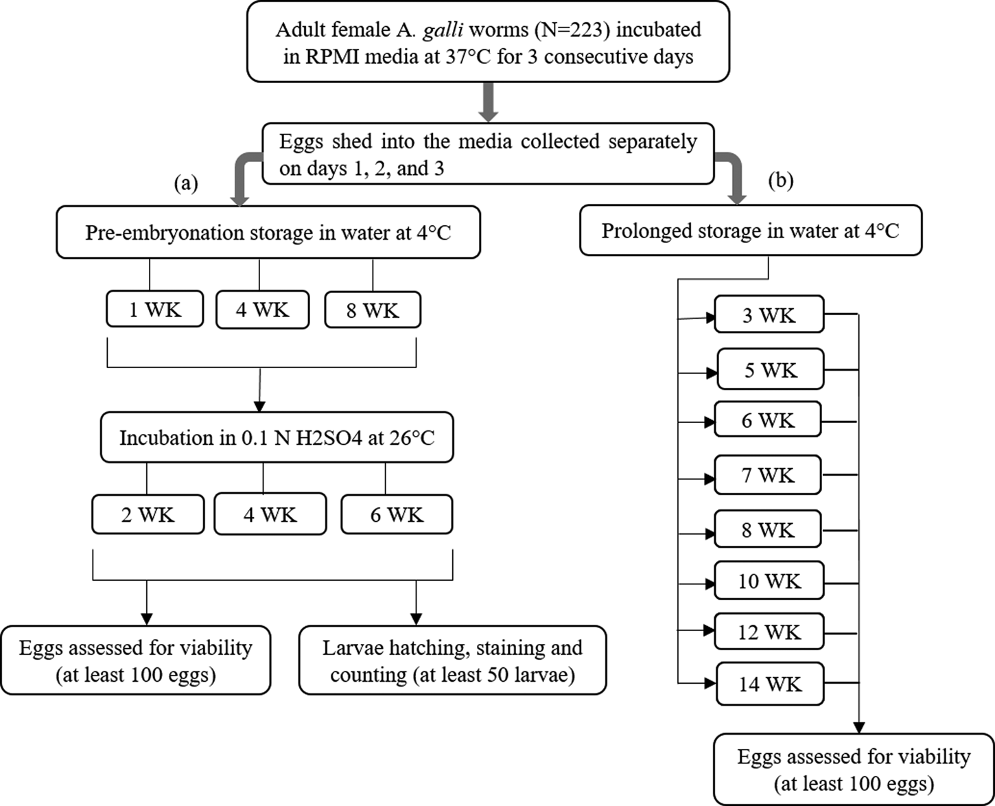
The experimental subjects of this study were A. galli eggs stored at 4°C for 1 week. Eggs separately recovered on 3 consecutive days (days 1, 2 and 3) were subjected to either (1) a 3 × 3 factorial arrangement of storage in water at 4°C (1, 4 or 8 weeks) followed by aerobic incubation in 0.1 N H2SO4 at 26°C (2, 4 or 6 weeks) or (2) prolonged storage in water at 4°C for 14 weeks: a factorial arrangement of day of egg collection (days 1, 2 or 3) × storage period (3, 5, 6, 7, 8, 10, 12 or 14 weeks) (fig. 1). There were three replicates of each treatment combination. An egg suspension in sterile distilled water (boiled and cooled) was prepared from the sample of each collection day. A total of 162 Eppendorf tubes (1.5 ml) were filled with 1 ml of an A. galli egg suspension containing approximately 500 eggs and then the tube lids sealed. These samples were then subjected to each condition, and, after the relevant treatment periods, eggs were monitored for viability and development as detailed below. To ensure aerobic conditions during embryonation, tube lids were left open and samples were aerated manually for 5 min three times per week.
Experimental design. Ascaridia galli eggs recovered after 1, 2 or 3 days of in vitro culture of adult female worms were subjected to two treatment conditions: (a) storage in water at 4°C (1, 4 or 8 weeks) followed by incubation in 0.1 N H2SO4 at 26°C (2, 4 or 6 weeks); or (b) prolonged storage at 4°C (up to 14 weeks). Egg development and viability was assessed by microscopic examination of at least 100 eggs. At the end of each incubation period at 26°C, embryonated eggs were subjected to induced larvae hatch followed by vital staining and microscopic examination of at least 50 larvae for viability assessment.

Monitoring of egg development and viability
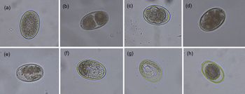
The development of eggs was evaluated by examining egg morphology under a compound binocular microscope equipped with a digital Nikon H550S camera (Nikon Corporation, Tokyo, Japan). A minimum of 100 eggs per Eppendorf tube were examined and the proportion of eggs at different stages of development (undeveloped, early development, vermiform and embryonated) or damaged/dead were recorded for each category using the morphological classifications described elsewhere (Tarbiat et al., Reference Tarbiat, Jansson and Höglund2015; Thapa et al., Reference Thapa, Thamsborg, Meyling, Dhakal and Mejer2017) and shown in fig. 2. Undeveloped eggs (fig. 2a) contained a single cell, which almost completely filled the eggshell and appeared granulated. Eggs undergoing mitosis (two cells and above) without signs of differentiation were classified as early development stages (fig. 2b–d). The vermiform stage (fig. 2e, f) was characterized by a non-motile (tadpole-like) embryo which almost filled the entire capsular space with high terminal opacity. Embryonated eggs that had completed development contained coiled slender motile larvae (fig. 2g). Eggs with an abnormal intra-capsular mass, disrupted eggshell and a shrunken internal embryonic mass were considered dead (fig. 2h).
Morphological characteristics of Ascaridia galli eggs at different developmental stages (original magnification 200×): undeveloped (a); early development (b–d); vermiform (e and f); embryonated (g) and dead (h).

In vitro larvae hatch and viability test
At the end of each incubation period at 26°C, embryonated eggs were subjected to in vitro hatching, liberating the larvae using a modification of the method developed by Dick et al. (Reference Dick, Leland and Hansen1973). Ascaridia galli eggs were placed in a solution containing equal parts of 4% NaHO (Chemsupply Pty Ltd, Gillman, Australia) and 4% NaClO (Pental Limited, Shepparton, Australia) for 24 h at 25°C. After this treatment, the samples were incubated in 0.2% Tween-80 (Redox Pty Ltd, Auburn, Australia) for 1 h and washed three times in distilled water by centrifugation at 425g for 3 min. Liberation of the larvae was enforced by centrifugation at a speed of 930g for 10 min. The liberated larvae were tested for viability using the methylene blue (Fronine Pty Ltd, Melbourne, Australia) exclusion method, where uptake of dye indicates cell death and inactivation as described earlier (Shafir et al., Reference Shafir, Sorvillo, Sorvillo and Eberhard2011). Hatched larvae in solution were mixed 1:1 with a 1:10,000 dilution of methylene blue. Viable larvae remained motile and unstained (fig. 3a, b), whereas non-viable larvae absorbed the methylene blue stain (fig. 3c, d). At least 50 liberated larvae were counted per sample and their viability recorded by microscopic examination at 100× magnification.
Hatched A. galli larvae stained with methylene blue (original magnification 200×). (a) Viable larvae demonstrating intact membrane and impermeability to methylene blue with stained remnant of egg shell debris. (b) Fully liberated viable motile larvae demonstrating intact membrane and impermeability to methylene blue. (c) Non-viable larvae demonstrating uptake of methylene blue with remnant of egg shell debris. (d) Fully liberated nonviable larvae demonstrating uptake of methylene blue.

Statistical analysis
Data were statistically analysed using JMP® software version 14.3.0 (SAS Institute Inc., Cary, NC, USA). Distributions of the data and model residuals were assessed for compliance with the assumptions of ANOVA. No data transformations were required. Two analyses were carried out to evaluate effects of study factors on egg viability and embryonation capacity. In the first analysis, day of egg collection (1, 2 or 3), pre-embryonation storage period at 4°C (1, 4 or 8 weeks), and incubation period at 26°C (2, 4 or 6 weeks) and their interactions were fitted as fixed effects in a linear model. In the second analysis, the day of egg collection (1, 2 or 3), storage period at 4°C (3–14 weeks) and their interaction were fitted as fixed effects in a linear model. In order to evaluate overall changes in egg viability, the percentage viable egg stages was defined as the proportion of eggs in developmental stages other than dead. Percentages were based on triplicate counts of 100 eggs so a total of 300 observations. For larval measurements a one-way ANOVA was performed to test the effect of incubation period on viability of hatched larvae. Percentages were based on triplicate counts of 50 eggs or 50 larvae, so a total of 150 observations. All post hoc comparisons were performed using Tukey's HSD test and data were presented using least-squares means (LSM) and standard errors (SE). Linear regression was used to test the association between egg/in ovo larvae viability and storage or incubation periods. The significance was set at P < 0.05 throughout the analysis.
Results
Rate and quality of egg recovery by day of worm incubation
A total of 223 mature female worms were recovered from 50 Hy-Line Brown hens humanely killed on the same day and used to collect eggs for 3 consecutive days. The size of the worms was not measured but all were viable and structurally intact. A total of 1.35 × 10⁶ eggs were collected with 49.2, 38.5 and 12.3% recovered on days 1, 2 and 3 of in vitro worm incubation respectively. The mean egg yield per female worm per day was 2973 ± 125, 2326 ± 14 and 745 ± 69 on days 1, 2 and 3 of collection, respectively (P < 0.0001) and a single female worm produced 6,044 eggs on average. The percentage of intact (undamaged) eggs at the time of recovery was 99% with no difference between eggs collected on different days (P = 0.69).
Effects of experimental factors on egg viability and embryonation capacity
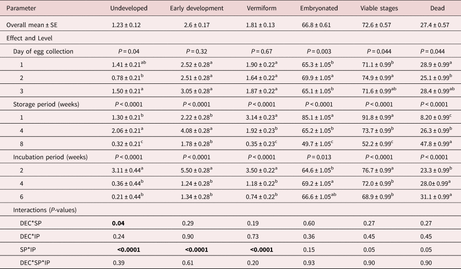
Table 1 summarizes the effects of day of egg collection, storage period at 4°C and incubation period at 26°C on the percentage of A. galli eggs in different developmental stages. Comparison of overall egg viability status between egg collection days revealed small differences, with eggs from day 2 having the highest viability (P = 0.044). Pre-embryonation storage period at 4°C had a marked effect on egg viability. Eggs stored for 1 week had a significantly higher percentage of viable stages (91.8%) than eggs kept for 4 weeks (73.7%) which in turn had a higher percentage than those stored for 8 weeks (52.2%) (P < 0.0001). Percentage viable stages also declined significantly with incubation period. Eggs incubated for 2 weeks had the highest overall viability (76.9%) followed by those incubated for 4 (72.6%) or 6 (65.3%) weeks (P < 0.0001). There was a 2.0% (y = 80.4 – 2x) loss in egg viability every week of aerobic incubation at 26°C. There were no significant interactions (P > 0.05) between treatment effects for total viable stages.
Viability and developmental profile of A. galli eggs under treatment condition 1. Percentages (LSM) of different A. galli egg developmental stages following pre-embryonation storage at 4°C (1–8 weeks) and subsequent aerobic incubation at 26°C (1–6 weeks).

Values are LSM ± SE; LSM, least square mean; SE, standard error; DEC, day of egg collection; SP, storage period; IP, incubation period. Significance levels (P-values) arising from both main and interaction effects are shown in the table. Different superscripts (a, b, c) along columns indicate a significant difference (P < 0.05) for each measurement and factor. Values in bold represent statistically significant interactions between treatment effects.
Irrespective the duration of the incubation period, the vast majority of eggs had either reached the coiled embryo stage (66.8%) or were dead (27.4%) when being stored at 26°C (table 1). Treatment effects on percentage embryonation were thus similar to those for total viable stages presented above. There was a small but significant difference (P = 0.003) in the percentage of embryonated eggs between collection days. Eggs collected on day 2 appeared to exhibit slightly higher overall embryonation (69.9%) than those from day 1 (65.3%) and day 3 (65.1%). Percentage embryonation showed a sharp decline with storage period at a rate of 5.0% (y = 88.5 – 5x) per week (P < 0.0001) but not incubation period. The proportions of undeveloped, early development and vermiform stages diminished drastically (P < 0.0001) with pre-embryonation storage period and incubation period. There were significant interactions between the effects of storage period and embryonation period on the early stages of development (table 1). These were largely due to unusually high values for these stages in samples stored for 4 weeks and incubated for 2 weeks.
Viability of hatched larvae
The induced egg hatch resulted in liberation of 96.3–98.5% of larvae from the egg shell. Incubation period at 26°C had a highly significant effect on the percentage viability of liberated larvae (P = 0.0007). A significantly higher percentage of viable larvae were observed in eggs incubated for 2 (88.2%, P = 0.0002) or 4 (86.4%, P = 0.0095) than for 6 (82.6%) weeks. Larvae viability declined by 1.4% (y = 91.3 – 1.4x) for every week of incubation at 26°C between 2 and 6 weeks.
Effect of prolonged storage at 4°C on egg developmental stages and mortality rate
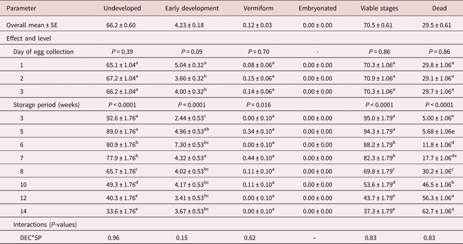
Following storage at 4°C the majority of eggs either remained undeveloped but viable (66.2%) or dead (29.5%) with the former decreasing and the latter increasing with storage time (table 2). Only a small proportion (4.2%) entered the early development stage, a negligible proportion (0.12%) reached the vermiform stage and no egg was fully embryonated. Proportions of all egg developmental stages as well as egg mortality rate did not differ (P > 0.05) between the egg collection days. The percentage of viable eggs diminished significantly with refrigeration period by 6.2% per week (y = 119.9 – 6.2x, P < 0.0001). In contrast, egg mortality sharply increased with storage period (P < 0.0001) reaching 62.7% after 14 weeks of storage. There was no significant interaction (P > 0.05) between day of egg collection and storage period on the observation of any of the egg developmental stages and mortality.
Viability and developmental profile of A. galli eggs under treatment condition 2. Percentages (LSM) of different A. galli egg developmental stages following prolonged storage at 4°C (3–14 weeks).

Values are LSM ± SE; LSM, least square mean; SE, standard error; DEC, day of egg collection; SP , storage period. Significance levels (P-values) arising from both main and interaction effects are shown in the table. Different superscripts (a, b, c, d, e) along columns indicate a significant difference (P < 0.05) for group LSM comparison at each level.
Discussion
The present study provides quantitative data on the rate of in vitro egg production by mature A. galli female worms cultured in artificial media, and subsequent changes in egg viability under different storage and incubation conditions. We showed that per capita egg yield declined with time of worm incubation but the morphological quality of eggs harvested did not differ over the first 3 days of egg collection. However, day of egg recovery did have minor effects on subsequent egg development and embryonation ability. Much larger effects on egg viability were induced by differences in the duration of storage at 4°C and following incubation at 26°C. Overall viability of eggs declined at different rates at these two temperatures. A high rate of loss of egg viability (6.2% per week) was observed after storage at 4°C whereas a smaller but significant rate of decline in egg (2.0%) and in ovo larval (1.4%) viability (3.4% in aggregate) per week of incubation period at 26°C was observed.
Our first hypothesis that the rate of egg production and quality of egg harvested will decline with time of incubation of mature worms was supported by our findings in the case of egg production, but not egg quality. The mean egg yield per female worm per day significantly declined by the day of worm incubation. Overall, a single female worm produced about 6,044 eggs over 3 days with the highest proportion (49.2%) recovered on day 1 and the least (12.4%) on day 3. Given that A. galli is a large and highly fecund nematode, the mean (2,015) per capita egg production per day in the current in vitro system, appeared to be substantially lower than estimates of in vivo egg production. While it is accepted that worm size, parasite density and host–parasite interaction can affect fecundity of A. galli, in vivo per capita egg production rates in excreta ranging from 9,000 (Daş et al., Reference Daş, Hennies, Sohnrey, Rahimian, Wongrak, Stehr and Gauly2017) to as high as 40,000 (Wongrak et al., Reference Wongrak, Gauly and Daş2015) per day were reported. A likely reason for the lower egg yield in vitro is the artificial environment and lack of food resources such that by the third day of incubation the majority of the worms were immobile (but intact) indicating probable death. However, the morphological quality of A. galli eggs harvested by the current procedure was not affected by the duration of worm incubation in RPMI media. The proportion of morphologically abnormal eggs at the time of recovery was very low (≤1%) irrespective of the day of collection.
Subsequent egg development and embryonation ability was mainly affected by other factors rather than the day of egg collection although there was a small but significant effect of day of collection (P = 0.044) with the most favourable development in eggs collected on day 2. Our finding that a high proportion of harvested eggs embryonate following shorter storage and incubation periods (>80%) affirms the observation that mature female worms lay mature eggs which can complete development (Kim et al., Reference Kim, Pyo, Hwang, Park, Hwang, Chai and Shin2012). Overall, this method of egg collection generated a large number of eggs of high concentration and quality with minimal effort once worms were collected. It did not require separation from excreta or macerated worm tissues nor did it suffer from a low or variable percentage of viable eggs. It can be concluded that the current egg collection approach can be considered as the most favourable for assays involving A. galli eggs where initial viability and embryonation ability are a priority.
The second hypothesis was that the viability of stored eggs will decline at different rates following storage without embryonation at 4°C and after embryonation at 26°C was supported by the data with a higher rate of loss of viability observed during storage at 4°C. Storage of fresh unembryonated eggs at 4°C caused high rate of loss in viability as a function of the refrigeration period. The main adverse effect of such storage was the death of the eggs prior to development. This was evident in both the short term (up to 8 weeks, table 1) and relatively prolonged (up to 14 weeks, table 2) storage conditions of the current study. In the 14-week storage experiment without inducing embryonation, morphological assessment showed a linear decline in overall egg viability of 6.2% every week of storage at 4°C with percentage viability reducing to only 37% after 14 weeks. As 4°C is a commonly used preservation temperature for laboratory storage of nematode eggs, our data indicate that refrigerated A. galli eggs may experience complete loss of viability by around 20 weeks of storage despite the fact that these eggs have resistant thick shells and have been shown to survive extreme low temperatures for shorter periods (Cruthers et al., Reference Cruthers, Weise and Hansen1974; Tarbiat et al., Reference Tarbiat, Jansson and Höglund2015). In the 8-week experiment, the rate of loss of total viable stages assessed following embryonation was somewhat lower at around 5.7% per week of storage at 4°C, and the ability to embryonate was reduced at a rate of 5.0% per week. In agreement with these observations, Tarbiat et al. (Reference Tarbiat, Rahimian, Jansson, Halvarsson and Höglund2018) observed that the embryonation ability of A. galli eggs declined linearly with the duration of aerobic refrigeration in water at a rate of approximately 4.4% per week over a 72-day storage period. However, when eggs were stored in water at 4°C under anaerobic conditions the embryonation capacity declined by only about 0.15% per week. These suggest that presence of oxygen during storage at 4°C had a major adverse effect on the viability of eggs. In the present experiment anaerobic conditions were approximated by storing eggs in boiled and cooled water with lids sealed but this appears not to have been sufficient.
As expected, egg development was very limited at 4°C with undeveloped viable eggs or dead eggs being dominant. The percentage of eggs that entered early development stages never exceeded 5% during storage at this temperature. This observation is in accordance with the report of Tarbiat et al. (Reference Tarbiat, Jansson and Höglund2015) who recorded that A. galli egg development was highly limited at 5°C. This result is also similar to the reported inhibition of development of Toxocara canis eggs at 4°C (Gamboa, Reference Gamboa2005) and Ascaris suum eggs at 5°C (Kim et al., Reference Kim, Pyo, Hwang, Park, Hwang, Chai and Shin2012).
The rate of decline in egg viability following incubation at 26°C for 2–6 weeks comprised two components. The first was a decline in morphological normality of eggs and the second a decline in the viability of larvae artificially hatched from morphologically normal eggs. The former showed a rate of decline of 2.0% per week during the 6-week period at 26°C coupled with a decline in larval viability of 1.4% per week. The additive effects of these two rates of loss (3.4%) was suggestive of a slower rate of loss of viability during storage at 26°C than at 4°C (5.7–6.2%). These indicate that a storage period of at least 40 weeks at 26°C would elapse before a total loss of viability occurred compared to the estimated 20 weeks at 4°C. Findings from early studies support our observation that A. galli eggs can survive for prolonged period at embryonation temperatures. For instance, Elliot (Reference Elliott1954) observed that embryonated A. galli eggs stored aerobically in water for 36 weeks at 28°C were infective to chickens. Similarly, Butler & Christenson (Reference Butler and Christenson1942) reported that cultures of embryonated A. galli eggs remained viable for nearly 2 years as assessed by an in ovo larval motility testing method. However, some other researchers were of the opinion that infectivity may not be maintained in A. galli egg cultures stored at room temperature (26–30°C) beyond 8–10 months (Ackert et al., Reference Ackert, Cooper and Dewhirst1947; Todd et al., Reference Todd, Crowdus and Wyant1952; Elliott, Reference Elliott1954). In the present study, antimicrobial agents were not used during incubation at 26°C and it is possible that reduction in egg viability over time may be attributed to microbial degradation. It has been shown in vitro that bacterial (Bottjer et al., Reference Bottjer, Bone and Gill1985; Tian et al., Reference Tian, Yang and Zhang2007) and fungal (Terrill et al., Reference Terrill, Larsen, Samples, Husted, Miller, Kaplan and Gelaye2004; Thapa et al., Reference Thapa, Meyling, Katakam, Thamsborg and Mejer2015b) activities can damage eggs of parasitic nematodes. Anecdotally, this was infrequently observed during microscopic observation of the samples in the current study probably due to the embryonation medium (0.1 N H2SO4) limiting microbial growth. These findings raise the possibility that aerobic storage of eggs at 26°C in the embryonated stage may prolong the effective storage time of A. galli eggs relative to storage in the unembryonated stage at 4°C. Aerobic conditions are important if storing at this temperature as Tarbiat et al. (Reference Tarbiat, Rahimian, Jansson, Halvarsson and Höglund2018) demonstrated rapid loss of viability of eggs stored at 26°C under anaerobic conditions. The combined results of the present study and that of Tarbiat et al. (Reference Tarbiat, Rahimian, Jansson, Halvarsson and Höglund2018) suggest that under anaerobic conditions, storage at 4°C maximizes the duration of viability, whereas under aerobic conditions, storage at 26°C maximizes the duration of viability.
Our third hypothesis that larvae liberated from morphologically normal vermiform eggs will have high viability as assessed by motility and vital staining was supported by the findings. The present effort is the first to assess viability of fully embryonated A. galli larvae by inducing in vitro larvae hatch followed by viability dye exclusion. The hatching assay adopted in this study was reported to have 97–98% hatch efficiency resulting in liberation of 92–93% viable larvae (Dick et al., Reference Dick, Leland and Hansen1973). In consonance with this report, in the current study, 96–98% of embryonated eggs were induced to hatch as observed by the vital staining coupled with microscopic observation immediately after hatching. The analysis result revealed that the viability of hatched larvae decreased by 1.4% for every week of incubation at 26°C up to 6 weeks with values of 88.2, 86.4 and 82.6% following 2, 4 and 6 weeks of incubation respectively. Our results indicate that for optimum yield of embryonated eggs, a 2-week incubation period is preferable to longer periods.
Previous studies have established that A. galli eggs recovered from worm uteri are not good sources of infective material because of maturity differences and low embryonation potential (Rahimian et al., Reference Rahimian, Gauly and Daş2016). Likewise, excreta eggs are also not always a safe source of infective material especially for inducing A. galli-specific infections because of the morphological similarity with eggs of H. gallinarum (Thienpont et al., Reference Thienpont, Rochette and Vanparijs1986). In the present study the key finding is that eggs oviposited in vitro are shed in large numbers and have high and similar embryonation ability with fully matured excreta eggs reported in other studies (Rahimian et al., Reference Rahimian, Gauly and Daş2016; Tarbiat et al., Reference Tarbiat, Rahimian, Jansson, Halvarsson and Höglund2018). However, using A. galli eggs recovered by the current approach is not always feasible given the absolute requirement of chicken necropsy. Other clear advantages of this method include less risk of egg damage, good mature egg yield, less labour intensive and resolves the problem of mixed infections. Future studies should compare the infectious capacity of A. galli eggs recovered from excreta and by the in vitro collection system evaluated herein.
In conclusion, this study showed that high-quality A. galli eggs could be recovered from mature female worms incubated at 37°C in RPMI media for at least 3 consecutive days. Total egg production was 6,044 per female worm with 49.2, 38.5 and 12.3% recovered on days 1, 2 and 3 of incubation respectively. Egg viability and subsequent embryonation ability was minimally affected by the day of egg recovery but strongly affected by the storage and incubation conditions. Overall viability of eggs declined at different rates following pre-embryonation storage at 4°C and after incubation at 26°C being 5.7–6.2% and 3.4%, respectively. A novel combination of hatch assay followed by vital staining revealed that storage duration had significant but relatively small adverse effects on the viability of larvae hatched from morphologically normal embryonated eggs. From a practical point of view, the high rate of loss of viability during storage observed in this study has clear implications for work involving the maintenance of A. galli strains or experiments requiring sufficient numbers of viable eggs. In the absence of standardized methods for in vitro maintenance of viable A. galli eggs, our findings and those of others would indicate that under aerobic conditions storage at 26°C may be preferable to storage at 4°C whereas the converse appears to be true under anaerobic conditions. Further studies on optimization of storage methods to maximize the duration of viability of A. galli eggs are warranted.
Acknowledgements
The authors thank Michael Raue and Danielle Smith, staff at University of New England, Australia, for their technical support during the study.
Financial support
This research was funded by the Australian Eggs (Project 1BS003). Teka Feyera was supported by a University of New England postgraduate scholarship.
Conflicts of interest
None