Introduction
Both the assembly of transcriptional machinery and organization of appropriate chromatin structure are critical for establishing the programme of early mouse development. This process begins shortly after fertilization when transcription factors are loaded into pronuclei. Zygotic gene activation (ZGA) then occurs in two phases: a minor ZGA is initiated in the S/G2 mitotic transition of the 1-cell embryo and a major ZGA in the G2 phase of the 2-cell embryo (Schultz, Reference Schultz1993, Reference Schultz2002; Aoki et al., Reference Aoki and Worrad1997). ZGA is imposed by a chromatin-mediated transcriptionally repressive state that is established in the 2-cell embryo and ensures appropriate embryonic gene expression (Schultz, Reference Schultz2002).
Modification of chromatin is a fundamental regulatory process during development. This task is carried out primarily by two classes of protein complexes. The first class contains histone acetyltransferase and histone deacetylase enzymes (Imhof & Wolffe, Reference Imhof and Wolffe1998; Varga-Weisz & Becker, Reference Varga-Weisz and Becker1998). The second class consists of ATP-dependent chromatin-remodelling complexes that use energy generated by ATP hydrolysis to locally alter histone/DNA interactions and thus regulate the accessibility of various factors to chromatin and higher-order chromatin structures. These remodelling complexes can be divided into three subfamilies: SWI/SNF, ISWI and Mi-2/NuRD. Each of theses complexes contains multiple subunits that interact with each other (Li, Reference Li2002).
SRG3, Brg1 and Ini1 are the three core subunits of the SWI/SNF complex. SRG3 or Smarcc1, is a mouse homologue of yeast SWI3 and human BAF155, while Ini1 is the mouse homologue of SNF5. Deficiencies in SRG3, Brg1 or Ini1 result in peri-implantation lethality, indicating that these factors play essential roles during preimplantation development (Bultman et al., Reference Bultman and Gebuhr2000; Klochendler-Yeivin et al., Reference Klochendler-Yeivin and Fiette2000; Guidi et al., Reference Guidi and Sands2001; Kim et al., Reference Kim and Huh2001). SRG3 mRNA is expressed in germinal vesicle (GV) stage oocytes and preimplantation embryos in rhesus monkey (Zheng et al., Reference Zheng and Patel2004). However, the precise spatiotemporal expression and subcellular localization of these factors in early development has not been systematically examined.
An understanding of the expression profiles of SRG3, Brg1 and Ini1 during early mouse development will provide a base for understanding the function of these remodelling factors in early embryogenesis and the mechanisms by which remodelling may influence other developmental events. In this study, we report the dynamic expression and subcellular localization of SRG3 in growing oocytes and in preimplantation embryos. We also compare the SRG3 expression pattern with that of Brg1 and Ini1. Possible roles of these remodelling factors in early embryogenesis are discussed.
Materials and Methods
Oocyte and embryo collection and culture
Kunming white mice were obtained from the Center for Experimental Animals (Chinese Academy of Sciences, Shanghai) and were used throughout the study. Cumulus cell-free, fully grown oocytes were obtained from PMSG-primed 3-week-old to 4-week-old female mice by puncturing ovarian follicles with a 30 gauge needle and were maintained in HEPES-buffered Eagle's MEM containing 3 mg/ml bovine serum albumin (BSA). Female Kunming mice were superovulated by injection of 10 IU PMSG followed 48 h later by 10 IU hCG. Metaphase II-arrested (MII) eggs were collected from superovulated female mice 13–14 h after injection of hCG. Superovulated females were mated to male mice and embryos collected. Embryos were cultured in 50 μl droplets of KSOM medium under mineral oil at 37 °C in a humidified atmosphere of 5% CO2. To minimize differences in developmental speed, in vivo fertilized embryos were synchronized according to a previously published strategy (Ram & Schultz, Reference Ram and Schultz1993). Briefly, embryos were collected 16 h after hCG and cultured as described above. Embryos with visible pronuclei were discarded. The remaining embryos were examined every 45 min and those that formed pronuclei were collected, cultured to appropriate developmental stages and used for western blot analysis or treated with inhibitors.
Animal tissue preparations and immunohistochemistry
Ovaries were fixed in 4% paraformaldehyde, dehydrated, embedded in paraffin and sectioned (5 μm) using a Leica Histoslide RM 2135 Slide microtome (Leica Microsystems). Immunohistochemical staining was performed as follows. Tissue samples were de-waxed, rehydrated, incubated with 3% hydrogen peroxide in methanol for 10 min to quench endogenous peroxidase activity and then heated at 97 °C in 10 mM sodium citrate buffer, pH 6.0 for 15 min to expose epitopes. Tissue samples were incubated with PBS/BSA (3 mg/ml) for 20 min, then with an anti-SRG3 antibody (1:50 in PBS/BSA, Santa Cruz Biotechnology) overnight at 4 °C. Tissue samples were then washed in PBS/BSA for 15 min and incubated with FITC-conjugated secondary antibody (Jackson ImmunoResearch) (1:100) in PBS/BSA for 60 min at room temperature in the dark. Tissue samples were then stained for 5 min with DAPI and mounted in DABCO mounting solution (Sigma). Oocytes were staged according to established standards (Klinger & De Felici, Reference Klinger and De Felici2002; Matzuk et al., Reference Matzuk and Burns2002).
Intracytoplasmic sperm injection (ICSI)
Female Kunming mice (6–10 weeks old) were superovulated by injection of 10 IU PMSG, followed 48 h later by 10 IU hCG. Oocytes were collected from oviducts at approximately 14–15 h following hCG injection. Oocytes were freed from cumulus cells by treatment with 0.1% hyaluronidase in HEPES–CZB for 5 min. Oocytes were rinsed thoroughly and kept in KSOM at 37 °C under 5% CO2.
Spermatozoa were obtained from Kunming male mice (3–6 months old). The caudal epididymis was isolated and placed in HEPES–CZB and large tubules were cut in several places to allow the spermatozoa to escape into the medium. Soon after spermatozoa dispersed into the medium, a drop of concentrated sperm suspension was transferred to 0.5 ml HEPES–CZB and incubated for 10 min at 37 °C. The upper 200 μl of the supernatant containing actively motile spermatozoa was collected for intracytoplasmic sperm injection (ICSI). The procedure was performed according to published methods with minor modifications (Kimura & Yanagimachi, Reference Kimura and Yanagimachi1995). Briefly, 2 μl of sperm suspension was mixed thoroughly with 10 μl of HEPES–CZB medium containing 12% PVP-360. Part of this mixture was transferred to a micromanipulation chamber on a microscope stage. Motile sperm were drawn tail first into the injection pipette and moved back and forth until the head–midpiece junction (i.e. the neck) was at the opening of the injection pipette. The head was separated from the midpiece and tail by applying one or more piezo pulses. After discarding the entire tail, the head was redrawn into the pipette and injected into an oocyte. The entire procedure was performed at 25 °C in HEPES–CZB within 10 min. Sperm-injected oocytes were cultured in 50 μl droplets of KSOM medium under mineral oil at 37 °C in a humidified atmosphere of 5% CO2. Embryos were subsequently used for immunofluorescence.
Inhibition tests
α-Amanitin (24 μg/ml) and cycloheximide (10 μg/ml) were used to inhibit specific events during preimplantation development. For inhibition tests, 1-cell embryos were transferred into KSOM medium containing the drug immediately following pronucleus formation and incubated until 10 h postpronuclear formation. Next, 1-cell embryos were treated with cycloheximide for 10 h. For the assays with 2-cell embryos, α-amanitin was added at S phase (6 h after pronucleus formation) and cycloheximide was added at the G2/M phase (12 h after pronucleus formation). Embryos were collected on the second day after incubation with the drug overnight. Embryos were treated with α-amanitin for 22 h and with cycloheximide for 16 h (Worrad et al., Reference Worrad and Ram1994).
Immunofluorescence and confocal laser-scanning microscopy
Embryos were collected and fixed for 1 h in 3.7% paraformaldehyde, washed three times in PBS/BSA, permeabalized in 0.5% Triton-X/PBS for 10 min, washed briefly in PBS/BSA and incubated in primary antibody (goat anti-SRG3, rabbit anti-Brg1, goat anti-Ini1, Santa Cruz Biotechnology, 1:50) in PBS/BSA overnight at 4 °C. Embryos were then washed in PBS/BSA for 15 min and incubated with FITC- or CY3-conjugated secondary antibody (Jackson ImmunoResearch) (1:100) in PBS/BSA for 60 min at room temperature in the dark. Embryos were then stained for 5 min with DAPI and mounted on glass slides in DABCO mounting solution (Sigma). Normal goat serum (Jackson ImmunoResearch) and normal rabbit IgG (Upstate) were used as negative controls for primary antibodies. No signal was detectable in nuclei of full grown oocytes or 1-cell embryos at 4, 6 and 10 h postfertilization, 2-cell, 4-cell, 8-cell, 16-cell and blastocyst stage embryos after incubation with goat serum or rabbit IgG (data not shown).
Fluorescence was detected using a laser-scanning confocal microscope (Fluoview 1000, Olympus) and the signal was quantified as previously described (Worrad et al., Reference Worrad and Ram1994). Briefly, the pixel value/unit area was measured from four different regions of the nucleus and four different regions of the cytoplasm and the average cytoplasmic fluorescence value was subtracted from the average nucleoplasm value. In each experiment, the fluorescence value from a given stage embryo was set at 100% and values obtained at other stages were expressed as percentages relative to this value. The significance of the differences between stages and conditions was evaluated using a t-test.
Western blot analysis
A total of 190 oocytes or embryos was collected in loading buffer (Harlow & Lane, Reference Harlow and Lane1988) and frozen at –80 °C until use. Whole-cell extracts from NIH3T3 cells were prepared using mammalian protein extraction reagent (Pierce) according to the manufacturer's protocol. Samples were boiled for 5 min and loaded onto 10% SDS-polyacrylamide gels. After electrophoresis, proteins were transferred onto a PVDF membrane (Amersham) for 1 h at 100 V in transfer buffer (25 mM Tris pH 8.3, 192 mM glycine and 20% methanol). Membranes were soaked for 1 h in blocking solution (5% non-fat milk in TBS) and incubated with the primary antibody in blocking solution overnight at 4 °C with gentle agitation. Following three washes in TBS containing 0.1% Tween-20 (TBST), membranes were incubated for 1 h in peroxidase-conjugated donkey anti-goat IgG (Jackson Immunoresearch) diluted 1:1000 in blocking solution and then washed as above. Immunoreactive proteins were visualized using ECL Plus reagents (Amersham) according to the manufacturer's instructions.
Results
SRG3 expression during mouse oogenesis
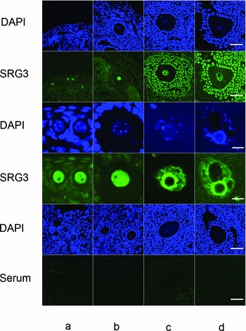
To determine the expression of SRG3 during oogenesis, we examined ovarian sections from 6–8-week-old mice by immunohistochemistry using an anti-SRG3 antibody. The anti-SRG3 antibody stained nuclei of primordial follicle (n = 167), primary follicle (n = 121), secondary follicle (n = 107) and preovulatory follicle oocytes (n = 67), whereas the normal goat serum did not (Fig. 1). Isolated fully grown oocytes also expressed high levels of SRG3 (see below, Fig. 2a).
Expression of SRG3 during oogenesis. Mouse ovarian sections were incubated with anti-SRG3 antibody (rows 1–4) or goat serum (rows 5, 6), followed by FITC-conjugated secondary antibody (green). Tissue sections were counterstained with DAPI (blue). (a) primordial follicles; (b) primary follicles; (c) secondary follicles; (d) preovulatory follicles. Images in rows 3 and 4 were enlarged from rows 1 and 2 respectively to show details of the nuclei. Bar = 50 μm in rows 1, 2, 5, 6; Bar = 10 μm in rows 3 and 4.

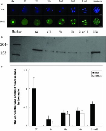
(a) Laser-scanning micrographs showing SRG3 expression in fully grown oocytes and embryos at 4, 6 and 10 h p.f. and at the 2-cell, 4-cell and blastocyst stages. Upper panels, DAPI staining (blue). Bottom panels, immunofluorescent staining with anti-SRG3 antibody (green). Bar = 50 μm. (b) Western blot analysis of total SRG3 protein in oocytes and early embryos. GV, fully grown oocytes; MII, metaphase II-arrested eggs; 6h, 6 h p.f.; 10 h, 10 h p.f.; 2-cell, 2-cell embryos; 3T3, whole-cell extracts from NIH3T3. (c) Quantitative analysis of SRG3 expression. SRG3-specific labelling was detected in the nucleus at GV, 4, 6 and 10 h p.f. and in the 2-cell embryo. GV, fully grown oocytes. The concentrations of SRG3 fluorescence in the nuclei at each stage were calculated using signals from GV (defined as 100%). Each column represents the mean ± SEM of five to eight embryos. The nuclear concentration of SRG3 in fully grown oocytes is significantly higher than that in 1-cell embryos at 4 h p.f. (p < 0.001) and at 6 h p.f. is significantly higher than 4 h p.f. (p < 0.01, t-test)

Expression of SRG3 protein in preimplantation embryos
In mouse embryos, SRG3 staining was observed in the pronuclei of the 1-cell embryo at 4, 6 and 10 h postfertilization, as well as in nuclei of the 2-cell, 4-cell, 8-cell and blastocyst stage embryos (Fig. 2a).
The temporal changes in nuclear levels of SRG3 in fully grown oocytes and in embryos were quantified by immunofluorescent confocal microscopy. Fluorescence was greatest in the germinal vesicle of the fully grown oocytes. SRG3 localized throughout the ooplasm after germinal vesicle breakdown. At 4 h postfertilization (p.f.), soon after the pronuclei had formed, nuclear SRG3 was detected, although levels were only slightly higher than cytoplasmic levels. As pronuclei migrated towards the centre and grew in size, the level of SRG3 increased continuously in the pronuclei. In most embryos, the male pronucleus was larger than the female pronucleus. Nuclear concentrations of SRG3 in the male pronucleus were greater than in the female pronucleus at all time points examined (Fig. 2a, c).
The total amount of SRG3 protein in fully grown oocytes and early embryos was examined by western blot. As shown in Fig. 2b, a high level of SRG3 protein was present in fully grown oocytes. The amount of protein was reduced dramatically, but still present in MII eggs, at 6 h p.f., 10 h p.f. and in the 2-cell embryos. Although nuclear concentrations of SRG3 increased from the 4-cell p.f. to the 2-cell stages (Fig. 2a, c), the total amount of the protein in the embryo remained relatively steady (Fig. 2b). These data suggest that the increase in the nuclear SRG3 results from translocation of SRG3 from the cytoplasm to the nucleus.
Composition analysis of SRG3 pool in the early embryo
Our immunofluorescence data indicates that SRG3 enters pronuclei shortly after formation and nuclear concentration increases as embryos develop. As this precedes ZGA, SRG3 protein must be maternally supplied. To investigate this possibility and to dissect components of SRG3 pool in early embryos, 1-cell embryos were treated with cycloheximide and α-amanitin, which inhibit protein synthesis and transcription, respectively.
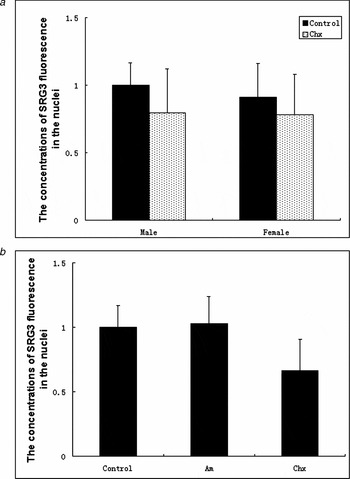
As predicted, in 1-cell embryos that had recently formed pronuclei, the protein synthesis inhibitor cycloheximide partially inhibited time-dependent increase in the nuclear concentrations of SRG3 in both 1-cell and 2-cell embryos (Fig. 3a, b). Note again that the concentration of SRG3 was greater in the male pronucleus than in the female pronucleus. In contrast, the transcriptional inhibitor α-amanitin did not substantially inhibit the increase in nuclear SRG3 that occurred between the 1-cell and 2-cell stages (Fig. 3a). These results confirm that a portion of the SRG3 is derived from translation of maternal mRNA.
(a) Inhibition of nuclear SRG3 in 1-cell embryos by the protein translation inhibitor cycloheximide. Quantification of nuclear fluorescence was performed as described in Materials and methods and data were expressed relative to the male PN (100%). Nuclear concentration of SRG3 protein was significantly lower in cycloheximide-treated embryos as compared to untreated embryos (p < 0.01). Chx, cycloheximide; male, male PN; female, female PN. Data are expressed as the mean ± SEM of 24–30 embryos. (b) Inhibition of nuclear SRG3 in 2-cell embryos by cycloheximide. As compared to untreated 2-cell embryos, cycloheximide significantly inhibited SRG3 expression in 2-cell embryos (p < 0.001) whereas α-amanitin did not (p > 0.05). Data were expressed as the mean ± SEM for 20 to 23 embryos. Am, α-amanitin; Chx, cycloheximide.

Co-expression of SRG3, Brg1 and Ini1 in preimplantation mouse embryos
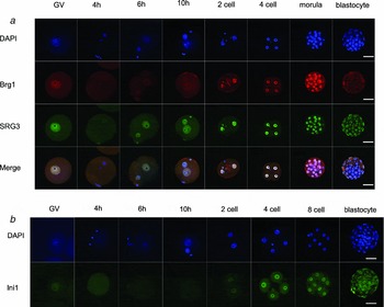
As SRG3, Ini1 and Brg1 are all core subunits of the SWI/SNF complex, they should be expressed in a similar time course and location in order to function in a same complex. Data from the immunofluorescence study indicated that Brg1 was expressed in fully grown oocytes and early embryos in a temporal and spatial pattern that completely coincides with SRG3 (Fig. 4a). Brg1 was detected at high levels in fully grown oocytes and at a lower level in pronuclei. Nuclear levels of Brg1 increased as embryos cleaved. In contrast, Ini1 was not detected in pronuclei at 10 h p.f. in 1-cell embryos. Nuclear localization of Ini1 was, however, apparent at the 2-cell stage and persisted in 4-cell, morula and blastocyst stages (Fig. 4b). Therefore, Ini1 was expressed in early mouse embryos at a time slightly later than SRG3 and Brg1.
(a) Co-localization of Brg1 and SRG3 in the fully grown oocyte and early preimplantation embryo. Panels in the first row were stained by DAPI (blue), in the second row by a Brg1-specific antibody (red) and in the third row by a SRG3-specific antibody (green). Panels in the fourth row correspond to merged images from panels of the threes previous rows. Embryos collected at GV (fully grown oocytes), 4, 6 and 10 h p.f., 2-cell, 4-cell, morula and blastocyst stages were processed for antibody staining. Bar = 50 μm. (b) Laser-scanning micrographs showing Ini1 expression in fully grown oocytes (GV) and embryos at 4, 6 and 10 h p.f. and 2-cell, 4-cell, 8-cell and blastocyst stages. Upper panels: DAPI staining (blue). Bottom panels: immunofluorescent staining (green) with an Ini1-specific antibody. Bar = 50 μm.

Discussion
Around the time of fertilization chromatin is remodelled globally in structure and epigenetic status (Vignon et al., Reference Vignon and Zhou2002). Such remodelling is essential for establishing the zygotic developmental programme. Our data show that SRG3, Brg1 and Ini1 proteins are expressed in the mouse embryo at early stages and are present in the nuclei of embryonic cells around the time that zygotic chromosome structure is being organized and transcription initiated. The expression profiles of these factors are consistent with their requirements during early development (Bultman et al., Reference Bultman and Gebuhr2000, Reference Bultman and Gebuhr2006; Klochendler-Yeivin et al., Reference Klochendler-Yeivin and Fiette2000; Guidi et al., Reference Guidi and Sands2001; Kim et al., Reference Kim and Huh2001), consistent with critical roles in early embryogenesis.
Transcription is highly active in growing oocytes. The level of transcription reaches its peak in secondary oocytes and declines thereafter (Moore et al., Reference Moore and Lintern-Moore1974). This activity ceases completely during chromatin condensation in later stage GV oocytes (Bouniol-Baly et al., Reference Bouniol-Baly and Hamraoui1999; De La Fuente et al., Reference De La Fuente and Viveiros2004; De La Fuente Reference De La Fuente2006). SRG3 is present persistently in the nuclei of oocytes before GVBD, neither the level nor the localization of SRG3 protein correlates with changes in transcription activity in oocytes. However, it is still possible that the functional status of SRG3 may change during the course of development.
The expression and subcellular localization of transcription factors change dynamically during oogenesis and early embryonic development and such changes correlate closely with the transcriptional activity of the early embryo (Ram & Schultz, Reference Ram and Schultz1993; Bouniol et al., Reference Bouniol and Nguyen1995; Aoki et al., Reference Aoki and Worrad1997). The expression patterns of SRG3, Brg1 and Ini1 during early embryogenesis are very similar to that of Sp1 and TBP (Worrad et al., Reference Worrad and Ram1994). With the exception of Ini1, these proteins are all detected in pronuclei shortly after their formation and their nuclear concentrations increase steadily as pronuclei migrate to the center of the embryo. As the embryo proceeds through early cleavage stages, nuclear concentrations of these factors and Ini1 increase. The closely related expression profiles of these transcription factors and remodelling factors may have functional implications. One common feature of all ATP-dependent chromatin-remodelling factors is the ability to enhance the accessibility of nucleosomal DNA to other proteins, such as transcription factors, by the ATP-dependent movement of nucleosomes in cis along a DNA fragment (Smith & Peterson, Reference Smith and Peterson2005). The co-expression of transcription and remodelling factors and the fact that development ceases at the peri-implantation stage in the presence of transcription factors, but absence of remodelling factors (Bultman et al., Reference Bultman and Gebuhr2000; Klochendler-Yeivin et al., Reference Klochendler-Yeivin and Fiette2000; Guidi et al., Reference Guidi and Sands2001; Kim et al., Reference Kim and Huh2001) suggest that remodelling factors are required for assembling and/or maintaining transcription factors in a functional state as the chromatin goes through a series of changes during early embryogenesis.
Male and female pronuclei differ in their ability to support transcription in 1-cell embryos. This difference is correlated with variations in the nuclear concentration of transcription factors (Worrad et al., Reference Worrad and Ram1994) and in chromatin structure (Nonchev & Tsanev, Reference Nonchev and Tsanev1990; Adenot et al., Reference Adenot and Mercier1997; Santos et al., Reference Santos and Hendrich2002). The observation that nuclear concentrations of SRG3 are consistently greater in the male pronucleus than in the female pronucleus provides an additional explanation for the differences in the transcriptional capacities of the male and female pronuclei: the male pronucleus contains not only higher concentrations of transcription factors, e.g. Sp1 and TBP (Worrad et al., Reference Worrad and Ram1994), but also higher concentrations of remodelling factors that should aid transcription factors in accessing their chromatin-binding sites.
Our results also suggest that inhibition of protein translation partially diminished nuclear levels of SRG3. One explanation for this result is that the initial SRG3 pool is composed of two fractions: a cycloheximide-insensitive maternal protein fraction and a cycloheximide-sensitive fraction representing newly translated SRG3 protein. During early stages, newly translated SRG3 comprises a small proportion of the total pool, which is likely to increase as development proceeds. Alternatively, cycloheximide may inhibited protein(s) required for transport of SRG3 into the nuclei and, therefore, blocked the transportation of SRG3. Considering that an inhibition in the transportation mechanism would more likely cause a complete, rather than a partial blockage of SRG3 in the nuclei, our data appear to favour the first explanation.
The correlated expression of SRG3, Brg1 and Ini1, the three core subunits of the SWI/SNF complex, in cleavage-stage embryos, suggest that these factors constitute a functional complex that regulates transcription and chromatin structure during development. Our data also indicate that whereas Brg1 and SRG3 are found in the pronuclei of the 1-cell embryo, Ini1 is not. The implications of these observations are not immediately clear. Previous studies have shown that a minimum catalytic core complex of three SWI/SNF components, Brg1, Ini1 and SRG3, can remodel both mononucleosome and nucleosome arrays (Phelan et al., Reference Phelan and Sif1999). Brg1 alone can substitute for the core complex, albeit at a lower efficiency. It will be interesting to examine whether, in the absence of Ini1, SRG3 alone or together with other subunit(s), can enhance the activity of Brg1 in the pronucleus.
Acknowledgements
We thank Tianlong Gao for his support in confocal microscopic analysis, Yong Lu and Ye Yang for literature collection and discussion and Youming Zhu, Feng Ying Li for excellent technical assistance and animal care. The study was supported by grants from National Basic Research Program of China (973 Program) (nos. 001CB509903, 001CB509904), Hi-Tech Research and Development Program of China (973 Program) (nos. 2001AA216121, 2004AA205010), National Natural Science Foundation of China (no. 30040003), Science and Technology Committee of Shanghai Municipality (nos. 99DJ14002, 00DJ1 4033, 01DJ14003, 03DJ14017), Chinese Academy of Science (no. KSCX-2–3–08), Shanghai Municipal Education Commission no. T0205 and Shanghai Jiao Tong University, School of Medicine.