Implications
In mature rams and bucks, but not bulls, nutrition exerts major effects on testicular mass. The change in mass reflects changes in the amount of seminiferous tissue and thus spermatogenic capacity. Spermatogenic efficiency is also altered, so the overall outcome is a major change (up to 250%) in the rate of production of spermatozoa. Except for extreme undernutrition, these effects are not linked to major changes in testosterone production or in sexual behaviour. Male small ruminants should thus be fed correctly for 2 months before mating. This is a critical component of ‘clean, green and ethical’ systems of animal production.
Introduction
The ruminants developed seasonal reproductive strategies in response to the pressures of natural selection imposed by their environment for millions of years, long before humans began to domesticate them in the pre-dawn of the agricultural age. Despite 11 000 years of environmental management and directed breeding, many of the characteristics of these original strategies are still with us. One of the most persistent is sensitivity to nutritional inputs, primarily because an adequate supply of nutrients, from body reserves or from diet, is absolutely essential for lactation. Avoiding nutritional scarcity during this critical stage of the mammalian reproductive process becomes a challenge because the supply of forage varies greatly throughout the year. In many regions, the periods of scarcity are predictable from year to year, but the actual season of scarcity might be winter (Temperate regions), autumn (Mediterranean regions) or spring (some Sub-tropical regions). Semi-arid and arid regions offer the greatest challenge because the timing is unpredictable for the peaks and troughs in forage supply. Considering this variety of environments and the fact that wild and domesticated ruminant species are found in all of them, it is no surprise that the problem of adequate nutrient supply at lactation has been solved by a variety of reproductive strategies, particularly when the duration of pregnancy varies as much as it does between the large and small ruminants.
In temperate regions, a spring birth has a very clear and consistent advantage, so photoperiodic time measurement, because it is the most reliable way to determine whether winter or summer is approaching, exerts a very powerful influence over reproductive activity (Lincoln and Short, Reference Lincoln and Short1980). However, the majority of the world’s sheep and goats live between 35 degree North and 35 degree South (Lindsay, Reference Lindsay1991), where the photoperiod-driven breeding patterns that characterise these species are often poorly co-ordinated with changes in forage supply (Sutama and Edey, Reference Sutama and Edey1985; Martin et al., Reference Martin, Hötzel, Blache, Walkden-Brown, Blackberry, Boukliq, Fisher and Miller2002). In these environments, many mammals have developed ‘opportunistic’ reproductive strategies that can cope with the problem (Bronson, Reference Bronson1985). The control systems of their reproductive axis respond to photoperiod, but they are more holistic and flexible than those of animals from temperate regions because they are heavily influenced by other inputs, particularly nutrition and socio-sexual stimuli.
Our studies extended the concept developed by Bronson (Reference Bronson1985) to two ruminants that have been introduced to Australia, both of which exhibit a considerable degree of opportunism – the Merino sheep (of Mediterranean origins) and the Australian cashmere goat (recently evolved from the feral goat population). The role of socio-sexual stimuli in the opportunistic breeding strategies of these animals has been reviewed elsewhere (Walkden-Brown et al., Reference Walkden-Brown, Martin and Restall1999). In this review, we will deal with the nutritional inputs into the processes that control reproductive function. We will focus primarily on the mature males and the endpoint will be the gametogenic and endocrine activity of the testes. Although our own studies are largely limited to the Australian goats and sheep, we will integrate them with work on other genotypes and environments, and we will also address related issues for cattle. In all cases, we will consider how this information affects animal industry.
Foetal life and puberty
In extensive management systems, pregnant females are often forgotten between mating and the final few weeks of pregnancy because it has traditionally been thought that there are no serious demands on their reserves until the foetus reaches a significant size. However, this ignores two major factors: (i) the role of metabolic inputs into embryo and foetal development (‘foetal programming’); (ii) the nutritional requirements for the development of the placenta (review: Bell, Reference Bell1984). In both cases, the consequences of nutritional mis-management may not become evident until much later, after birth or even after sexual maturity, but are associated with poor life-time performance (review: Bell, Reference Bell1984). The life-time productivity of the tissues that provide commodities (skin follicles; precursor muscle cells; mammary tissue) can be compromised, as can the development of reproductive tissues (review: Martin et al., Reference Martin, Milton, Davidson, Banchero Hunzicker, Lindsay and Blache2004). Thus, male lambs born to ewes that were underfed during the second half of pregnancy have fewer Sertoli cells in their testes at birth (Bielli et al., Reference Bielli, Pérez, Pedrana, Milton, Lopez, Blackberry, Duncombe, Rodriguez-Martinez and Martin2002), and retardation of intrauterine growth can delay the onset of puberty in male lambs (Da Silva et al., Reference Da Silva, Aitken, Rhind, Racey and Wallace2001). Further investigation is needed but it seems likely that the outcome in both cases would be a reduction in spermatogenic capacity in adult life.
The roles of postnatal nutrition in progress towards puberty in male farm animals has been reviewed by Brown (Reference Brown1994). We will focus on mature animals for the remainder of this review, but we would like to acknowledge the major contributions in recent years from the laboratory of John Kastelic who have studied metabolic input into pubertal development of young bulls, and have clearly demonstrated that superior calfhood nutrition, probably through the action of metabolic hormones, promotes gonadotrophin secretion and thus testicular growth, and an earlier onset of spermatogenesis (see Barth et al., Reference Barth, Brito and Kastelic2008). There is very broad agreement between these observations and our own on sexually mature male sheep, as we shall see below.
Mature males – testicular responses to nutrition
Nutritional influences on reproduction in farm animals were probably recognised soon after domestication. In the historical overview preceding his study, Clark (Reference Clark1934) was prepared to cite Aristotle and Darwin has having considered the concept, but gave Walter Heape the credit for providing the first experimental evidence of ‘flushing’ in English sheep in 1899. However, it was Clark (Reference Clark1934) himself who showed that flushing increased lambing rate by increasing ovulation rate. This phenomenon has long intrigued reproductive physiologists and continues to dominate research on the nutritional influences on reproduction to this day. By contrast, relatively little attention was paid to the male. It was recognised that the production of gametes by mature males is affected by nutrition but, after reviewing the ruminant literature, Moule (Reference Moule1963) was led to conclude: ‘…so far no workers seem to have attempted a systematic study of the overall effects of nutrition on semen production in male domestic animals’. There seemed to be no literature at all on male goats. For mature bulls, Moule missed the work of Flipse and Almquist (Reference Flipse and Almquist1961), leaving the study of a single 5-year-old bull by Mann and Walton (Reference Mann and Walton1953) who found that undernutrition reduced the concentrations of fructose and citrate in seminal plasma but did not affect the sperm output.
The most interesting of the earlier work on mature animals was perhaps that of Akira Mori, who carried out a controlled experiment on large groups of severely underfed mature rams in Japan during the Second World War. When his work was published many years later, it showed that semen quality and sperm production were improved within a month or 2 months by supplements that led to a weight gain (Mori, Reference Mori1959). Mori was convinced that protein shortage had caused the infertility in his flocks, but he did not test the concept formally with nutritionally rigorous diets. This sort of problem, and the worry expressed by Moule (Reference Moule1963) that many experiments on ‘nutrition’ concerned only pathological effects, requires us to define the term as we shall use it in this review. Deficiencies in trace elements (e.g. zinc, Martin et al., Reference Martin, White, Markey and Blackberry1994b) or vitamins (Robinson et al., Reference Robinson, Ashworth, Rooke, Mitchell and McEvoy2006), heavy metal toxicities, or imbalances in essential amino or fatty acids, have long been known to affect male reproductive function adversely (Leatham, Reference Leatham1975), but for the present review, we need to constrain the discussion as much as possible to shifts in energy or protein balance of similar magnitude and duration to those that would be experienced by animals grazing natural forage. Generally, we will avoid the extremes of this range too, some of which lead to total gonadal dysfunction (the animals studied by Mori (Reference Mori1959) must have been close to this). Similarly, we will not consider responses to fasting because of the confounding effects of stress.
Nutrition and the production of spermatozoa
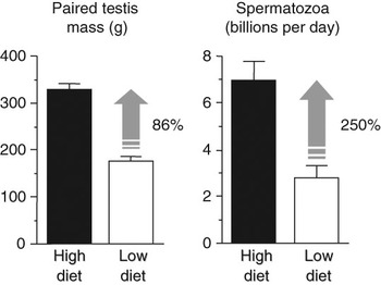
The study by Mori (Reference Mori1959) included Merino rams and his data were soon supported by two further studies with this breed. Salamon (Reference Salamon1964) used high-frequency semen collections and Setchell et al. (Reference Setchell, Waites and Lindner1965) used a histological assessment of the seminiferous tubules and epididymis, but they both came to the same conclusion – the daily rate of sperm production is reduced by underfeeding and improved by refeeding. Importantly, the quality of the semen produced, measured as sperm count and sperm motility, is also decreased by undernutrition for a period of time greater than the 7-week duration of spermatogenesis (Parker and Thwaites, Reference Parker and Thwaites1972; Robinson et al., Reference Robinson, Ashworth, Rooke, Mitchell and McEvoy2006). Goats were studied by Hiroe and Tomizuka (Reference Hiroe and Tomizuka1965), who also found that loss of body mass was associated with reductions in the output of spermatozoa. There were some negative findings too (Tilton et al., Reference Tilton, Warnick, Cunha, Loggins and Shirley1964), but subsequent studies (Parker and Thwaites, Reference Parker and Thwaites1972; Braden et al., Reference Braden, Turnbull, Mattner and Moule1974; Oldham et al., Reference Oldham, Adams, Gherardi, Lindsay and Mackintosh1978; Alkass et al., Reference Alkass, Bryant and Walton1982; Martin et al., Reference Martin, Sutherland and Lindsay1987; Cameron et al., Reference Cameron, Murphy and Oldham1988) led to the wide acceptance of strong, direct relationships between plane of nutrition, testicular mass and the number of spermatozoa available for ejaculation, for the small ruminants at least (Figure 1).
Effect of nutrition on testicular growth and the production of spermatozoa in 1-year-old Merino rams (P < 0.01 for both). The diets led to liveweight gains of 17.2 kg (High) and −5.8 kg (Low) after 9 weeks (P < 0.001). Semen was collected daily with an artificial vagina and the number of spermatozoa per ejaculate was averaged for the ninth week. The rate of production of spermatozoa was calculated from numbers of stages VI, VII and VIII spermatids in testicular homogenates following castration at the end of the experiment. Testicular mass was determined after slaughter. Redrawn after Cameron et al. (Reference Cameron, Murphy and Oldham1988).

For cattle, the picture is more complex. Most of the older literature describes experiments with young Bos taurus bulls that are up to 2 years old and post-pubertal but still growing rapidly (review Brown, Reference Brown1994). In such animals, nutrition clearly affects testicular development and the production of spermatozoa in much the same way as it does in mature sheep and goats (Davies et al., Reference Davies, Mann and Rowson1957; Flipse and Almquist, Reference Flipse and Almquist1961; Van Demark and Mauger, Reference Van Demark and Mauger1964; Van Demark et al., Reference Van Demark, Fritz and Mauger1964; Gauthier and Berbigier, Reference Gauthier and Berbigier1982). The same applies to Bos indicus bulls (Rekwot et al., Reference Rekwot, Oyedipe, Akerejola and Kumi-Diaka1987; Tegegne et al., Reference Tegegne, Entwistle and Mukasa-Mugerwa1992). As mentioned above, the Kastelic laboratory has added an extra level of sophistication to our understanding of the effects of nutrition over the first 18 months of life (review: Barth et al., Reference Barth, Brito and Kastelic2008). However, after the period of rapid growth has ended, there appears to be little or no response to nutrition (Flipse and Almquist, Reference Flipse and Almquist1961; Van Demark and Mauger, Reference Van Demark and Mauger1964; Van Demark et al., Reference Van Demark, Fritz and Mauger1964; Coulter et al., Reference Coulter, Carruthers, Amann and Kozub1987). Caution is needed with these older studies on mature bulls. For example, in the experiment by Van Demark and colleagues, the ‘low’ plane of nutrition had little effect on body weight, as well as testicular size, even over 12 months, although production of spermatozoa was reduced (Van Demark and Mauger, Reference Van Demark and Mauger1964; Van Demark et al., Reference Van Demark, Fritz and Mauger1964). In the other cases, the ‘high’ planes seemed to constitute overfeeding, as the sperm production was markedly decreased compared to ‘medium’ or ‘low’ planes (Coulter et al., Reference Coulter, Carruthers, Amann and Kozub1987; Mwansa and Makarechian, Reference Mwansa and Makarechian1991). In addition, the energy and protein balance of the experimental diets requires clarification, as both have been manipulated in these studies, but not compared in the one experiment.
Nutritional effects on spermatogenic tissues
In rams, changing nutrition alters not only the total mass of testicular tissue, but also the efficiency with which the gametes are produced by that tissue. This is evident from the fact that proportional changes in the sperm production are consistently greater than changes in testicular size. For example, Oldham et al. (Reference Oldham, Adams, Gherardi, Lindsay and Mackintosh1978) found that a 25% increase in testicular size led to an 81% increase in the production of spermatozoa and Cameron et al. (Reference Cameron, Murphy and Oldham1988) found that an 86% increase in testicular size led to a 250% increase in the production of spermatozoa (Figure 1). The number of ejaculated spermatozoa is not affected until 7 weeks after a change in diet, suggesting that the effects on spermatogenic efficiency are exerted after the last spermatogonial division. This is similar to the effects of stimulatory photoperiod in more seasonal breeds of sheep, where efficiency is increased by reducing the rate of degeneration (presumably apoptosis) of germ cells following the mitotic and meiotic divisions of the spermatogenic cycle (Hochereau-de Reviers et al., Reference Hochereau-de Reviers, Perreau and Lincoln1985). From histological studies it was clear that nutrition affects the diameter of the seminiferous tubules, the relative proportion of the testis occupied by the seminiferous tubules and the proportion of the seminiferous tubule occupied by the seminiferous epithelium (Setchell et al., Reference Setchell, Waites and Lindner1965; Oldham et al., Reference Oldham, Adams, Gherardi, Lindsay and Mackintosh1978).
In a detailed study, Hötzel et al. (Reference Hötzel, Markey, Walkden-Brown, Blackberry and Martin1998) also showed that seminiferous tubule length and diameter were higher in well-fed rams than in underfed rams. Their most challenging finding was that the number of Sertoli cell nuclei per testis was also affected (well-fed 120 ± 6×10⁸; underfed 77 ± 7×10⁸; P < 0.001), whereas the proportion of testis occupied by Sertoli cell nuclei was not. This is consistent with changes in the spermatogenic function of the testis, particularly the increase in spermatogenic efficiency, but it is in conflict with the notion that Sertoli cell number is established before puberty in all species, including the ram (Monet-Kuntz et al., Reference Monet-Kuntz, Hochereau de Reviers and Terqui1984) and goat (Hochereau-de Reviers et al., Reference Hochereau-de Reviers, Perreau, Delouis, Chemineau and Courot1986). However, there had been several reports of changes in Sertoli cell number after puberty (see Hötzel et al., Reference Hötzel, Markey, Walkden-Brown, Blackberry and Martin1998) and, recently, Sarah Meachem and her colleagues have addressed this question directly. For hamsters, they provided solid evidence for seasonal changes and have proposed that adult Sertoli cells are not terminally differentiated. Their view is that Sertoli cells can enter a transitional state exhibiting features common to both undifferentiated and differentiated Sertoli cells (Tarulli et al., Reference Tarulli, Stanton, Lerchl and Meachem2006).
Nutritional effects on testicular interstitial tissues
Hötzel et al. (Reference Hötzel, Markey, Walkden-Brown, Blackberry and Martin1998) found that the number of Leydig cells per testis in the ram was not affected by diet, but the total volume of Leydig cells per testis was 30% higher in well-fed rams than in underfed rams, indicating a change in the volume of individual cells. This is in broad agreement with the study by Setchell et al. (Reference Setchell, Waites and Lindner1965) and suggests that the rate of production of testosterone should also be affected. This should be reflected in circulating testosterone concentrations, especially when combined with changes in the testicular blood flow. Blood flow through the testis is closely related to testis mass and thus changes in response to nutrition (Setchell et al., Reference Setchell, Waites and Lindner1965). The relative volume of blood and lymph vessels was not affected by diet in the study by Hötzel et al. (Reference Hötzel, Markey, Walkden-Brown, Blackberry and Martin1998), but their absolute volumes increased or decreased as a function of the change in testicular size. These changes in the vasculature and the associated changes in blood flow, accommodate the transport of testosterone to the peripheral circulation (Setchell, Reference Setchell1986 and Reference Setchell1990). Thus, the present data are in agreement with a previous nutritional study in mature rams (Setchell et al., Reference Setchell, Waites and Lindner1965) and suggest that total blood flow was affected while there was probably little or no change in blood flow per unit mass of testis. As a consequence, larger amounts of testosterone produced by the testis would have reached the peripheral circulation in rams fed with the high diet than in those fed with the low diet. In assessing the literature on this topic, an important consideration is the severity and duration of the dietary treatments. For example, Setchell et al. (Reference Setchell, Waites and Lindner1965) compared rams that were severely underfed for 3 months (body fat was reduced to less than 12% of live mass) with rams that were arguably obese (25% to 49% fat). In the study by Hötzel et al. (Reference Hötzel, Markey, Walkden-Brown, Blackberry and Martin1998), similar conditions were imposed. Mature cashmere bucks require about 4 months of dietary treatment before testosterone production is affected (Walkden-Brown et al., Reference Walkden-Brown, Restall, Norton, Scaramuzzi and Martin1994). Differences in severity and duration explain the disagreements between these studies and other study showing no effect of nutritional treatments on peripheral testosterone concentrations (Ritar et al., Reference Ritar, Adams and Sanders1984; Martin et al., Reference Martin, Tjondronegoro and Blackberry1994a).
In bulls, the effects of nutrition on testosterone production were clear from the earliest studies based on measurement of fructose and citrate concentrations in seminal plasma (indicators of stimulation of the seminal vesicles by testosterone). From their study of a single mature bull, Mann and Walton (Reference Mann and Walton1953) concluded that undernutrition reduced testosterone production and this was supported by Davies et al. (Reference Davies, Mann and Rowson1957), although their data was derived from younger animals. Subsequently, these effects in growing bulls were confirmed by direct measurement of testicular testosterone content (Mann et al., Reference Mann, Rowson, Short and Skinner1967) and plasma testosterone concentrations (Gauthier and Berbigier, Reference Gauthier and Berbigier1982).
Nutrition and the expression of sexual behaviour in mature males
Despite the clear, mechanistic evidence of links between nutrition and reproductive endocrinology, there is some controversy on the effect of undernutrition on the expression of sexual behaviour of male sheep, mainly due to the methods used to assess libido and the frequency of observations (Parker and Thwaites, Reference Parker and Thwaites1972). If feed restriction is sufficiently long and severe, such that over 30% of body mass is lost, the expression of normal sexual behaviour is compromised (Parker and Thwaites, Reference Parker and Thwaites1972). However, sexual behaviour requires fairly intensive motor activity (Banks, Reference Banks1964) so a decline in libido could be caused by a general weakness in underfed rams rather than energy restriction (Tilbrook and Cameron, Reference Tilbrook and Cameron1990). In fact, during mating, motor activity is greater in rams than in ewes, because of the very active part taken by the rams during courtship (Banks, Reference Banks1964). Interestingly, it is also greater in rams with smaller testes than in rams with larger testes (Raadsma and Edey, Reference Raadsma and Edey1985), suggesting that the cost of reproduction is greater in animals that produce less spermatozoa. Overall, it appears that libido is more sensitive to undernutrition than sperm production but increases in the plane of nutrition stimulate sperm production before affecting libido.
Energy balance is, of course, a reflection of energy expenditure as well as energy intake. Thus, exercise affects testicular size (Thwaites, Reference Thwaites1995) and rams mating under field conditions show a loss of testicular weight, a reduction in the sperm production, and increase in proportion of intertubular tissue, a decrease in proportion of spermatocytes, round spermatids and decrease in number of spermatozoa in the epididymus (Knight et al., Reference Knight, Gherardi and Lindsay1987). This might explain why rams have better reproductive performance when kept in intensive systems than when kept in extensive systems (Fourie et al., Reference Fourie, Schwalbach, Neser and Van der Westhuizen2004). However, overfed males show reduced sexual activity possibly because they are overweight and have difficulty expressing courtship or mounting (Okolski, Reference Okolski1975). Thus, regular exercise can increase the fertilising ability of Merino rams mating under field conditions (Combrink and Schoeman, Reference Combrink and Schoeman1993).
Physiological mechanisms
Nutrition affects male reproductive function in two types of physiological process: (a) through the metabolic and reproductive centres of the brain, leading to a change in the output of gonadotrophin-releasing hormone (GnRH); (b) through pathways that seem to be independent of changes in GnRH secretion (Hötzel et al., Reference Hötzel, Walkden-Brown, Blackberry and Martin1995). The outcome of the combined pathways is the change in testicular mass, primarily seminiferous tissue and in the efficiency of spermatogenesis as described above. We now have a strong understanding of the metabolic inputs to the brain-gonadal axis (nutrients, metabolites and substrates) as well as the endocrine signals from metabolic and storage tissues.
Setchell et al. began unravelling the physiological processes by describing the dependence of testicular metabolism on glucose and found that underfeeding reduced the metabolic activity and blood flow of the testis (Setchell et al., Reference Setchell, Waites and Lindner1965; Setchell and Hinks, Reference Setchell and Hinks1967). They also found that testosterone output was reduced and supported the suggestion from work on rats that the pituitary gonadotrophins had a role in nutritional responses (the ‘pseudohyophysectomy’ mechanism of the 1940s). However, few other data were available when we began work in this area, arguably no surprise considering how few studies had been done and the limitations of the endocrine techniques of the time.
Progress depended on the availability of an efficient experimental model. We were greatly aided by the availability of an acute nutritional stimulus that would elicit physiological responses – lupin grain, a highly digestible source of energy and protein that had low concentrations of soluble carbohydrates so could be acutely fed to ruminants in large quantities (e.g. 1 kg/day) without causing acidosis. We combined this treatment with the mature Merino ram, an animal that showed rapid, repeatable testicular responses to changes in food supply at all times of the year. Finally, we are able to use the strong linear relationship between testicular size and the rate of production of spermatozoa to estimate the gametogenic potential of the testis (Amann, Reference Amann1970; Knight, Reference Knight1977).
A multi-dimensional control system based on energy homeostasis
Here, we deal with the relationship between metabolic status and the activity of the system that controls the central driver of reproductive function, pulsatile GnRH secretion. We reflect on these relationships within the framework of a model that comprises four interdependent ‘dimensions’: (i) genetic, (ii) structural, (iii) communicational and (iv) temporal. To illustrate these concepts, we will mainly use the male sheep that has been acutely placed on a high plane of nutrition. Most of the processes involved are autonomic by nature but, to aid clarity of expression, we will occasionally risk the use of anthropomorphic language (e.g. ‘decision to reproduce’). These concepts have been presented in detail previously in a cross-model comparison in which we used information from mature male sheep and post-partum dairy cows (Blache et al., Reference Blache, Chagas and Martin2007). Therefore, here, we will cover the topics briefly.
Bioenergetics of reproduction – the concept of ‘metabolic status’
Energy costs are attached to all components of the reproductive process, from the expression of specific behaviours, such as sexual or maternal behaviour, to the production of morphological elements, such as gametes, foetuses and milk (for a full review, see Martin et al., Reference Martin, Blache and Williams2008). Considering the energetic requirements, the relationship between the metabolic and the reproductive regulatory systems needs to be highly tuned if the probability of success is to be reasonable. Thus, the regulatory processes that link nutrition and reproduction are largely the same regulatory processes that control energy homeostasis.
At any given time, for any given animal, the amount of energy available for reproduction depends on the difference between the amount of energy expended, including the demands for maintenance, and the pool of disposable energy. The pool of disposable energy includes the energy derived from feed intake plus the energy stored in body tissues, especially adipose tissue, liver and muscle. Most authors refer to ‘energy balance’ but we prefer ‘metabolic status’ because it includes a strong integrative dimension.
Dimension 1 – the genetic dimension
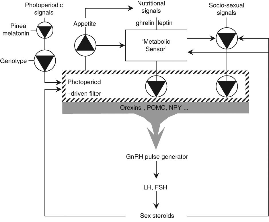
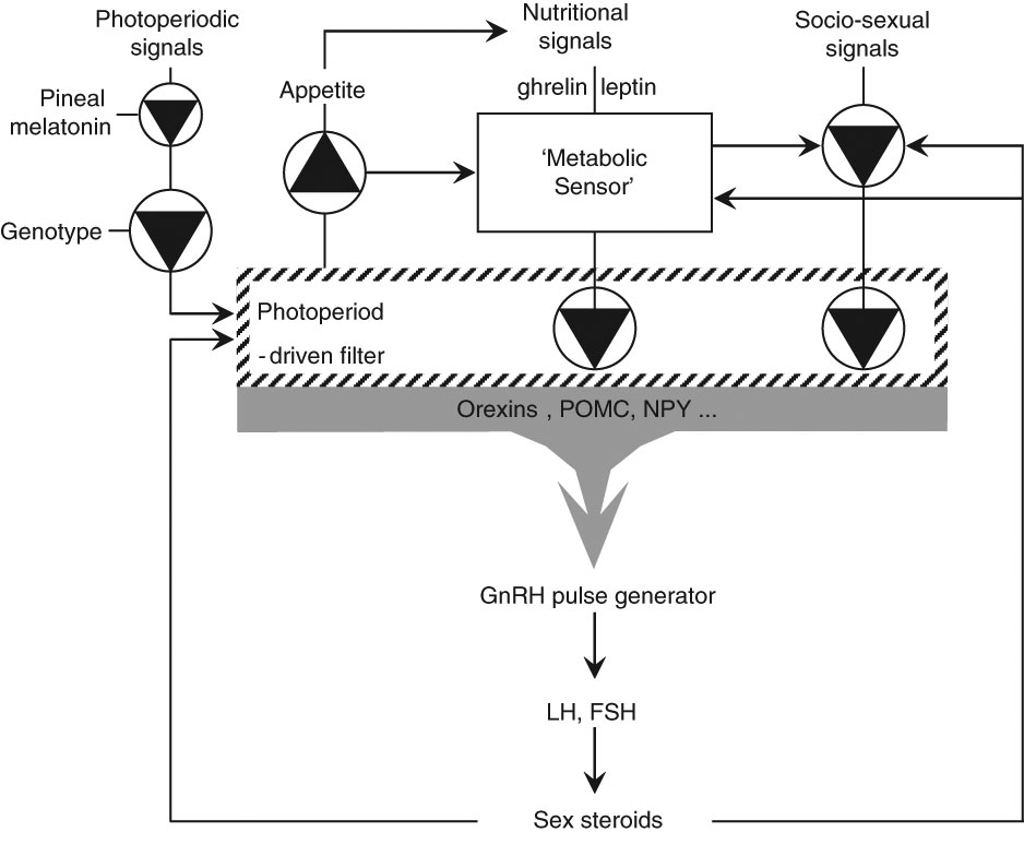
In male sheep, the effects of metabolic status and dietary manipulation on the reproductive axis differ between genotypes. This genetic dimension accounts for variations in the responses to all environmental inputs. We have previously suggested that photoperiod acts as a ‘filter’ of the effect of nutrition on the reproductive system and that this filtering effect depends on the genotype of the animal (Blache et al., Reference Blache, Zhang and Martin2003; Figure 2). This is because, in the ram, the effect of nutrition on the reproductive endocrine axis is smaller in breeds that are very responsive to photoperiod (e.g. Suffolk) than in breeds (e.g. Merino) that are less responsive (Martin et al., Reference Martin, Tjondronegoro, Boukhliq, Blackberry, Briegel, Blache, Fisher and Adams1999 and Reference Martin, Hötzel, Blache, Walkden-Brown, Blackberry, Boukliq, Fisher and Miller2002; Hötzel et al., Reference Hötzel, Walkden-Brown, Fisher and Martin2003). Interestingly, two lines of evidence suggest that this genetic influence is exerted at the very top of the hierarchy of the reproductive control system – first, pineal function does not differ greatly between Suffolk and Merino rams (Martin et al., Reference Martin, Tjondronegoro, Boukhliq, Blackberry, Briegel, Blache, Fisher and Adams1999 and Reference Martin, Hötzel, Blache, Walkden-Brown, Blackberry, Boukliq, Fisher and Miller2002); second, studies with monozygotic twin sheep suggest that most variation in the activity of the hypothalamic-pituitary axis has environmental causes (Celi et al., Reference Celi, Walkden-Brown, Széll, Blache, Wilkinson and Martin2007).
A schema describing the proposed relationships between photoperiodic, nutritional and social cues and the ways that they interact with genotype and steroid feedback in the control of hypothalamo-pituitary-testicular axis in the male sheep. Nutritional input is via ‘metabolic status’, a reflection of the difference between energy expenditure and the sum of energy available from food intake and from energy reserves, as measured by a hypothetical ‘metabolic sensor’. Redrafted after Blache et al. (Reference Blache, Zhang and Martin2003).
Dimension 2 – the structural dimension
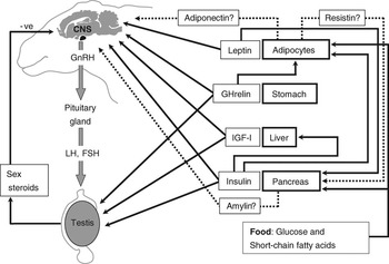
The brain and the gonads have always been seen as the primary targets for nutritional input into reproductive function but we now accept major roles for the pancreas, liver and adipose tissue (Figure 3). For adipose tissue, this transformation has been spectacular, as it has been elevated from a passive storage site to a vital endocrine organ that produces a number of signals (detailed below). Similarly, the digestive system is also now implicated in the regulatory processes through which nutrition affects reproduction as its endocrine output has been detected and identified. The digestive system also produces very direct metabolic information in the form of energy metabolites (glucose, fatty acids) and amino acids (detailed below). Effectively, every tissue involved in the regulation of metabolic status is also involved in the communication dimension of the system that regulates reproductive activity.
Schematic summary of the potential relationships among the endocrine and neural inputs into the systems that control the reproductive system and mediate the responses to change in metabolic status. For clarity, we have omitted some hormonal systems (growth hormone (GH), prolactin and oxytocin) that are also involved in the control of the metabolic status but do not appear to exert any direct action on gonadotrophin-releasing hormone (GnRH) secretion.

Dimension 3 – the communication dimension
The systems that regulate the reproductive axis need to be able to respond accurately to changes in metabolic status. This is accomplished through communication processes that are endocrine, neural and nutrient-based.
The GnRH neuroendocrine system – the fundamental controller of reproduction: the secretion of GnRH by the hypothalamus, and consequently luteinizing hormone (LH) and follicle-stimulating hormone (FSH) from the pituitary gland, are central to the reproductive process (review: Martin, Reference Martin1984). There is a threshold frequency of GnRH pulses above which males will produce sperm and females will ovulate so, effectively, the ‘decision’ of an animal to reproduce or not is implemented through pathways, as yet not described, that control the production of GnRH. The GnRH neuroendocrine system is also the final common pathway via which gonadal activity is usually influenced by external factors, including socio-sexual signals and photoperiodic cues, as well as metabolic status (Figure 2; reviews: Blache et al., Reference Blache, Chagas, Blackberry, Vercoe and Martin2000, Reference Blache, Adam and Martin2002, Reference Blache, Zhang and Martin2003 and Reference Blache, Zhang and Martin2006). In mature male sheep, an acute increase in the intake of energy and protein induces, first, an increase in the frequency of pulses of GnRH and LH, and then an increase in the tonic secretion of FSH; an acute reduction of feed intake has the opposite effects (Martin et al., Reference Martin, Tjondronegoro and Blackberry1994a). That said, it is important to note that tissues central to the control of reproduction, including the pituitary gland and gonads, can also respond independently to metabolic inputs, if not to the extent of switching the reproductive process on or off.
Insulin
Insulin is affected by energy balance and food intake and seems to be involved in the control of reproduction in male sheep because: (i) following an acute increase in dietary allowance, the increase in insulin secretion coincides with the increase in LH pulse frequency (Zhang et al., Reference Zhang, Blache, Blackberry and Martin2004); (ii) a high plane of nutrition leads to high concentrations of insulin in both plasma and cerebrospinal fluid (CSF) (Miller et al., Reference Miller, Blache, Boukhliq, Curlewis and Martin1998, Zhang et al., Reference Zhang, Blache, Blackberry and Martin2004 and Reference Zhang, Blache, Blackberry and Martin2005); (iii) with a restricted diet or with diabetes, a low dose of insulin infused into the third ventricle increases LH pulse frequency to values similar to those seen in well-fed animals (Miller et al., Reference Miller, Blache and Martin1995 and Reference Miller, Findlay, Morrison, Raver and Adam2002; Tanaka et al., Reference Tanaka, Nagatani, Bucholtz, Ohkura, Tsukamura, Maeda and Foster2000); (iv) insulin receptors are present in the ram hypothalamus (Blache et al., Reference Blache, Adam and Martin2002); (v) When GnRH/LH secretion is stimulated by an increase in nutrition, the response is associated with increased amounts of circulating insulin entering the brain, a decrease in hypothalamic expression of the orexigenic neuropeptides, neuropeptide-Y and agouti-related peptide, and an increase in the expression of the anorexigenic peptides pro-opiomelanocortin and cocaine- and amphetamine regulated transcript (Miller et al., Reference Miller, Harrison, Bennett, Findlay and Adam2007). A role for this group of regulatory pathways is supported by studies in other species showing that insulin receptors in the hypothalamus co-localize with all four peptides (Schwartz, Reference Schwartz2006).
Growth hormone (GH) and insulin-like growth factor-I (IGF-I)
Energy balance certainly affects plasma concentrations of GH under a wide variety of conditions but, in male sheep at least, an increase in nutrition induces a decrease in plasma GH concentrations (Miller et al., Reference Miller, Blache, Boukhliq, Curlewis and Martin1998). Circulating concentrations of IGF-I are also affected by diet, but concentrations of IGF-I in CSF are not (Miller et al., Reference Miller, Blache, Boukhliq, Curlewis and Martin1998) and we have not been able to demonstrate that IGF-I infusion into the third ventricle affects LH pulse frequency (Blache et al., Reference Blache, Chagas, Blackberry, Vercoe and Martin2000). It therefore seems likely that, in male sheep at least, IGF-I is not heavily involved in responses of the critical reproductive centres to changes in nutritional status.
Thyroid hormones
In the mature male sheep, the concentrations of thyroid hormones in plasma and CSF are not affected by the acute elevations of nutrition that increase LH pulse frequency, suggesting that they play no role in the stimulation of GnRH activity (Miller et al., Reference Miller, Blache, Boukhliq, Curlewis and Martin1998; Zhang et al., Reference Zhang, Blache, Blackberry and Martin2004 and Reference Zhang, Blache, Blackberry and Martin2005).
Adipocyte hormones
Adipose tissue was once considered as a passive energy reserve but is now seen as playing an active role in the regulation of food intake, metabolism, immunity, thermoregulation and cardiovascular function, as well as reproduction. Of the 20 or more endocrine products of adipose tissue, leptin appears to exert the most influence over the reproductive axis in ruminants (review: Chilliard et al., Reference Chilliard, Delavaud and Bonnet2005), although future research might reveal key roles for the others. Both short- and long-term changes in metabolic status alter the expression and release of leptin and the sensitivity of gonadal and brain tissues to it, and numerous experiments in female and male sheep have shown that leptin can affect the reproductive neuroendocrine control system (review: Adam et al., Reference Adam, Archer and Miller2003; Chilliard et al., Reference Chilliard, Delavaud and Bonnet2005). However, the consensus seems to be that leptin plays permissive rather than triggering roles.
Two other adipose hormones, adiponectin and resistin, have yet to receive the same interest as leptin but perhaps should do so because they are exclusively produced by adipose tissue. In contrast to leptin, adiponectin stimulates energy expenditure without any effect on feed intake in rats when it is infused into the cerebral ventricle (Ahima, Reference Ahima2005). In male rat pituitary cells in culture, adiponectin reduces the expression of GnRH receptor and decreases the secretion of LH (Malagon et al., Reference Malagon, Rodriguez-Pacheco, Martinez-Fuentes, Tovar, Pinilla, Tena-Sempere, Dieguez and Castano2006). Resistin has been named as such it seems to mediate insulin resistance (Steppan et al., Reference Steppan, Bailey, Bhat, Brown, Banerjee, Wright, Patel, Ahima and Lazar2001) and the gene encoding for it has been localised to the rat testis, where its expression is controlled by nutrition, suggesting a potential direct action on the gonads (Nogueiras et al., Reference Nogueiras, Gallego, Gualillo, Caminos, Garcia-Caballero, Casanueva and Dieguez2003 and Reference Nogueiras, Barreiro, Caminos, Gaytan, Suominen, Navarro, Casanueva, Aguilar, Toppari, Dieguez and Tena-Sempere2004). With respect to ruminants, the resistin gene has been localised to the mammary gland and adipocytes of the cow (Komatsu et al., Reference Komatsu, Itoh, Mikawa and Hodate2003). It has been proposed that neither resistin nor adiponectin act directly on the GnRH cells or the neuropeptide systems that control GnRH secretion. Instead, resistin inhibits insulin secretion while adiponectin stimulates insulin secretion (Figure 3). Therefore, the balance of secretion of these two adipocyte hormones controls inputs by insulin, and consequently leptin, into the neuropeptidergic regulation of both food intake and reproduction (review: see Budak et al., Reference Budak, Fernández Sánchez, Bellver, Cerveró, Simón and Pellicer2006).
Gastro-instestinal hormones
A most interesting potential candidate in the regulation of reproduction by metabolic status is ghrelin, a gut hormone that is the endogenous ligand of the growth hormone secretagogue (GHS) receptor. In rodents, the distribution of GHS receptors overlaps that of GnRH in the arcuate nucleus (St-Pierre et al., Reference St-Pierre, Wang and Tache2003) and ghrelin decreases the secretion of LH (Fernandez-Fernandez et al., Reference Fernandez-Fernandez, Tena-Sempere, Aguilar and Pinilla2004). However, in sheep, ghrelin secretion is inhibited by an increase in feed intake and stimulated by fasting (Sugino et al., Reference Sugino, Hasegawa, Kurose, Kojima, Kangawa and Terashima2004). Interestingly, ghrelin and GHS receptors are expressed in human and rat testis (Tena-Sempere, Reference Tena-Sempere2005) and ghrelin is found in most reproductive tissues in both male and female sheep (Miller et al., Reference Miller, Harrison, Brown, Doyle, Lindsay, Adam and Lea2005). Clearly, further work is required.
Direct nutrient inputs
Many products of digestion enter the circulation and can act as signals in their own right, regulating the secretion of GnRH pulses or perhaps acting directly on the gonad to promote gamete and hormone production. In rams, for example, the volatile fatty acids, a major currency in energy transactions in ruminants, stimulate GnRH secretion (Boukhliq and Martin, Reference Boukhliq and Martin1997; Boukhliq et al., Reference Boukhliq, Martin, White, Blackberry and Murray1997). The role of glucose is less clear – we detected no GnRH response to intra-abomasal or intravenous infusions of glucose in rams (Miller et al., Reference Miller, Blache and Martin1995; Boukhliq et al., Reference Boukhliq, Miller and Martin1996 and Reference Boukhliq, Martin, White, Blackberry and Murray1997). This suggests that there are nutritional inputs, perhaps some form of ‘nutrient sensing or signalling’ that are not associated with whole body energy status but can nevertheless stimulate the hypothalamic-pituitary-gonadal axis. At gonadal level, the ovarian follicles of sheep respond strongly to direct inputs from glucose, fatty acids and a number of metabolic hormones, and there are local intra-follicular nutrient sensing and integrative pathways and mechanisms that modulate gonadotrophin-stimulated folliculogenesis and regulate ovulation rate (review: Scaramuzzi et al., Reference Scaramuzzi, Campbell, Downing, Kendall, Khalid, Munoz-Gutiérrez and Somchit2006). This concept has not been tested for the testis of the sheep.
Interactions among the elements of the communication dimension
At any given time, metabolic status depends on the status of all three compartments of energy balance – intake, storage and expenditure – and the various hormonal systems that they control or influence (Figure 3). These interactions are perhaps best exemplified by leptin secretion because it is affected by intake and expenditure as well as storage (mass of adipose tissue). In turn, leptin can stimulate the activity of three other endocrine systems that are involved in controlling the reproductive axis: (i) pancreatic insulin (Zieba et al., Reference Zieba, Amstalden, Macie, Keisler, Raver, Gertler and Williams2003); (ii) pituitary GH (Henry et al., Reference Henry, Goding, Tilbrook, Dunshea and Clarke2001) and (iii) thyroid hormones (Flier et al., Reference Flier, Harris and Hollenberg2000). In addition, leptin secretion is affected by other inputs, such as the products of digestion and absorption, and autonomic neural activity (Pénicaud et al., Reference Pénicaud, Cousin, Leloup, Lorsignol and Casteilla2000).
It is important to note that the ultimate level of integration lies within the central nervous system. The brain mechanisms involved in the sensing of metabolic status and in the connection of metabolic status to GnRH neuronal activity are poorly understood and the list of candidate neurochemical links can be bewildering. Receptors for leptin and insulin will always be a focus of attention. The orexins, despite their relevance being questioned (Blache et al., Reference Blache, Zhang and Martin2003) continue to be of interest because the expression of orexin receptor 2 is dramatically inhibited by high intakes of energy and protein (Blache et al., Reference Blache, Zhang and Martin2006). We need to test whether this change in sensitivity to orexins is linked to a change in reproductive activity or a change in food intake. At this stage, it seems unlikely that μ-receptor opioidergic systems are involved (Celi et al., Reference Celi, Miller, Blache and Martin2009) but, as outlined above, the regulatory system does include the neuropeptides that are involved in the control of food intake (Miller et al., Reference Miller, Harrison, Bennett, Findlay and Adam2007).
More recently, the neuropeptide, kisspeptin, has drawn a lot of attention. In female sheep, it has been shown to stimulate GnRH and FSH secretion, to respond to sex steroids, and to play a role in the seasonality of ovulation (Messager et al., Reference Messager, Chatzidaki, Ma, Hendrick, Zahn, Dixon, Thresher, Malinge, Lomet, Carlton, Colledge, Caraty and Aparicio2005; Smith et al., Reference Smith, Clay, Caraty and Clarke2007 and Reference Smith, Coolen, Kriegsfeld, Sari, Jaafarzadehshirazi, Maltby, Bateman, Goodman, Tilbrook, Ubuka, Bentley, Clarke and Lehman2008). Importantly, in male rats, the kisspeptin receptor is found in over 75% of GnRH neurons (Irwig et al., Reference Irwig, Fraley, Smith, Acohido, Popa, Cunningham, Gottsch, Clifton and Steiner2004). Interestingly, the ligand acts through a membrane receptor so it could mediate the rapid responses of the GnRH system to acute changes in metabolic status. Kisspeptin could play an important integrative role because research in rodents has suggested that Kiss1 neurons could be the primary transducers of internal and environmental cues that regulate the neuroendocrine reproductive axis (review: Popa et al., Reference Popa, Clifton and Steiner2008). The interactions among peripheral signals (insulin, leptin, ghrelin, adiponectin and resistin) and the neuroendocrine systems (kisspeptin, neuropeptides involved in the control of food intake) need to be revealed if we are to understand the integrative processes that lead the brain to the ‘decision to reproduce’.
Dimension 4 – the temporal dimension
There is a range of dynamic aspects in the reproductive responses to nutritional inputs – the effects of time per se, as seen in the effects of photoperiod and foetal programming, and the concept of ‘metabolic memory’ that allows for nutritional information to be carried forward beyond the life of the original metabolic stimulus.
In the ram, the response of the GnRH neurons to an abrupt change in nutrition is initially rapid and robust, but then fades over the next few weeks (Martin et al., Reference Martin, Tjondronegoro and Blackberry1994a; Zhang et al., Reference Zhang, Blache, Blackberry and Martin2004). In contrast, the long-term effect of nutrition on the ram testis, measured on a scale of several weeks, seems to be independent of changes in the primary, GnRH-based, control system (Hötzel et al., Reference Hötzel, Walkden-Brown, Blackberry and Martin1995). On an annual timescale, the role of nutritional inputs, as well as the types of response to those inputs, can vary substantially, especially in genotypes that experience seasonal changes in appetite (Rhind et al., Reference Rhind, Archer and Adam2002). Suggested inputs into the reproductive control systems that might implement these strategies include actions of leptin or ghrelin in testis, brain expression of neuropeptides affected by photoperiod, innervation of adipocytes by the sympathetic and parasympathetic systems, and autonomic nervous projections from the adipocytes to the brain.
Previous metabolic status influences the reproductive response to an increase in energy availability. With respect to adipose stores, mature rams in low body condition, but not rams in high body condition, show a robust and repeatable increase in LH pulse frequency in response to an increase in intake. In low body condition rams, the leptin response is also blunted but the response to insulin is not (Zhang et al., Reference Zhang, Blache, Blackberry and Martin2005). These observations suggest that (i) neither insulin nor leptin are necessary for inducing an increase in GnRH pulse frequency in response to an increase in food intake and (ii) leptin secretion does not always respond to an influx of nutrients. Together with studies in dairy cattle (review: Blache et al., Reference Blache, Chagas and Martin2007), these observations are consistent with the notion of a ‘metabolic memory’ that modulates the stimulatory effect of nutrient intake according to the level of either energy reserves or energy expenditure. Recently, the processes that might underpin ‘metabolic memory’ have been investigated in sheep and cattle and the data suggest that leptin and insulin are central to the concept (Chilliard et al., Reference Chilliard, Delavaud and Bonnet2005). In addition, growth factors, such as insulin and growth hormones, also affect leptin secretion in whole animal studies and in isolated adipocytes (Chilliard et al., Reference Chilliard, Delavaud and Bonnet2005), and could therefore also be part of ‘metabolic memory’. Other endocrine factors secreted by adipose tissue or by the digestive system might also be involved: for example, several metabolic effects have been described for adiponectin in rodents, such as increased insulin resistance and fatty acid oxidation, and reduced glucose output by the liver (review: Diez and Iglesias, Reference Diez and Iglesias2003). These interactions between endocrine systems and nutrient supply may effectively act as a sort of peripheral integratory mechanism that complements the brain’s integratory mechanisms, with the combination being responsible for ‘metabolic memory’.
Practical application
Around the world, there is an increasing demand for animal products that are ‘clean, green and ethical’ (CGE). ‘Clean’ involves minimising the use of drugs, chemicals and hormones; ‘green’ involves minimising the impact of the industry on the environment, including the production of greenhouse gases by ruminants; ‘ethical’ has an obvious focus on animal welfare, but ethical judgement needs to be applied to all practices in the rest of the supply chain, not just the farmers. CGE principles are relevant to all forms of animal production, from low-input extensive grazing systems to high-input intensive systems involving confinement of the animals.
The nutrition-reproduction relationship is encapsulated in the CGE principle through ‘focus feeding’, a cost-effective way to feed energy at critical points in the reproductive process (Martin et al., Reference Martin, Milton, Davidson, Banchero Hunzicker, Lindsay and Blache2004). For example, producers can use focus feeding to boost the sperm production before mating, maximise potential litter size by increasing the ovulation rate and maximise postnatal survival and development, and minimise non-productive periods caused by delays in puberty or long post-partum anoestrus. Reproductive inefficiencies have always been seen as limiting profitability but there is now an extra imperative in the ‘green’ aspect of ruminant production systems – females that are not breeding are still producing methane, increasing the amount of greenhouse gases per unit of production of carcass or milk. This issue magnifies the consequences of, in particular, post-partum anoestrus in dairy cattle and offspring mortality in the sheep industry (Martin et al., Reference Martin, Durmic, Kenyon and Vercoe2009).
For each period of focus feeding, we need to consider both the composition and duration of the diet, and we could use conserved or stored feed, or we could shift the entire reproductive process so that the critical periods are better aligned with peaks and troughs in the availability of forage. As we have discussed above, in balanced diets, this generally concerns the supply of energy. Often, supplements can achieve their aims by supplying a high-intensity stimulus for a very short period (e.g. ‘flushing’ for a few days to increase ovulation rate in sheep) effectively achieving the same effect as a mild dose of exogenous hormone. We use the term ‘nutritional pharmacology’ to describe such phenomena (Martin et al., Reference Martin, Blache and Williams2008) and we have now extended the concept to include forage plants that provide beneficial secondary compounds for directly managing methane production and animal health (e.g. gastro-intenstinal parasites, acidosis), thus further reducing the use of drugs.
The male in extensive management systems
It should now be a common practice to feed supplements to males for 8 weeks before mating to ensure that their testicular size and sperm production are maximal (review: Martin et al., Reference Martin, Milton, Davidson, Banchero Hunzicker, Lindsay and Blache2004). Producers should have their males healthy and ‘fit not fat’ (Combrink and Schoeman, Reference Combrink and Schoeman1993) and should take steps to prevent them from being stressed or losing any condition during the 8-week period leading up to mating. This can be done by grazing management or by supplementation. Quality nutrition for a few males is a reliable, cheap way to guarantee maximum sperm supplies. Some issues need research: (i) Is constant good condition better than an annual 8-week supplement with respect to sperm quantity and quality? (ii) How are sperm supplies affected if animals are losing weight but still in relatively good body condition at mating? (iii) What is the ideal condition score? (iv) We need to extend the focus feeding concept to males of other breeds besides the Merino sheep and the cashmere goat.
The male in intensive management systems, including artificial insemination (AI) centres
In seasonal systems, it should still be common practice to feed supplements to males for 8 weeks before the breeding period to ensure that their testicular size and sperm production are maximal. Unfortunately, it also seems to be common practice to overfeed males in the interests of maximising fertility and body size, especially in AI centres and studs, yet there is little doubt that this, as well as a lack of physical exercise, is detrimental to their performance (Okolski, Reference Okolski1975; Combrink and Schoeman, Reference Combrink and Schoeman1993). Again, more research is needed to extend the ‘focus feeding’ concept to males of other ruminant genotypes and to ascertain if there is any advantage in maintaining males at a constant body mass throughout the year rather than using an 8-week period of pre-mating supplementation.
Conclusions
Recent years have seen many advances in our understanding of the effects of nutrition on the reproductive system of mature male ruminants. Responses in testicular size due to changes in the size of the seminiferous tubules are also associated with alterations in the efficiency of spermatogenesis, all of which lead to changes in the rate of production of spermatozoa. These effects are rarely accompanied by similar changes in the endocrine function of the testis, as measured by the production of testosterone or inhibin. In rams, changes in nutrition affect gonadotrophin secretion for only a few weeks, whereas testicular growth is affected for several months. In mature male goats during the non-breeding season, nutrition-induced testicular growth does not seem to be associated with any gonadotrophin response. It therefore seems likely that nutrition-driven testicular growth is at least partly independent of changes in gonadotrophin secretion. The energetic components of the diet, rather than the protein content, seem to be responsible for affecting gonadotrophin secretion in the ram. Variations in the expression of the nutritional responses between sexes, breeds and species probably reflect variations in the role of this environmental factor as a modulator of reproductive function.
Future research should concentrate on the intra-testicular systems mediating the effects of nutrition on the production of spermatozoa, as well as the neural and neuroendocrine processes leading to changes in gonadotrophin secretion. These pursuits will be greatly aided if we can determine how metabolic factors affect the GnRH-dependent and – independent effects on testicular growth. Finally, we need a serious research effort in the area of novel forages that can be used to improve health and also cut down greenhouse gas emissions. All this research will help us develop ‘natural systems’ for controlling and improving the productivity of farmed ruminants, at the same time greatly improving the image of ruminant industries in society and thus the marketplace.
Acknowledgements
We would like to thank Ms Margaret Blackberry for her endless toil in the laboratory. In addition, these studies, done over many years, could not have been contemplated without the generous assistance of the students and staff in Animal Science at The University of WA. Funding was supplied by the National Health and Medical Research Council, the Australian Research Council, Meat and Livestock Australia, CSIRO, the Australian Wool Corporation and the University of WA.