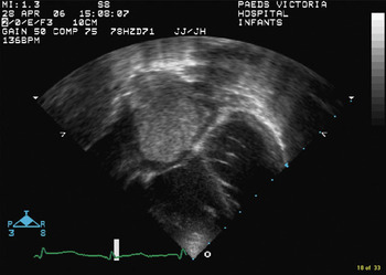
Apreviously well 25 month-old male presented with a 4 week history of low-grade fever, anorexia, pallor, decreased energy, and irritability. Based on the levels of haemoglobin, but without further investigation, the patient was started on supplementation for iron-deficient anemia. Shortly thereafter, he presented to the emergency department with respiratory distress. Clinical evaluation revealed reduced entry of air to the right chest, pallor, and bilateral, pre-tibial pitting oedema. Computed tomography and magnetic resonance imaging confirmed the presence of a pleural effusion, but also revealed a hepatic mass, and a mass occupying the right atrium. On echocardiography, this was clearly seen in the four-chamber view (Fig. 1). The inferior caval vein was completely obstructed, and the right atrium was almost completely filled by an extension of the large hepatic tumour, with minimal residual flow of blood round the mass from the superior caval vein to the tricuspid valve. Hepatic biopsy confirmed a diagnosis of hepatoblastoma. This is an uncommon tumour in children, which rarely invades the inferior caval vein or the right atrium.Reference Wang, Chen, Chuang, Yang, Chang and Wu1
Figure 1.

Chemotherapy included vincristine, 5-flurouracil, and cisplatinum, with the later addition of adriamycin in subsequent cycles. This resulted in reduction of the intracardiac tumour, eventually allowing normal flow of blood from the superior caval vein into the tricuspid valve. The inferior caval venous obstruction, while improved, did not resolve completely.