INTRODUCTION
In all living organisms, the 5S ribosomal RNA (rRNA) is an integral component of the ribosome. It forms part of the large ribosomal subunit, together with the 28S and 5·8S rRNAs and around 50 proteins. Although the 5S rRNA has been studied for more than 50 years, its precise function in protein synthesis has not been totally elucidated. However, recent data indicate that the 5S rRNA links together and coordinates all of the functional centres of the ribosome (Dinman, Reference Dinman2005). 5S rRNA also regulates the signalling pathways that couple cell proliferation with ribosome production (Sloan et al. Reference Sloan, Bohnsack and Watkins2013), and may function as a wrench-like RNA chaperone before completely integrating into the native 60S subunit as a structural entity (Leidig et al. Reference Leidig, Thoms, Holdermann, Bradatsch, Berninghausen, Bange, Sinning, Hurt and Beckmann2014).
Eukaryotic organisms usually contain from 100 to 1000 copies of the 5S rRNA genes (Torres-Machorro et al. Reference Torres-Machorro, Hernandez, Cevallos and Lopez-Villasenor2010). However, Plasmodium falciparum possesses only three copies, whereas the ciliated protozoan Euplotes eurystomus contains about a million copies (Roberson et al. Reference Roberson, Wolffe, Hauser and Olins1989). In most cases, 5S rRNA genes are organized as tandem head-to-tail arrays in one or several loci, but 5S rRNA genes can also be randomly scattered in the genome. RNA polymerase III (Pol III) is responsible for the synthesis of 5S rRNA, while RNA polymerase I (Pol I) produces the 18S, 5·8S and 28S rRNA molecules. 5S rRNA genes possess a type 1 intragenic promoter, which in Xenopus laevis and other organisms consists of a box A, an intermediate element (IE) and a box C. These three elements constitute the internal control region (ICR) (Schramm and Hernandez, Reference Schramm and Hernandez2002). The ICR is recognized by transcription factor TFIIIA, a protein that contains nine zinc fingers. The TFIIIA–DNA complex recruits TFIIIC, which in turn promotes the binding of TFIIIB and Pol III (Acker et al. Reference Acker, Conesa and Lefebvre2013; Moir and Willis, Reference Moir and Willis2013). Transcription of the 5S rRNA gene terminates within a T tract located downstream of the coding region. Consequently, the 3′ end of the transcript contains several U residues that have to be removed in order to generate a mature and functional 5S rRNA (van et al. Reference van, Lennertz and Parker2000; van Spaendonk et al. Reference van Spaendonk, Ramesar, van, Eling, Beetsma, van Gemert, Hooghof, Janse and Waters2001). As it is transcribed, the 5S rRNA acquires a distinctive secondary structure that is highly conserved throughout nature. Immediately after transcription, the La protein interacts transiently with the U residues present in the 3′ end of the 5S rRNA, to protect it from exonuclease digestion (Wolin and Cedervall, Reference Wolin and Cedervall2002). Then, ribosomal proteins L5 and L11 associate with the 5S rRNA to form the 5S ribonucleoprotein (RNP), which migrates to the nucleolus to participate in large ribosomal subunit assembly. In Xenopus oocytes, 5S rRNA is stored in the cytoplasm as 7S RNPs that contain TFIIIA, or as 42S RNPs that contain tRNAs and the thesaurin proteins (Viel et al. Reference Viel, le, Philippe, Morales, Mazabraud and Denis1991).
Little is known about 5S rRNA and ribosome biogenesis in the early-diverging eukaryote Leishmania major, the causative agent of cutaneous leishmaniasis in the Old World. The genome of this trypanosomatid parasite contains only 11 copies of the 5S rRNA gene, which are interspersed and associated with other Pol III-transcribed genes (Ivens et al. Reference Ivens, Peacock, Worthey, Murphy, Aggarwal, Berriman, Sisk, Rajandream, Adlem, Aert, Anupama, Apostolou, Attipoe, Bason, Bauser, Beck, Beverley, Bianchettin, Borzym, Bothe, Bruschi, Collins, Cadag, Ciarloni, Clayton, Coulson, Cronin, Cruz, Davies and De2005). Interestingly, out of the three transcription factors required for 5S rRNA transcription, only TFIIIB has been identified in Leishmania and other trypanosomatids (Ivens et al. Reference Ivens, Peacock, Worthey, Murphy, Aggarwal, Berriman, Sisk, Rajandream, Adlem, Aert, Anupama, Apostolou, Attipoe, Bason, Bauser, Beck, Beverley, Bianchettin, Borzym, Bothe, Bruschi, Collins, Cadag, Ciarloni, Clayton, Coulson, Cronin, Cruz, Davies and De2005; Schimanski et al. Reference Schimanski, Nguyen and Gunzl2005a ; Velez-Ramirez et al. Reference Velez-Ramirez, Florencio-Martinez, Romero-Meza, Rojas-Sanchez, Moreno-Campos, Arroyo, Ortega-Lopez, Manning-Cela and Martinez-Calvillo2015). Here we report that the number and genomic organization of 5S rRNA genes are conserved among different Leishmania species. In contrast to most organisms, in L. major 5S rRNA genes are disseminated all over the nucleus and do not seem to be associated with the nucleolus. A similar distribution of 5S rRNA genes was observed in related parasite Trypanosoma cruzi. By contrast, 5S rRNA transcripts in L. major were localized within the nucleolus, and dispersed in the cytoplasm.
MATERIALS AND METHODS
Bioinformatic analysis
Information for sequence analysis and synteny maps of the different species of Leishmania (L. major, L. mexicana, L. braziliensis, L. infantum and L. tarentolae) was obtained from the TriTrypDB databases (http://tritrypdb.org/tritrypdb/) (Release 26). The basic local alignment search tool (BLAST) was used to identify non-annotated 5S rRNA genes and pseudogenes. Sequence comparisons were performed using the DNAman program (version 7). To locate or verify the presence and identity of tRNA genes, the tRNAscan-SE program was used (http://lowelab.ucsc.edu/tRNAscan-SE/). The consensus secondary structure of 5S rRNA was predicted with the RNAalifold web server (http://rna.tbi.univie.ac.at/cgi-bin/RNAalifold.cgi).
Northern blot analysis
Northern blot experiments were performed as previously described (Rojas-Sánchez et al. Reference Rojas-Sánchez, Figueroa-Angulo, Moreno-Campos, Florencio-Martínez, Manning-Cela and Martínez-Calvillo2016). The 5S rRNA probe corresponds to oligonucleotide 5S rRNA-R1 (5′-GAGTACGGCACTCAGGGTT), and the antisense probe corresponds to oligonucleotide 5SrRNA-F1 (5′-GAGTACGACCACACTTGAGTG).
Plasmid constructs
Three DNA fragments from L. major were amplified by polymerase chain reaction (PCR) and cloned into pGEM-T Easy vector (Promega, Fitchburg, USA). For plasmid p5SrRNA, a 118 bp fragment (from +1 to +118 in relation to the transcription start site of the 5SrRNA genes) was amplified with oligonucleotides 5SrRNA-F1 (5′-GAGTACGACCACACTTGAGTG) and 5SrRNA-R1 (5′-GAGTACGGCACTCAGGGTT). For the p5SrRNA344 vector, a 344 bp fragment (from −31 to + 313 in relation to the transcription start site of the LmjF15·5SrRNA.01 gene) was amplified with oligonucleotides Lm15-rRNA5S 5′ (5′- GAAAGCATCTCTGTGGGTTCGA) and Lm15-rRNA5S 3′ (5′- CCCGGGGTCCTGCAAATG). For vector pRegionE, a 120 bp fragment from LmjF.06·0600 (that encodes a hypothetical protein) was amplified with oligonucleotides Lm15-rRNA5S 5′ (5′-GAAAGCATCTCTGTGGGTTCGA) and Lm15-rRNA5S 3′ (5′-CCCGGGGTCCTGCAAATG). The PTP tag consists of Protein A (ProtA) and Protein C (ProtC) epitopes separated by a tobacco etch virus protease cleavage site (Schimanski et al. Reference Schimanski, Nguyen and Gunzl2005b ). To obtain the pB6-PTP vector, the calmodulin-binding site (CBS) present in pB6-TAP (Martinez-Calvillo et al. Reference Martinez-Calvillo, Saxena, Green, Leland and Myler2007) was replaced with the ProtC epitope. To do so, pB6-TAP was digested with XbaI and NheI and the CBS was eliminated by agarose gel electrophoresis. The ProtC sequence was obtained by annealing primers ProtC-sense (5′-CTAGAGGTTCCGACGGCGAAGATCAGGTGGATCCTCGTCTTATTGATGGGAAATATGATATTCCAACTACTG) and ProtC-antisense (5′-CTAGCAGTAGTTGGAATATCATATTTCCCATCAATAAGACGAGGATCCACCTGATCTTCGCCGTCGGAACCT), which contain XbaI and NheI restriction sites at the ends. The ProtC fragment was then cloned into the pB6-TAP backbone. To obtain vector pElp3b-PTP, the RPB6 gene present in pB6-PTP was replaced with the L. major Elp3b gene (LmjF.23·1350). To prepare the pB6-PTP backbone, the vector was digested with XmaI and XbaI and the RPB6 gene fragment was eliminated by agarose gel electrophoresis. The Elp3b gene was PCR-amplified from genomic DNA with oligonucleotides ELP3-AgeI-5′ (5′-ATACCGGTATGTCCGACCACGCCGACAT) and ELP3-XbaI-3′ (5′-ATTCTAGAGGGTGTGTTCTTGTGGAACC). The PCR product was cloned into pGEM-T Easy vector (Promega, Fitchburg, USA), digested with AgeI and XbaI and ligated into the pB6-PTP backbone. To obtain plasmid pCold-LmNop56, the entire Nop56 gene from L. major (LmNop56) was amplified with primers LmNop56-KpnI-F (5′-GGTACCATGTCGAGAACGCTGTACA) and LmNop56-XbaI-R (5′-TCTAGACCCGGACTCAGAGGCGGC), cloned into pGEM-T Easy and then subcloned into the KpnI and XbaI restriction sites of the pCold1 expression vector (Takara Bio Inc., Shiga, Japan). All constructs were verified by sequencing.
Cell culture and transfection
Promastigotes from L. major MHOM/IL/81/Friedlin (LSB-132·1) were grown in BM medium (1 × M199 medium pH 7·2 containing 10% heat-inactivated fetal bovine serum, 9·5 g L−1 brain heart infusion broth (Sigma, St. Louis, USA), 40 mm 4-(2-hydroxyethyl)-1-piperazineethanesulfonic acid (HEPES), 0·01 mg mL−1 haemin, 0·0002% biotin, 100 IU mL−1 penicillin, 100 µg mL−1 streptomycin and 1 × L-glutamine) at 26 °C and harvested in the mid-log phase. Epimastigotes of T. cruzi CL Brener were maintained in LIT (liver infusion-tryptose) medium containing 10% heat-inactivated fetal bovine serum, 50 IU mL−1 penicillin, 50 µg mL−1 streptomycin and 0·025 mg mL−1 haemin at 28 °C. To obtain an L. major cell line that expresses a PTP-tagged version of the nucleolar protein Elp3b, wild-type L. major cells were transfected by electroporation with the pElp3b-PTP vector. Electroporation and cell plating were performed as previously described (Beverley and Clayton, Reference Beverley and Clayton1993). Briefly, 4 × 10⁷ promastigotes in 0·4 mL electroporation buffer (25 mm HEPES, 120 mm KCl, 0·15 mm CaCl2, 5 mm MgCl2, 10 mm KH2PO4, 10 mm K2HPO4, 2 mm EDTA, pH 7·6) were transfected with 10 µg of pElp3b-PTP by electroporation at 1600 V, 50 µF and 25 Ω (BTX Electro Square Porator ECM 830, Holliston, USA). Cells were grown for 24 h before spreading on plates containing 0·7% Seaplaque GTG agarose (FMC Bioproducts) in BM medium with 50 µg mL−1 G418. Some of the isolated colonies were selected for further analysis.
5' Rapid Amplification of cDNA Ends (5'-RACE) analysis
In order to locate the transcription start site of the L. major 5S rRNA genes, 5′-RACE experiments were performed with 4 µg of total RNA with a kit from Life Technologies, Inc. The first strand cDNA was synthesized with primer 5SrRNA-R1 (5′- GAGTACGGCACTCAGGGTT), and the PCR amplifications were performed with the nested primer 5SrRNA-R2 (5′-CCCGAGTCATCACTGACCTCAG) and nested Abridged Anchor Primer, AAP (5′-GGCCACGCGTCGACTAGTACGGGIIGGGIIGGGIIG). The nested PCR products were cloned into the pGEM-T Easy vector (Promega, Fitchburg, USA) and sequenced.
RT–PCR analysis
Transcription termination sites for 5S rRNA genes were mapped by poly(A) tailing of total RNA. For this purpose, 4 µg of total RNA were mixed with 1 µL of 25 mm ATP, 2 µL of 5 × poly (A) polymerase reaction buffer (USB, Cleveland, USA) and 1200 units of yeast poly (A) polymerase (USB, Cleveland, USA) in a final volume of 20 µL. The mixture was incubated for 20 min at 37 °C, and the reaction was terminated by heating at 65 °C for 10 min. The cDNA was prepared with oligonucleotide Nested(dT) (5′-CCTCTGAAGGTTCACGGATCCACATCTAGATTTTTTTTTTTTTTTTTTVN). The first PCR amplification was done with primers 5SrRNA-F1 and primer B1 (5′-CCTCTGAAGGTTCACGGAT), and the second PCR with primers 5SrRNA-F2 (5′-TGAAAACACCATATCCCGT) and primer B2 (5′-CACGGATCCACATCTAGAT). The nested PCR products were cloned into the pGEM-T Easy vector (Promega, Fitchburg, USA) and sequenced.
DNA-fluorescent in situ hybridization (FISH) combined with indirect immunofluorescence
For the experiments with L. major, a cellular line that expresses the nucleolar recombinant protein Elp3b-PTP was used. For the DNA-FISH assay, 1 × 10⁶ mid-log cells were harvested, washed twice with phosphate buffer solution (PBS) and spread into a poly-L-lysine coated glass slide. Cells were fixed with 4% paraformaldehyde for 30 min at 4 °C, permeabilized with 0·1% Triton X-100 for 5 min at room temperature, and washed with PBS. The DNA probe was denatured in hybridization buffer (50% deionized formamide, 300 mm NaCl, 20 mm NaH2PO4, 2 mm EDTA, 10% dextran sulphate, 250 µg mL−1 herring salmon sperm DNA) at 95 °C for 10 min. Finally, sealed in a plastic frame, the probe was incubated for 10 min at 80 °C, and then for 16 h at 37 °C. The seal was removed and the slides were first washed with 50% formamide and 2× saline sodium citrate (SSC) buffer for 30 min at 37 °C; and then with 1×, 2× and 3× SSC buffer for 10 min at 37 °C each. SSC buffer 20× contains 3 m NaCl in 0·3 m sodium citrate (pH 7·0). The slides were incubated with streptavidin Alexa Fluor 568 conjugate (1:1500 dilution, Life Technologies, Carlsbad, CA, USA) in blocking buffer (150 mm NaCl, 100 mm maleic acid, 0·1% Tween-20) for 30 min. Finally, two washes with PBS and 0·05% Tween-20 for 5 min and one wash with PBS were made prior the mounting with Vectashield with 4′, 6-diamidino-2-phenylindole (DAPI) solution (Vector Laboratories, Burlingame, CA). DNase or RNase control treatments were done after permeabilization using 6 U of DNase I (RNase-free) (Thermo Scientific, Waltham, USA) for 1 h at 37 °C; or 20 µg RNase A (Promega, Fitchburg, USA) for 30 min at 37 °C. The 344-bp probe (from −31 to + 313 in relation to the transcription start site of the LmjF15·5SrRNA.01 gene) was amplified by PCR from the p5SrRNA344 plasmid with oligonucleotides Lm15-rRNA5S 5′ and Lm15-rRNA5S 3′ and was labelled with Biotin-High Prime (Roche). To continue with the indirect immunofluorescence, after the final wash with PBS, cells were fixed with 4% paraformaldehyde for 30 min at 4 °C, washed twice with PBS and permeabilized with 0·1% nonidet P-40. Cells were blocked with 2% bovine serum albumin (BSA) for 30 min and incubated with rabbit anti-Protein C polyclonal antibody (Delta Biolabs, Muraoka Drive Gilroy, CA, USA) diluted at 1:25 with blocking solution for 1 h at room temperature, and washed with PBS. Next, cells were incubated with a secondary goat anti-rabbit antibody conjugated with Alexa Fluor 488 (Life Technologies, Carlsbad, CA, USA) diluted at 1:300 with blocking solution at room temperature for 1 h, and washed with PBS for 10 min. Finally, cells were mounted with Vectashield-DAPI solution (Vector Laboratories, Burlingame, CA). Images were obtained using a Carl Zeiss AX10 microscope and analysed with the ZEN 2012 software (Blue edition). The experiments with T. cruzi were performed as described above, using wild-type epimastigotes, the LmNop56 polyclonal antibody (diluted at 1:100) and goat anti-mouse antibody conjugated with Alexa Fluor 488 (Life Technologies, Carlsbad, CA, USA) (diluted at 1:300).
RNA-FISH combined with indirect immunofluorescence
An antisense RNA probe, complementary to the 5S rRNA, was generated by in vitro transcription using the MEGAshortscript T7 kit (Ambion) with biotin-11-UTP (Ambion) following the manufacturer's instructions. The template DNA was a PCR product obtained from vector p5SrRNA with oligonucleotide 5SrRNA-F1 and a T7-promoter primer. Promastigotes from the L. major cellular line that expresses the recombinant protein Elp3b-PTP were harvested, spread in glass slides, permeabilized and washed as indicated in the ‘DNA-FISH’ section. The RNA probe (4 ng) was denatured at 70 °C for 5 min in hybridization buffer with 40 U of RiboLock RNase Inhibitor (Thermo Scientific, Waltham, USA). Slides were sealed in plastic frames and hybridized for 16 h at 40 °C. The slides were washed with 50% formamide and 2 × SSC for 15 min at 42 °C, and were incubated with streptavidin Alexa Fluor 568 conjugate (1:1000 dilution, Life Technologies, Carlsbad, CA, USA) in PBS for 1 h at room temperature. Finally, the slides were washed in 2 × SCC for 10 min and were mounted in Vectashield-DAPI solution (Vector Laboratories, Burlingame, CA). DNase and RNase control treatments were also performed after permeabilization. In some experiments, indirect immunofluorescence was performed before the RNA-FISH assay. In such cases, cells were harvested, spread in glass slides, permeabilized and washed as indicated in the ‘DNA-FISH’ section. Cells were then incubated with rabbit anti-Protein C polyclonal antibody (Delta Biolabs, Muraoka Drive Gilroy, CA, USA) diluted at 1:20 in 0·5% BSA for 30 min at room temperature and washed with PBS. Cells were then incubated with a secondary goat anti-rabbit antibody conjugated with Alexa Fluor 488 (Life Technologies, Carlsbad, CA, USA) diluted at 1:300 with blocking solution at room temperature for 30 min washed twice with PBS to continue with the hybridization with the RNA probe.
Production of LmNOP56 polyclonal antibody
Escherichia coli BL21 (DE3) competent cells were transformed with the pCold-LmNop56 construct. Expression of LmNop56 recombinant protein (LmNop56r) was induced with 1 mm IPTG (isopropyl β-D-1-thiogalactopyranoside) at 37 °C for 18 h. The LmNop56r protein was purified by affinity chromatography with Ni-Sepharose 6 Fast Flow matrix (GE Healthcare, Little Chalfont, England) following the manufacturer's instructions. Six-week-old male BALB-C mice were immunized intravenously with purified LmNop56r (100 µg) mixed with TiterMax Gold adjuvant (Sigma, St. Louis, USA) at a 1:1 (ratio). Pre-immune normal mouse serum was collected before inoculation. Serum was collected 6 weeks after antigen immunization. The specificity of the anti-LmNop56 polyclonal antibody was confirmed by the Western blot analysis against LmNop56r and parasite protein extracts.
RESULTS
Sequence and genomic analysis of 5S rRNA genes in L. major
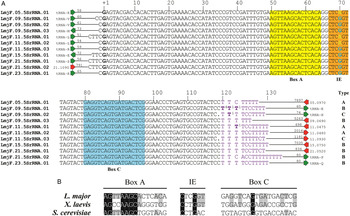
The L. major genome possesses only 11 copies of the 5S rRNA gene, which are interspersed on six different chromosomes (5, 9, 11, 15, 21 and 23) and associated with other Pol III-transcribed genes (Ivens et al. Reference Ivens, Peacock, Worthey, Murphy, Aggarwal, Berriman, Sisk, Rajandream, Adlem, Aert, Anupama, Apostolou, Attipoe, Bason, Bauser, Beck, Beverley, Bianchettin, Borzym, Bothe, Bruschi, Collins, Cadag, Ciarloni, Clayton, Coulson, Cronin, Cruz, Davies and De2005). At their 5′ end, 10 of the 5S rRNA genes are flanked by a tRNA gene located at a short distance (between 51 and 73 bp) (Fig. 1A). The remaining 5S rRNA gene (LmjF.21·5SrRNA.02) contains a protein-coding gene at its 5′ end. At their 3′ end, most 5S rRNA genes are flanked by a protein-coding gene, which is separated from the 5S rRNA gene by long distances ranging from 698 to 7897 bp (Fig. 1A).
Sequence analysis of 5S rRNA genes in L. major. (A) Sequence alignment of the 11 5S rRNA genes present in L. major, as annotated in the TriTrypDB databases. Flanking tRNA genes (green arrows) and protein-coding genes (red arrows) are indicated. The numbers specify the distance (in bp) between the 5S rRNA gene and flanking genes. The G shown in bold type (position +1) corresponds to the transcription start site mapped by 5′-RACE analysis. At the 3′ end, the clusters of Ts are shown in purple. T residues in bold type denote the transcription termination sites located by RT–PCR, specifying the number of clones found at each position. The locations of putative box A, intermediate element (IE) and box C are shown. Genes were classified as types A, B and C, as indicated. (B) Sequence alignment of putative internal control elements (box A, IE and box C) in L. major, X. laevis and S. cerevisiae. Nucleotides conserved in the three species are denoted by black shading, while nucleotides conserved between L. major and X. laevis or S. cerevisiae are indicated by grey shading.

According to the TriTrypDB databases, the length of the eleven L. major 5S rRNA genes fluctuates from 119 to 123 bp. A sequence alignment showed that these size variations are due to differences in the assigned first nucleotide of the genes (Fig. 1A). Also, two of the genes lack a C in the 3′ end of the gene. The rest of the sequence is identical, with the exception of base +70, which can be either a G or an A (Fig. 1A). In order to determine if there is actually variation in the 5′ end of the 5S rRNA genes, a 5′-RACE experiment was carried out. All the clones analysed started at the G that is labelled as +1 in Fig. 1A, which strongly suggests that transcription of the 5S rRNA genes initiates in that nucleotide. Transcription of 5S rRNA genes in T. cruzi and Trypanosoma brucei is reported to initiate at a G located in the same position (Lenardo et al. Reference Lenardo, Dorfman, Reddy and Donelson1985; Hernandez-Rivas et al. Reference Hernandez-Rivas, Martinez-Calvillo, Romero and Hernandez1992). Thus, the eleven L. major 5S rRNA genes can be classified into three groups: type A genes (119 bp long) contain an A at position 70 and two Cs in the 3′ end; type B genes (119 bp long) possess a G at base 70 and two Cs in the 3′ end; and type C genes (118 bp long) contain a G at position 70 and one C in the 3′ end (Fig. 1A).
A cluster of several T residues acts as a signal to terminate Pol III transcription (Moir and Willis, Reference Moir and Willis2013). Sequence analysis revealed that all the 5S rRNA genes in L. major contain a run of 6–11 Ts at the 3′ end (Fig. 1A). One gene (LmjF.09·5SrRNA.03) has a continuous cluster of six T residues, while the rest of the genes contain discontinuous T tracts that are interrupted by one or several Cs. A common feature in Pol III-transcribed genes is the presence of a second cluster of Ts that acts as a potential ‘backup’ termination signal (Braglia et al. Reference Braglia, Percudani and Dieci2005). In L. major, only one of the 5S rRNA genes (LmjF.21·5SrRNA.02) possesses a backup T-run (Figs 1A and S1).
To determine the transcription termination sites on 5S rRNA genes in L. major, RT–PCR was performed. As expected, transcription of the 5S rRNA genes ends within the cluster of Ts located downstream of the genes (Fig. 1A). Several clones ended at the C located immediately before the T cluster, most likely representing the mature 3′ end of the L. major 5S rRNA. In T. brucei, the mature 3′ terminus of the 5S rRNA was mapped in a similar position (Hitchcock et al. Reference Hitchcock, Zeiner, Sturm and Campbell2004). Interestingly, we identified transcripts from the three types of 5S rRNA genes present in L. major, which showed that at least one copy of types A, B and C genes is transcribed in exponentially growing promastigotes.
The ICR in 5S rRNA genes in X. laevis and Saccharomyces cerevisiae consists of a box A, an IE and a box C (White, Reference White2011; Dieci et al. Reference Dieci, Bosio, Fermi and Ferrari2013). Sequence analysis of the 5S rRNA genes in L. major allowed us to identify putative homologues of these three promoter elements (Fig. 1B). Leishmania major box A is very similar to S. cerevisiae box A, as nine out of 15 nucleotides are conserved. In contrast, the IE from L. major is more similar to the IE from X. laevis than to the one from S. cerevisiae: four out of six nucleotides are conserved between L. major and X. laevis, while two bases are conserved between L. major and S. cerevisiae. The least conserved promoter element is box C, as seven out of 18 bases are conserved between L. major and X. laevis, and only two bases are conserved between L. major and S. cerevisiae (Fig. 1B). Notably, base +70, which is the only internal nucleotide that is variable among 5S rRNA genes, lies within the IE (Fig. 1A). Thus, the occurrence of a change within the ICR in 5S rRNA genes suggests that differential regulation of Pol III transcription is feasible in L. major.
It was reported that Leishmania and T. brucei produce natural antisense RNAs complementary to 18S, 5·8S and 28S rRNA molecules. Although their function is not known, they associate with polyribosomes, and programmed cell death induces their fragmentation (Padmanabhan et al. Reference Padmanabhan, Samant, Cloutier, Simard and Papadopoulou2012). Also, chloroplasts from Arabidopsis thaliana and other plants produce an antisense 5S rRNA that inhibits 5S rRNA maturation (Sharwood et al. Reference Sharwood, Hotto, Bollenbach and Stern2011). To explore if antisense 5S rRNA transcripts are generated in L. major, Northern blot analyses with strand-specific oligonucleotides were performed. Interestingly, while an abundant 5S rRNA of the expected size (~120 bases) was observed, no antisense 5S rRNA transcripts were detected (data not shown). Thus, unlike other rRNA molecules in Leishmania and 5S rRNA in chloroplasts, stable antisense RNA complementary to 5S rRNA is not detected in L. major.
Synteny of 5S rRNA genes in Leishmania species
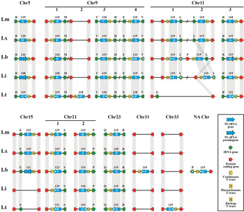
To determine the number of 5S rRNA genes in other species of Leishmania, we analysed the genome databases of L. mexicana U1103, L. braziliensis M2904, L. infantum JPCM5 and L. tarentolae Parrot-TarII (Peacock et al. Reference Peacock, Seeger, Harris, Murphy, Ruiz, Quail, Peters, Adlem, Tivey, Aslett, Kerhornou, Ivens, Fraser, Rajandream, Carver, Norbertczak, Chillingworth, Hance, Jagels, Moule, Ormond, Rutter, Squares, Whitehead, Rabbinowitsch, Arrowsmith, White, Thurston, Bringaud and Baldauf2007; Downing et al. Reference Downing, Imamura, Decuypere, Clark, Coombs, Cotton, Hilley, de, Maes, Mottram, Quail, Rijal, Sanders, Schonian, Stark, Sundar, Vanaerschot, Hertz-Fowler, Dujardin and Berriman2011; Rogers et al. Reference Rogers, Hilley, Dickens, Wilkes, Bates, Depledge, Harris, Her, Herzyk, Imamura, Otto, Sanders, Seeger, Dujardin, Berriman, Smith, Hertz-Fowler and Mottram2011; Raymond et al. Reference Raymond, Boisvert, Roy, Ritt, Legare, Isnard, Stanke, Olivier, Tremblay, Papadopoulou, Ouellette and Corbeil2012). Eleven 5S rRNA genes are anotated in L. mexicana, seven genes in L. infantum and six genes in L. braziliensis. In L. tarentolae no single 5S rRNA gene is annotated. However, BLAST searches in the genome databases allowed us to identify several 5S rRNA genes in L. tarentolae and additional genes or pseudogenes in the other species (Fig. 2).
Synteny of 5S rRNA genes in different species of Leishmania. The genomic context of 5S rRNA genes is presented for L. major (Lm), L. mexicana (Lx), L. braziliensis (Lb), L. infantum (Li) and L. tarentolae (Lt). Orthologous genes are joined by grey lines. Chromosome location is indicated on top of each individual synteny map, except for a 5S rRNA gene in Lb, whose chromosomal location has not been assigned yet (NA Chr). The number above each 5S rRNA gene (blue arrows) denotes the length of the gene (in bp). The type of 5S rRNA gene (A-S) is indicated inside each blue arrow. Putative 5S rRNA pseudogenes are shown as blue arrows bordered by dashed lines. The type of T cluster at the 3′ end of each 5S rRNA gene is indicated: continuous T tract (C), discontinuous T tract (D) and backup T tract (B). For tRNA genes (green arrows), the corresponding amino acid is indicated (R=Arg, M=Met, V=Val, H=His, E=Glu, L=Leu, S=Ser, P=Pro, G=Gly and Q=Gln). Protein-coding genes are shown as red arrows. Genes that are not annotated in the TriTrypDB databases are labelled with an asterisk (*). Figure is not to scale.

Sequence comparisons showed that 5S rRNA genes are conserved in the different Leishmania species (Fig. S1, panels A and B). As the G at position +1 is present in all the genes, we considered this base as the first nucleotide for all the genes. The last base of each 5S rRNA gene was established as the nucleotide located right before the T cluster. With these parameters, we found that the length of the genes ranged from 108 to 125 bases. However, five of the annotated genes seem to actually correspond to pseudogenes, since they lack parts of the ICR and/or present several changes all along the sequence: the gene located on Chr5 in L. infantum (108 bp), and four genes in L. braziliensis (the gene on Chr5, gene 1 on Chr9, gene 3 on Chr11 and the gene on Chr15) (Fig. S2). Thus, the number of authentic 5S rRNA genes seems to be: 11 genes in L. major and L. mexicana, 10 genes in L. tarentolae and nine genes in L. braziliensis and L. infantum.
Overall, the sequence of the 5S rRNA genes in Leishmania is highly conserved (Fig. S1, panel A). Remarkably, the most variable position is nucleotide +70, which is located within the IE. As mentioned earlier, in L. major it can be either a G or an A. In L. mexicana, L. braziliensis and L. tarentolae nucleotide +70 is always an A, while in L. infantum it is a C in five genes and an A in four genes (Fig. S1, panel A). The order of the 5S rRNA genes is conserved between L. major and L. mexicana, as they are flanked by syntenic protein-coding genes and tRNA genes (Fig. 2). L. infantum does not contain the gene located on Chr15 in the other species. The majority of the 5S rRNA genes in L. braziliensis show synteny with the other species of Leishmania. However, L. braziliensis possesses two 5S rRNA genes that are not present in any other Leishmania species: one located on Chr31 and one whose chromosomal location has not been determined yet (Fig. 2). L. tarentolae does not contain genes 1 and 2 on Chr11 and the gene on Chr15, but possesses additional 5S rRNA genes on Chr9 (gene 2) and Chr33 (Fig. 2).
The vast majority of 5S rRNA genes in the different Leishmania species contain a tRNA gene at their 5′ end. It is worth noting that several non-annotated tRNA genes were identified in these loci with the tRNAscan-SE search server (marked with an asterisk in Fig. 2). All the 5S rRNA genes in the different species of Leishmania contain a cluster of several T residues at the 3′ end (Fig. S1, panels A and B). Most genes have discontinuous T tracts that are interrupted by up to three nucleotides, which predominantly are Cs. Interestingly, four copies of the 5S rRNA genes in L. infantum have a backup T cluster.
5S rRNA genes are located in the nuclear periphery
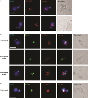
To investigate the nuclear distribution of 5S rRNA genes in promastigotes from L. major, FISH assays were performed with a biotin-labelled dsDNA probe, and incubating with streptavidin Alexa Fluor 568 conjugate. The probe clearly detected the 5S rRNA genes as several bright spots localized in the nuclear periphery or scattered throughout the nucleus (which was stained with DAPI) (Fig. 3A). In order to determine the localization of 5S rRNA genes with respect to the nucleolus, an L. major cell line that expresses a PTP-tagged version of the nucleolar protein Elp3b (Alsford and Horn, Reference Alsford and Horn2011) was obtained (see the ‘Materials and Methods’ section). Conjugated FISH and indirect immunofluorescence experiments were carried out in this cell line. These experiments showed that in most cells 5S rRNA genes are not associated with the nucleolus (Fig. 3B). In contrast, nucleolar colocalization of 18S rRNA genes and Elp3b was observed, as expected (Fig. 3C). DNase and RNase treatment controls were used to confirm the specific binding of the probe to DNA. As expected, after DNase treatment 5S rRNA-gene signal was abolished (as well as DAPI signal), but after RNase treatment the fluorescent spots were still clearly detected. Thus, these data show that the 5S rRNA genes present in L. major are mainly distributed in the nuclear periphery (Fig. 3B).
5S rRNA genes in L. major are located in the nuclear periphery. (A) DNA-FISH experiments using a 5S rRNA-gene probe (red). Nuclei (N) and kinetoplast (K) were stained with DAPI (blue). Cells from two independent experiments are shown. (B) DNA-FISH coupled with immunofluorescence using a 5S rRNA-gene probe (red), and an anti-protein C antibody to detect a PTP-tagged version of the nucleolar protein Elp3b (green). Control treatments with DNase and RNase are shown. (C) DNA-FISH coupled with immunofluorescence using an 18S rRNA-gene probe (red) and an anti-protein C antibody to detect the recombinant protein Elp3b-PTP (green). Size bars represent 2 µm.

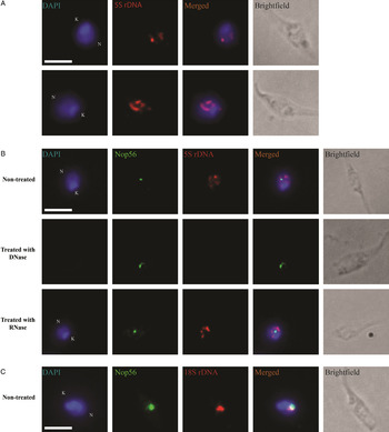
In T. cruzi, 5S rRNA genes are organized as tandem arrays in two different chromosomes (Hernandez-Rivas et al. Reference Hernandez-Rivas, Martinez-Calvillo, Romero and Hernandez1992). To determine the nuclear distribution of these genes in epimastigotes, we performed FISH experiments with a biotin-labelled dsDNA probe. Similarly to L. major, several bright spots were distributed throughout the nucleus (Fig. 4A). To analyse the localization of 5S rRNA genes with respect to the nucleolus, conjugated FISH and indirect immunofluorescence experiments were carried out with an antibody directed against the nucleolar protein 56 (Nop56). As shown in Fig. 4B, 5S rRNA genes in T. cruzi epimastigotes do not seem to associate with the nucleolus. Thus, regardless of whether 5S rRNA genes are scattered in the genome or organized as tandem arrays, they are apparently not associated with the nucleolus in trypanosomatids.
5S rRNA genes in T. cruzi are dispersed in the nucleus. (A) DNA-FISH experiments using a 5S rRNA-gene probe (red). Nuclei (N) and kinetoplast (K) were stained with DAPI (blue). Cells from two independent experiments are shown. (B) DNA-FISH coupled with immunofluorescence using a 5S rRNA-gene probe (red), and an antibody against the nucleolar protein Nop56 (green). Control treatments with DNase and RNase are shown. (C) DNA-FISH coupled with immunofluorescence using an 18S rRNA-gene probe (red), and an antibody to detect Nop56 (green). Size bars represent 2 µm.

5S rRNA is located in the nucleolus and cytoplasm
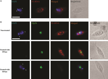
The subcellular localization of L. major 5S rRNA was determined by FISH analysis. In these experiments, a biotinylated antisense riboprobe was transcribed in vitro and coupled with fluorescent streptavidin to detect the 5S rRNA transcripts. The hybridization produced a discrete and large spot in the nucleus, as well as strong fluorescence dispersed throughout the cytoplasm (Fig. 5A). By combining FISH and immunofluorescence assays we demonstrated that the 5S rRNA and Elp3b signals colocalize, showing that the 5S rRNA transcripts are localized in the nucleolus (Fig. 5B). The observed cytoplasmic signal, which is stronger in the areas around the nucleus, most likely corresponds to 5S rRNA present in ribosomes that are distributed all over the cytoplasm. As expected, 5S rRNA signal was not affected in control FISH experiments with cells pretreated with DNase. By contrast, no signal was detected in the control treatment with RNase (Fig. 5B).
5S rRNA transcripts are located in the nucleolus and the cytoplasm. (A) RNA-FISH assay using a biotinylated antisense riboprobe (red) complementary to the 5S rRNA. Nuclei (N) and kinetoplast (K) were stained with DAPI (blue). (B) RNA-FISH coupled with immunofluorescence using an antisense 5S rRNA riboprobe (red), and an anti-protein C antibody to detect a PTP-tagged version of the nucleolar protein Elp3b (green). Control treatments with DNase and RNase are shown. Size bars represent 2 µm.

DISCUSSION
Our analysis indicates that the genus Leishmania is characterized by the presence of a small number of 5S rRNA genes, which ranges from nine genes in L. braziliensis and L. infantum to 11 genes in L. major and L. mexicana. Interestingly, in the species that lack a 5S rRNA gene there is in some cases a degenerate sequence (pseudogene) in the corresponding region of synteny (Fig. 2), as has been reported previously for protein-coding genes in Leishmania (Peacock et al. Reference Peacock, Seeger, Harris, Murphy, Ruiz, Quail, Peters, Adlem, Tivey, Aslett, Kerhornou, Ivens, Fraser, Rajandream, Carver, Norbertczak, Chillingworth, Hance, Jagels, Moule, Ormond, Rutter, Squares, Whitehead, Rabbinowitsch, Arrowsmith, White, Thurston, Bringaud and Baldauf2007; Rogers et al. Reference Rogers, Hilley, Dickens, Wilkes, Bates, Depledge, Harris, Her, Herzyk, Imamura, Otto, Sanders, Seeger, Dujardin, Berriman, Smith, Hertz-Fowler and Mottram2011). It is worth noting that the orientation of all 5S rRNA genes is conserved across Leishmania species, with the exception of gene 2 on Chr11, which is inverted in L. braziliensis and L. infantum (Fig. 2). While in Leishmania the 5S rRNA genes are dispersed as independent copies throughout the genome, in the different species of the genus Trypanosoma they are tandemly repeated, and in some species they are linked to the spliced-leader genes, transcribed by Pol II (Torres-Machorro et al. Reference Torres-Machorro, Hernandez, Cevallos and Lopez-Villasenor2010; Beauparlant and Drouin, Reference Beauparlant and Drouin2014). It was estimated that T. brucei and T. cruzi possess approximately 1500 copies of the 5S rRNA gene (Hasan et al. Reference Hasan, Turner and Cordingley1984; Hernandez-Rivas et al. Reference Hernandez-Rivas, Martinez-Calvillo, Romero and Hernandez1992). Thus, the number and genomic organization of 5S rRNA genes is highly variable between Leishmania and Trypanosoma. In order to determine the genomic context of 5S rRNA genes in other trypanosomatids, we analysed the genome databases of Crithidia fasciculata, Leptomonas seymouri and Endotrypanun monterogeii. While only one 5S rRNA gene was found in C. fasciculata, no single gene was identified in L. seymouri, which probably indicates that these genome databases are incomplete. On the other hand, 14 different 5S rRNA genes were found in E. monterogeii. Interestingly, some of these 5S rRNA genes are dispersed as single genes (such as in Leishmania), whereas other genes are organized in tandem arrays that contain up to four genes (such as in Trypanosoma) (data not shown). Analyses of genome sequences from other trypanosomatids, not available at present time, will help understand the evolution of 5S rRNA genes in this group of early-branched eukaryotes.
In L. major, 5S rRNA genes can be grouped into three types (A–C), and RT–PCR analysis showed that at least one gene of each type is transcribed in exponentially growing promastigotes (Fig. 1A). A total of 19 different types (A to S) of 5S rRNA genes are present in the five Leishmania species analysed (showing 3–6 types per species) (Figs 2 and S1). Type A is the most widespread, as it is represented by eight genes in L. mexicana, three genes in L. major and three genes in L. infantum. Type G corresponds to six genes in L. braziliensis and four genes in L. tarentolae (Fig. S1, panel C). Most of the other types are represented by a single 5S rRNA gene. Prediction of the consensus secondary structure of the 5S rRNA genes in Leishmania shows that it consists of five helices (I–V), two hairpin loops (C and D), two internal loops (B and E) and a hinge region (A), which are organized in a three-helix junction (Fig. S3), as reported in other organisms (Szymanski et al. Reference Szymanski, Zielezinski, Barciszewski, Erdmann and Karlowski2016). The variable nucleotide +70 is positioned within helix V, and it base-pairs with nucleotide +105. In most genes, an A at position +70 base-pairs with a U at position +105. However, it is interesting to note that two 5S rRNA genes in L. infantum that possess a C at nucleotide +70 contain a G at base +105, so that base complementarity is maintained. Position +12 is also variable, but it is located in loop A (Fig. S3).
Conserved internal promoter elements most likely control transcription of 5S rRNA genes in Leishmania (Fig. 1). Interestingly, the vast majority of 5S rRNA genes in these parasites are preceded by a tRNA gene located at a short distance. Thus, it is tempting to hypothesize that, in addition to the internal promoter elements, boxes A and B from the neighbour tRNA gene might participate in the transcriptional regulation of 5S rRNA, as has been reported for snRNA genes in trypanosomatids (Nakaar et al. Reference Nakaar, Dare, Hong, Ullu and Tschudi1994; Rojas-Sánchez et al. Reference Rojas-Sánchez, Figueroa-Angulo, Moreno-Campos, Florencio-Martínez, Manning-Cela and Martínez-Calvillo2016). A cluster of T residues of variable length, frequently followed by a backup T-cluster, was found on every single 5S rRNA gene in the different species of Leishmania analysed (Fig. S1). A second T stretch is also common in tRNA genes in trypanosomatids (Padilla-Mejia et al. Reference Padilla-Mejia, Florencio-Martinez, Figueroa-Angulo, Manning-Cela, Hernandez-Rivas, Myler and Martinez-Calvillo2009). In other organisms, transcripts that end within the backup T tract contain long 3′ trailer regions, whose processing to create mature RNAs generates small RNA molecules that may play functional roles. In human and other vertebrates, for instance, small RNAs derived from tRNA 3′ trailers accumulate in the cytoplasm, where they participate in the regulation of gene silencing and might be involved in other biological functions (Haussecker et al. Reference Haussecker, Huang, Lau, Parameswaran, Fire and Kay2010; Liao et al. Reference Liao, Ma, Guo, Zhang, Zhou, Shao, Chen and Qu2010). It would be interesting to determine if stable 5S rRNA and/or tRNA 3′ trailers are produced in Leishmania and if they participate in gene regulation.
Combined DNA-FISH and indirect immunofluorescence experiments showed that the 11 5S rRNA genes present in L. major are mainly located at the nuclear periphery (Fig. 3). In T. cruzi, 5S rRNA genes were observed as bright spots distributed throughout the nucleus (Fig. 4). Interestingly, most genes in L. major and T. cruzi do not seem to be located within the nucleolus or at the nucleolar periphery. A similar result was reported for 5S rRNA genes in T. brucei (Ersfeld and Gull, Reference Ersfeld and Gull1997). However, this is different from what has been found in S. cerevisiae and Dictyostelium, where 5S rRNA genes are linked to the rRNA units (that contain the 18S, 5·8S and 28S rRNA genes) and are consequently nucleolar (Hofmann et al. Reference Hofmann, Winckler, Hanenkamp, Bukenberger, Schumann, Marschalek and Dingermann1993; Thompson et al. Reference Thompson, Haeusler, Good and Engelke2003). Nevertheless, association of 5S rRNA genes and the nucleolus has been observed in several species where the 5S rRNA genes are not linked to the rRNA unit in the linear DNA (Haeusler and Engelke, Reference Haeusler and Engelke2006). For instance, in the plant Pisum sativum, which has around 5000 copies of the 5S rRNA genes organized in six tandem arrays, a clear association of at least one of the arrays with the nucleolar periphery was observed (Highett et al. Reference Highett, Beven and Shaw1993). Also, 5S rRNA genes in human cells were often located at the nucleolar periphery (Matera et al. Reference Matera, Frey, Margelot and Wolin1995). It has been hypothesized that the 5S rRNA genes that are associated with the nucleolus are most actively transcribed, producing transcripts at the nucleolus where they are needed (Highett et al. Reference Highett, Beven and Shaw1993). However, in Drosophila melanogaster, 5S rRNA genes do not seem to be located in the vicinity of the nucleolus (Hochstrasser and Sedat, Reference Hochstrasser and Sedat1987). It is worth noting that the observed distribution of 5S rRNA genes in L. major resembles the nuclear spreading reported for BRF1, a subunit of Pol III transcription factor TFIIIB, in T. brucei (Velez-Ramirez et al. Reference Velez-Ramirez, Florencio-Martinez, Romero-Meza, Rojas-Sanchez, Moreno-Campos, Arroyo, Ortega-Lopez, Manning-Cela and Martinez-Calvillo2015).
For the purpose of associating with pre-ribosomes, 5S rRNAs are expected to locate at the nucleolus at some point. In accord with this fact, combined RNA-FISH and indirect immunofluorescence assays demonstrated that the 5S rRNA colocalizes with the nucleolar protein Elp3b (Fig. 5), which shows that the 5S rRNA transcripts are localized in the nucleolus in L. major. Nevertheless, 5S rRNA was also found to be scattered in the cytoplasm, especially in the areas near the nucleus, where mature ribosomes are located. A similar nucleolar and cytoplasmic distribution has been reported for 5S rRNA and 28S rRNA in vertebrates (Matera et al. Reference Matera, Frey, Margelot and Wolin1995; Weisenberger and Scheer, Reference Weisenberger and Scheer1995), and for 18S rRNA in T. brucei (Chaves et al. Reference Chaves, Zomerdijk, Dirks-Mulder, Dirks, Raap and Borst1998). It is worth noting that there is a substantial difference between the patterns observed for the 5S rRNA and the Elp3b protein (Fig. 5). In trypanosomatids, the nucleolus has a bipartite structure, as it is composed of a dense fibrillar component (that seems to include the fibrillar centres) and a granular component (Ogbadoyi et al. Reference Ogbadoyi, Ersfeld, Robinson, Sherwin and Gull2000; López-Velázquez et al. Reference López-Velázquez, Hernández, López-Villaseñor, Reyes-Vivas, Segura-Valdez and Jiménez-García2005). While 5S rRNA is expected to locate into the granular component, where pre-ribosome assembly occurs, Elp3b would be localized in the dense fibrillar component, the sites of active Pol I transcription in trypanosomatids.
In conclusion, in the present study we have shown that the genus Leishmania presents a small number of 5S rRNA genes, whose sequence and genomic organization is conserved. Our results also demonstrate that 5S rRNA genes in L. major are mainly located at the nuclear periphery, and not associated with the nucleolus, as has been reported in most species. A similar localization was observed for the tandemly repeated 5S rRNA genes in T. cruzi. By contrast, 5S rRNA transcripts in L. major were localized within the nucleolus, and scattered throughout the cytoplasm. Thus, our results significantly increase the knowledge of 5S rRNA genes and Pol III transcription in an early-branched eukaryote.
SUPPLEMENTARY MATERIAL
The supplementary material for this article can be found at http://dx.doi.org/10.1017/S0031182016001712
ACKNOWLEDGEMENTS
We thank Javier R. Ambrosio, Luis F. Jiménez-García and Rosaura Hernández-Rivas for fruitful discussions and Ana M. Cevallos-Gaos for critical reading of the manuscript. We also thank Rossana Arroyo and Jaime Ortega López for the pCold1 vector. This work is one of the requirements to obtain the PhD degree in Posgrado en Ciencias Biológicas (UNAM) for Rodrigo Moreno-Campos, who was the recipient of doctoral fellowships from CONACyT (Fellowship 257038, CVU 288471) and CEP/UNAM (302114072).
FINANCIAL SUPPORT
This work was supported by grants 251831 from CONACyT, IN210712 and IN214715 from PAPIIT (UNAM) to S. Martínez-Calvillo and by grants 132312 and 139898 from CONACYT to R. Manning-Cela.