

Introduction
Panaeolus (Fr.) Quél. is a small agaric genus comprising approximately 77 species (Kirk, Reference Kirk, Roskov, Orrell, Nicolson, Bailly, Kirk, Bourgoin, DeWalt, Decock, De Wever, Nieukerken, van, Zarucchi and Penev2018). The family includes species with very wide distributions, because most members are associated with dung of several herbivores, especially cattle and horses (Gerhardt, Reference Gerhardt1996). The genera Anellaria P. Karst., Copelandia Bres. and Panaeolina Maire are considered distinct by some mycologists (Singer, Reference Singer1986; Young, Reference Young1989), whereas others consider the species placed in these genera to be members of Panaeolus s.l. under a broader concept (Ola’h, Reference Ola’h1969; Watling & Gregory, Reference Watling and Gregory1987). Classified by Singer (Reference Singer1986) in the family Coprinaceae Overeem & Weese, more recently the genus was suggested to belong in the Bolbitiaceae Singer (Matheny et al., Reference Matheny, Curtis, Hofstetter, Aime, Moncalvo, Ge, Yang, Slot, Ammirati, Baroni, Bougher, Hughes, Lodge, Kerrigan, Seidl, Aanen, DeNitis, Daniele, Desjardin, Kropp, Norvell, Parker, Vellinga, Vilgalys and Hibbett2006), but in the MycoBank database it is placed in the Psathyrellaceae Vilgalys, Moncalvo & Redhead. Although the systematic position of the genus remains unclear, the available data indicate that Panaeolus and its allies may be best placed in a new family, with an intermediate position between the Bolbitiaceae and Psathyrellaceae (Tóth et al., Reference Tóth, Hausknecht, Krisai-Greilhuber, Papp, Vágvölgyi and Nagy2013).
In Brazil, twelve Panaeolus names are reported, mainly from the south of the country, although only a few studies have focused on the genus and its allies (Stijve & De Meijer, Reference Stijve and De Meijer1993; Alves & Cavalcanti, Reference Alves and Cavalcanti1996). Rick (Reference Rick1961) reported nine Panaeolus taxa from Rio Grande do Sul State, but in most cases these names are misidentifications or synonyms or lack specimens (e.g. P. atro-sanguineus Henn. and P. phalarnarum Fr.). De Meijer (Reference De Meijer2006) listed the following taxa from Paraná State: Panaeolus antillarum (Fr.) Dennis, P. cinctulus (Bolton) Sacc., P. papilionaceus var. parvisporus Ew. Gerhardt and P. cf. reticulatus Overh. De Meijer (Reference De Meijer2006) also reported Panaeolus foenisecii (Fr.) J. Schröt, currently a member of the genus Paneolina, and Panaeolus cambodginiensis Oláh & R. Heim, P. cyanescens (Berk. & Broome) Sacc., and P. aff. tropicalis Oláh, which are currently members of the neurotropic genus Copelandia Bres (Singer, Reference Singer1986; Young, Reference Young1989). More recently, Copelandia mexicana Guzmán, a non-coprophilous species, was reported from the western region of Paraná State (Silva-Filho et al., Reference Silva-Filho, Seger and Cortez2018a).
Considering the limited knowledge of the mycobiota, and the agaricoid fungi in particular, for this study we collected specimens from selected areas in western Paraná State (Silva-Filho et al., Reference Silva-Filho, Coelho and Cortez2016; Silva-Filho & Cortez, Reference Silva-Filho and Cortez2017; Silva-Filho et al., Reference Silva-Filho, Seger and Cortez2018a, Reference Silva-Filho, Teixeira-Silva and Cortez2018b). Our aim is to provide information on the richness, taxonomy and distribution of mushrooms found in that region, and the results dealing with Panaeolus are presented here, including full descriptions of collected materials and a key for species reported from the state.
Materials and Methods
Specimens were collected in two localities, comprising fragments of seasonal semideciduous forest (Atlantic Forest biome) in the western region of Paraná State: Reserva Nacional do Patrimônio Nacional (RPPN) Fazenda Açu, in the Terra Roxa municipality, and São Camilo State Park, in the Palotina municipality.
Morphological analysis (macro- and microscopic) followed standard procedures for agaricoid fungi (Largent et al., Reference Largent, Johnson and Watling1977; Largent, Reference Largent1986). Colour names and codes used in the macroscopic descriptions are based on Kornerup & Wanscher (Reference Kornerup and Wanscher1978). The morphological terminology follows that used by Gerhardt (Reference Gerhardt1996). In the description of basidiospores, size is given as length × width in face view × width in side view, Q is the quotient between the length and width, Qm is the medium value of Q, and n is the number of measured basidiospores/number of analyzed basidiomata/number of collections. All microscopic structures were observed from sections obtained from dried material, mounted in 5% KOH solution, while Melzer’s reagent was used to check amyloid reaction. Microscopic structures were measured at × 1000 magnification. Photographs were taken under an Olympus CX31 optical microscope with a Toup Cam FMA050 digital camera, and measurements taken using the software Toup Tek Toup View. All specimens were dried in the open air (± 38°C) and preserved at the mycological collection of the Herbarium of Campus Palotina (HCP), with the holotype housed at Universidade Federal do Paraná (UPCB).
Taxonomy
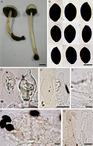
Panaeolus antillarum (Fr.) Dennis, Kew Bull. 15(1): 124 (1961). Fig. 1.
Panaeolus antillarum. A, Basidiomata; B, basidiospores; C, pleurocystidia; D, cheilocystidia; E, caulocystidia; F, pileipellis; G, pileocystidia. Scale bars: A, 10 mm; B–G, 10 μm. All photographs from A. G. S. Silva-Filho 801 (HCP 1152).

≡ Agaricus antillarum Fr., Elench. Fung. (Greifswald) 1: 42 (1828).
Pileus 29–39 mm in diameter, conic-campanulate, becoming conic-convex to broadly parabolic, surface slightly moist when young, at maturity dry, cracking, forming areolae at centre, smooth, margin regular and non-striate, pale grey (1B1), brownish orange (5C3) to orange grey (5B2) at the centre. Lamellae adnate, somewhat fleshy, crowded, with three sizes of lamelullae, edge paler and serrulate, brownish grey (5D2, 5E2), mottled. Stipe 85–100 × 6–7 mm, central, terete, equal to slightly bulbous at base, non-fistulose, surface slightly striated and subtly pruinose near the apex, consistency fleshy, yellowish white (1A2) to pale grey (1B1) (Fig. 1A). Context fleshy, 4–6 mm thick, pale grey (1B1). Veil absent. Spore print dark grey (1F1).
Basidiospores (15–)15.5–18 × (9–)9.5–10.5(–11) × 8.5–10.5 μm (Q = 1.47–1.72, Qm = 1.64, n = 50/1/1), broadly ellipsoid to subhexagonal in face view, ellipsoid in side view, smooth, thick-walled (up to 1.5 μm thick), apically truncate by a broad germ pore, 1.3–2.5 μm, dark brown (Fig. 1B), not bleaching in sulphuric acid. Basidia 24–34.5 × 12.5–15 μm, broadly clavate to cylindroclavate, tetrasporic, hyaline. Pleurocystidia 26.5–41 × 12.5–19.5 μm, as chrysocystidia-like sulphidia, non-abundant, clavate, fusoid, rarely pedicellate, some with a small mucronate apex, thin-walled, hyaline but with an irregular greyish green refractive body (Fig. 1C). Cheilocystidia 19.5–37 × 7–13(–17) μm, as leptocystidia, abundant, clavate, lageniform to fusoid, thin-walled, hyaline (Fig. 1D). Lamella edge sterile. Hymenophoral trama regular, made up of filamentous and inflated hyphae, 3.5–12 μm in diameter, smooth, thin-walled, hyaline. Pileipellis an epithelium of isodiametric to (sub)pyriform elements, 12.5–35.5 μm in diameter, smooth, thin-walled, hyaline (Fig. 1F). Pileocystidia 36.5–71 × 7–12.5 μm, scarce, forming fascicles at the pileus margin, cylindrical to lageniform, subcapitate, thin-walled, hyaline (Fig. 1F,G). Pileus trama made up of interwoven hyphae 4–14 μm in diameter, horizontally arranged, thin-walled, hyaline. Stipitipellis a cutis, composed of filamentous hyphae 1.5–6 μm in diameter, smooth, thin-walled, hyaline. Stipe trama regular, made up of hyphae 4.5–12.5 μm in diameter, smooth, thin-walled, hyaline. Caulocystidia 18.5–39 × 3.5–12 μm, leptocystidia, abundant, cylindrical, thin-walled, hyaline (Fig. 1E). Clamp connections infrequently in all tissues.
Specimen examined. Brazil. Paraná State: Palotina, São Camilo State Park, 332 m, 24°18′15.83′′S, 53°54′8.88′′W, 18 vi 2016, A. G. S. Silva-Filho 797 (HCP 1168).
Habitat and ecology. On pastures, solitary, growing on cow dung.
Distribution. Widely distributed on all continents (Halama et al., Reference Halama, Witkowska, Jasicka-Misiak and Poliwoda2014). In Brazil, it has been reported from the Atlantic Forest in the south (Rio Grande do Sul, Santa Catarina and Paraná States), in the southeast (São Paulo), in the northeast (Pernambuco) and from the Cerrado in the Central-West region (Goiás State) (Maia et al., Reference Maia, Carvalho Júnior, Cavalcanti, Gugliotta, Drechsler-Santos, Santiago, Cáceres, Gibertoni, Aptroot, Giachini, Soares, Silva, Magnago, Goto, Lira, Montoya, Pires-Zottarelli, Silva, Soares, Rezende, Luz, Gumboski, Wartchow, Karstedt, Freire, Coutinho, de Melo, Sotão, Baseia, Pereira, Oliveira, Souza, Bezerra, Araujo Neta, Pfenning, Gusmão, Neves, Capelari, Jaeger, Pulgarín, Menolli Junior, Medeiros, Friedrich, Chikowski, Pires, Melo, Silveira, Urrea-Valencia, Cortez and Silva2015). In Paraná State, it was recorded in dense ombrophilous forest (De Meijer, Reference De Meijer2006).
The robust, fleshy and greyish basidiomata, initially viscid and non-hygrophanous pileus, stipe with or without traces of a veil, smooth and opaque basidiospores with central and broad germ pore and presence of sulphidia are features that group Panaeolus antillarum in subg. Anellaria Gerhardt (Reference Gerhardt1996). This subgenus also includes Panaeolus semiovatus (Sowerby) S.Lundell & Nannfeldt and its varieties, which differ by the presence of a membranous veil and larger (up to 24 μm long) basidiospores (Gerhardt, Reference Gerhardt1996). Furthermore, Panaeolus antillarum is well distributed in tropical and subtropical areas, whereas P. semiovatus prefers temperate regions (Halama et al., Reference Halama, Witkowska, Jasicka-Misiak and Poliwoda2014). This mushroom is frequently found in pastures on cow dung, as one of the commonest coprophilous agarics in South Brazil.
Panaeolus antillarum is a species frequently misidentified by people looking for psychedelic fungi for recreational use, because it is very similar to Psilocybe species and Copelandia cyanescens, which are potentially psychoactive (Stamets, Reference Stamets1996), and grows on the same substrate types. The latter species is also a common coprophilous mushroom found in the same habitats in western areas of Paraná State (Silva-Filho et al., Reference Silva-Filho, Seger and Cortez2018a), and despite being macroscopically similar, the basidiomata of Copelandia cyanescens are smaller and turn bluish when touched. The absence of psilocybin, psilocin and baeocystin, or serotonin and 5-hydroxytryptophan, in Panaeolus antillarum was confirmed by Stijve & De Meijer (Reference Stijve and De Meijer1993) and Halama et al. (Reference Halama, Witkowska, Jasicka-Misiak and Poliwoda2014), based on Brazilian and Polish collections, respectively.
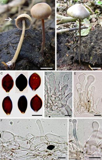
Panaeolus papilionaceus var. parvisporus Ew. Gerhardt, Bibl. Bot. 147: 58 (1996). Fig. 2.
Panaeolus papilionaceus var. parvisporus. A, Young basidiomata with remains of veil (arrow); B, mature basidioma; C, basidiospores; D, cheilocystidia; E, caulocystidia; F, pileipellis; H, pileocystidia. Scale bars: A and B, 10 mm; C–H, 10 μm. Photographs: A, A. G. S. Silva-Filho 801 (HCP 1152); B–H, A. G. S. Silva-Filho 798 (HCP 1167).

Pileus 12–31 mm in diameter, conic-campanulate to convex, sometimes umbonate, surface smooth, cracking at centre, margin non-striate, brownish grey (5C2) to light brown (5D4) at the disc, hygrophanous. Lamellae adnate, somewhat fleshy, crowded, with two sizes of lamelullae, edge even and concolorous with the sides, greenish grey (1D2) to medium grey (5E1), mottled. Stipe 33–110 × 2–5 mm, central, terete, equal to slightly bulbous at base, fistulose, surface smooth, yellowish white (1A2), pale grey (1B1) at apex, light brown (6D4) at centre, and light brown (6D5, 7D5) towards the base. Context fleshy, 1.5–3 mm thick, pale grey (1B1) (Fig. 2A,B). Veil present at pileus margin, forming appendiculate to dentate submembranous remnants, observed only in young specimens (Fig. 2A). Spore print dark grey (1F1).
Basidiospores (11–)11.5–14.5 × 7.5–9(–9.5) × 6–7.5 μm (Q = 1.36–1.68, Qm = 1.49, n = 40/4/4), limoniform in face view, elliptic in side view, smooth, thick-walled (up to 1.8 μm thick), apically truncate by a broad germ pore, 1.5–1.9 μm, dark brown (Fig. 2C), not bleaching in sulphuric acid. Basidia 20.5–25 × 9.5–13.5 μm, broadly clavate to cylindroclavate, tetrasporic, hyaline. Pleurocystidia absent. Cheilocystidia 20.5–33.5 × 4–7.5 μm, as leptocystidia, abundant, versiform, mostly cylindrical and ventricose, thin-walled, hyaline (Fig. 2D). Lamella edge sterile. Hymenophoral trama regular, made up of filamentous and inflated hyphae 3.5–8 μm in diameter, smooth, thin-walled, hyaline. Pileipellis an epithelium of (sub)isodiametric to pyriform elements, 10.5–19 μm in diameter, smooth, thin-walled, hyaline, rarely with light brownish content (Fig. 2F). Pileocystidia 19.5–44 × 7–12.5 μm, scattered, and in fascicles at the pileus margin, cylindrical to lageniform, with subcapitate apex, thin-walled, hyaline (Fig. 2F,G). Pileus trama consisting of interwoven and inflated hyphae, 10.5–20.5 μm in diameter, thin-walled, hyaline. Stipitipellis a cutis composed of filamentous hyphae 1.5–4.5 μm in diameter, smooth, thin-walled, hyaline. Stipe trama regular, composed of hyphae 3.5–16.5 μm in diameter, smooth, thin-walled, hyaline. Caulocystidia 18.5–56 × 5.5–10.5 μm, clavate, some with swollen apex, flexuous, thin-walled, hyaline (Fig. 2E). Clamp connections inconspicuous, present in all tissues.
Specimens examined. Brazil. Paraná State: Palotina, São Camilo State Park, 18 vi 2016, A. G. S. Silva-Filho 798 (HCP 1167), 799 (HCP 1166); ibid., 23 vi 2016, A. G. S. Silva-Filho 801 (HCP 1152), 803 (HCP 1170).
Habitat and ecology. Occurs on pastures growing on cow dung, gregarious to subcaespitose.
Distribution. Widely distributed (Gerhardt, Reference Gerhardt1996). In Brazil, it has been reported from Atlantic Forest in the south (Rio Grande do Sul and Paraná States) and the southeast (São Paulo) (Maia et al., Reference Maia, Carvalho Júnior, Cavalcanti, Gugliotta, Drechsler-Santos, Santiago, Cáceres, Gibertoni, Aptroot, Giachini, Soares, Silva, Magnago, Goto, Lira, Montoya, Pires-Zottarelli, Silva, Soares, Rezende, Luz, Gumboski, Wartchow, Karstedt, Freire, Coutinho, de Melo, Sotão, Baseia, Pereira, Oliveira, Souza, Bezerra, Araujo Neta, Pfenning, Gusmão, Neves, Capelari, Jaeger, Pulgarín, Menolli Junior, Medeiros, Friedrich, Chikowski, Pires, Melo, Silveira, Urrea-Valencia, Cortez and Silva2015). In Paraná State, De Meijer (Reference De Meijer2006) recorded it from dense ombrophilous forest.
The absence of metuloidal cystidia, the limoniform basidiospores in face view, the pileus with veil remnants in the margin and the non-bluing basidiomata place Panaeolus papilionaceus in subg. Panaeolus sect. Panaeolus in the sense of Gerhardt (Reference Gerhardt1996), along with Panaeolus venezolanus Guzmán and Panaeolus rubricaulis Petch. Gerhardt (Reference Gerhardt1996) states that Panaeolus papilionaceus var. papilionaceus is distinguished by the larger basidiospores (15–18 μm long) and distinctly appendiculate pileus margin. In contrast, Panaeolus papilionaceus var. parvisporus has basidiospores up to 15 μm long and possess a very fugacious veil at the pileus margin, sometimes absent. Our collections are characterised by basidiospores up to 14.5 μm long and fugacious traces of veil, observed only in young basidiomata, therefore we consider the South Brazilian specimens within the concept of Panaeolus papilionaceus var. parvisporus.
Panaeolus venezolanus, known from Venezuela and Mexico, has a membranous annulus on the stipe, which is absent in P. papilionaceus var. parvisporus (Guzmán, Reference Guzmán1978; Gerhardt, Reference Gerhardt1996). Panaeolus rubricaulis, described from Sri Lanka, differs in the presence of pleurosulphidia, which are also not found in P. papilionaceus var. parvisporus (Gerhardt, Reference Gerhardt1996).
Panaeolus papilionaceus falsely gained notoriety for its supposed hallucinogenic status after Schultes (Reference Schultes1939) doubtfully indicated Panaeolus campanulatus var. sphinctrinus (Fr.) Quél. to be the sacred teonanacatl used in Aztec ceremonies. Stamets (Reference Stamets1996) reported no effects after ingesting nearly thirty specimens of the species, although Guzmán et al. (Reference Guzmán, Allen and Gartz2000) considered Panaeolus papilionaceus var. papilionaceus sensu auct. non s. Ew. Gerhardt as a neurotropic species. However, the hallucinogenic or neurotropic status of that mushroom has not been supported by chemical analysis.
Panaeolus sylvaticus Silva-Filho & Cortez, sp. nov. – Type: Brazil, Paraná, Palotina, São Camilo State Park, 09 vi 2015, A. G. S. Silva-Filho 478 (holo UPCB). MycoBank no. 827918.
Basidiomata delicate with pileus less than 15 mm broad and stipe less than 3 mm broad, solitary on rotten wood, fallen leaves and small branches. Lamellae with inconspicuous pale droplet secretion on the edges. Basidiospores (8.5–)9–11.5(–12.5) × (5.5–)6–7.5 × 4–6.5 μm, limoniform in face view, ellipsoid in side view, smooth, truncate, brown to dark brown. Pleurocystidia absent. Cheilo-, pileo- and caulocystidia with yellowish droplets or content. Clamp connections present, inconspicuous. Fig. 3.
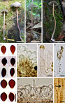
Panaeolus sylvaticus Silva-Filho & Cortez. A–C, Basidiomata; D, basidiospores; E, cheilocystidia; F, pileocystidia; G, caulocystidium; H, cross-section of pileus, showing the pileipellis with pileocystidia, I, stipite trama with oleipherous hyphae (arrow). Scale bars: A–C, 10 mm; D–I, 10 μm. Photographs: A and D–I, A. G. S. Silva-Filho 478 (UPCB – holotype); B, A. G. S. Silva-Filho 549 (HCP 1217); C, A. G. S. Silva-Filho 444 (HCP 1216).

Pileus 5–14 mm in diameter, broadly plane, umbonate, convex, conic-campanulate to broadly parabolic, surface smooth to slightly fibrillose, margin striate, rarely non-striate, occasionally undulate, colour varying from pale grey (1B1), light grey (1C1), grey (1D1), greyish yellow (1B4), yellowish grey (2B2) or greenish grey (1B2) to olive (2E3, 1F8) and olive brown (4F8). Lamellae adnate, thick membranous, crowded, with two sizes of lamelullae, first olive grey (1F8, 2F2), then dark grey (1F7) mottled, with even to slightly crenate and conspicuously paler edges, some with inconspicuous off-white secretory droplets. Stipe 33–52 × 1.5–3 mm, central, terete, equal, fistulose, with an insititious base, but occasionally with a white (1A1) to whitish basal mycelium, striated to slightly pubescent, consistency fleshy, yellowish white (1B1), greyish orange (5B4) or light orange (6A4) at apex, yellowish brown (5F8), brown (6E5, 6E4, 7E3) to dark brown (6F8) towards the base (Fig. 3A–C). Context fleshy, 1–2 mm thick, pale grey (1B1) to yellowish grey (1A2). Veil absent. Spore print dark grey (1F1).
Basidiospores (8.5–)9–11.5(–12.5) × (5.5–)6–7.5 × 4–6.5 μm (Q = 1.31–1.83, Qm = 1.47, n = 105/7/7), limoniform in face view, ellipsoid in side view, smooth, thick-walled (wall up to 1.2 μm thick), apically truncate by germ pore, 1.3–1.9 μm, orange brown to dark brown (Fig. 3D), not bleaching in sulphuric acid. Basidia 17–21(–28) × 7–10 μm, broadly clavate to cylindroclavate, predominantly tetrasporic, sometimes bisporic, hyaline. Pleurocystidia absent. Cheilocystidia 26–40 × 4.5–10 μm, as leptocystidia, clavate, clavate-pedicellate, cylindrical, cylindroclavate, flexuous, rarely ventricose, sometimes septate, abundant, thin-walled, hyaline, smooth or covered with numerous yellow-green droplets along the wall (Fig. 3E). Lamella edge sterile. Hymenophoral trama regular, made up of filamentous and inflated hyphae 2.5–15 μm in diameter, smooth, thin-walled, hyaline to yellowish brown. Subhymenium composed by filamentous hyphae, 2–4.5 μm in diameter, smooth, thin-walled, light brown. Pileipellis an epithelium composed of cylindrical, isodiametrical to pyriform cells, 11–33 μm in diameter, thin-walled, hyaline smooth or faintly covered by small yellow-green droplets (Fig. 3H). Subpellis a layer of filamentous hyphae, 2.5–4 μm in diameter, parallel, horizontally arranged, thin-walled, smooth, light brown. Pileocystidia 19.5–44 × 7–12.5 μm, cylindrical, clavate, lageniform, thin-walled, hyaline with yellow-green droplets, scattered at pileus centre and more evident (in fascicles) at pileus margin (Fig. 3F,H). Pileus trama composed of horizontally arranged, interwoven, filamentous and inflated hyphae 4.5–16.5 μm in diameter, hyphae thin-walled, hyaline to yellowish brown. Stipitipellis a cutis composed of smooth, thin-walled, light brown, filamentous hyphae 1.5–3.5 μm in diameter. Stipe trama regular, with hyphae 5–12 μm in diameter, smooth, thin-walled, hyaline and light brown. Caulocystidia 27–54(–57) × 3–11.5 μm, clavate, some with swollen apices, flexuous, thin-walled hyaline, with yellow-green droplets at apex (Fig. 3G). Clamp connections present, inconspicuous, observed only at the base of basidia and cystidia. Oleipherous hyphae observed in subpellis layer of pileipellis, lamella and stipite trama (Fig. 3I).
Additional specimens examined. Brazil. Paraná State: Palotina, São Camilo State Park, A. G. S. Silva-Filho 322 (HCP 1214); ibid., 30 ix 2015, A. G. S. Silva-Filho 636 (HCP 1215). Terra Roxa, RPPN Fazenda Açu, A. G. S. Silva-Filho 444 (HCP 1216); ibid., 20 vii 2015, A. G. S. Silva-Filho 549 (HCP 1217); ibid., 12 x 2016, A. G. S. Silva-Filho 650 (HCP 1218); ibid., 13 xii 2016, A. G. S. Silva-Filho 780 (HCP 1219).
Habitat and ecology. Occurs in seasonal semideciduous forest, solitary, growing on very rotten wood, fallen leaves and small branches.
Distribution. Known only from the type locality in Brazil.
Etymology. From Latin sylva (= forest), referring to the habitat of species.
Panaeolus sylvaticus is diagnosed by its small (less than 14 mm), grey to olive, smooth to slightly fibrillose pileus, weeping lamellae with off-white droplets, limoniform basidiospores in face view, and the absence of pleurocystidia, the presence of cheilo-, pileo- and caulocystidia with yellowish droplets or content, and the presence of clamp connections. Considering these distinctive morphological features and the specific ecology (substrate and habitat preferences), we propose Panaeolus sylvaticus as a new species.
By virtue of the smooth basidiospores, the absence of a membranous annulus, metuloids and chrysocystidia-like sulphidia, as well as the presence of cheilocystidia secreting yellowish droplets, Panaeolus sylvaticus belongs to Panaeolus subg. Panaeolus sect. Guttulati in the sense of Gerhardt (Reference Gerhardt1996). It was previously a monotypic section, comprising only Panaeolus guttulatus Bres., a species known from Europe and South Africa (Gerhardt, Reference Gerhardt1996; Pancorbo & Ribes, Reference Pancorbo and Ribes2010; Panchetti, Reference Panchetti2015).
Panaeolus guttulatus and P. sylvaticus share some morphological similarities: both present grey to olive colours in the pileus, pileocystidia and cheilocystidia with yellowish droplets, bi- and tetrasporic basidia, and the absence of pleurocystidia (Gerhardt, Reference Gerhardt1996). However, Panaeolus guttulatus has a darker and more robust pileus (10–30 mm in diameter) and smaller basidiospores (7–9 × 4–5 μm) with oil droplets. The production of the viscous substance by the cheilocystidia is a remarkable feature in Panaeolus guttulatus and can be observed even in dried specimens (Gerhardt, Reference Gerhardt1996), whereas in P. sylvaticus it is not so conspicuous. In addition, Panaeolus sylvaticus grows on rotting wood and leaf litter in seasonal semideciduous forest in the American subtropics, in contrast to P. guttulatus, which is found on sandy soil in temperate areas of Europe and South Africa (Gerhardt, Reference Gerhardt1996; Pancorbo & Ribes, Reference Pancorbo and Ribes2010).
Panaeolus fraxinophilus A.H.Sm., from North America, is another non-coprophilous species without pleurocystidia and with similar pileus size (8–15 mm) and basidiospores measuring 9–11 × 6.5–7 × 5–6 μm (Smith, Reference Smith1948). Despite these similarities, Panaeolus fraxinophilus has a dark grey to sordid yellowish brown pileus, not producing yellowish content in the cystidia, and the cheilocystidia are narrower (4–5 μm in diameter; Smith, Reference Smith1948). Moreover, Panaeolus fraxinophilus is a lignicolous species and grows only on wood of Fraxinus species (Smith, Reference Smith1948).
Finally, Panaeolus lignicola Rick (Reference Rick1930), described from Rio Grande do Sul State (South Brazil), is another non-coprophilous species with similar size of pileus (8 mm in diameter), stipe (20 × 2 mm) and basidiospores (10–12 × 8–9 μm; Rick, Reference Rick1961). Gerhardt (Reference Gerhardt1996) revised the alleged type collection of the species preserved at PACA Herbarium and reported much larger spores (12–14 × 8.5–9 × 7–7.5 μm). He noted that the obtained material is not a holotype but the ‘neotype’. Observing a relatively light yellow brownish colour of basidiospores and their olive brownish discoloration in KOH solution, Gerhardt (Reference Gerhardt1996) considered Rick’s specimen as a member of Psilocybe (Fr.) P.Kumm., “i.e. Deconica (W.G. Sm.) P.Karst.” and suggested a close morphological resemblance of the studied material to Deconica coprophila (Bull.) P.Karst., which, however, does not grow on wood. Neither the presence of droplets in the gill margin nor the presence of any kind of cystidia was reported by Rick (Reference Rick1930, Reference Rick1961) or Gerhardt (Reference Gerhardt1996). Given the differences in the basidiospore size and their colour, as mentioned by Rick (pale violaceous) and Gerhardt (light yellow brownish), it is very probable that the preserved ‘neotype’ does not correspond to original description of the species (Rick, Reference Rick1930), and therefore Panaeolus lignicola should be considered a doubtful name.
Key to Panaeolus Taxa Reported From Paraná State, Brazil
1a. Basidiomata small (less than 14 mm in diameter), growing in forests on very rotten wood, fallen leaves and small branches; cystidia with yellowish droplets or content P. sylvaticus
1b. Basidiomata over 14 mm in diameter, growing in pastures on cow dung or grass; cystidia without yellowish droplets or content _________________________________ 2
2a. Pleurocystidia present _________________________________ P. antillarum
2b. Pleurocystidia absent _________________________________ 3
3a. Basidiomata without veil, cheilocystidia often capitate P. cinctulus (De Meijer, Reference De Meijer2006)
3b. Basidiomata with remaining veil, cheilocystidia cylindrical P. papilionaceus var. parvisporus
Acknowledgements
We are grateful for the financial support of Conselho Nacional de Desenvolvimento Científico e Tecnológico (CNPq, Processo 483455/2013-3) and Fundação Araucária de Apoio ao Desenvolvimento Científico e Tecnológico do Estado do Paraná (Convênio 675/2014), Coordenação de Aperfeiçoamento de Pessoal de Nível Superior (CAPES), towards the M.Sc. scholarship of the first author, and thank the Universidade Federal do Paraná for access to facilities. The authors additionally thank the anonymous reviewers.