Kawasaki disease was first reported by Dr. Kawasaki in 1967 and is one of the most common acquired heart diseases in infants and children. It is characterised by a variety of symptoms and signs caused by systemic vasculitis, which mostly recover spontaneously within two weeks.Reference Kitamura1 In inflammatory coronary artery disease as sequelae of Kawasaki disease, an aneurysm is formed in 3–5% of treated patients, which regresses spontaneously over the year. In some patients, coronary artery occlusive lesions develop several years later regardless of the administration of high-dose γ-globulin; this leads to ischaemic heart disease.Reference Kitamura and Tsuda2 Obstructive lesions are most frequently observed at the inlet or outlet of coronary aneurysm, mainly the proximal part of the coronary artery.Reference Kitamura1,Reference Kitamura and Tsuda2 The number of patients requiring coronary revascularisation is very small in patients with Kawasaki disease, and the literature on coronary artery bypass grafting and percutaneous coronary intervention is lacking. Coronary artery lesions caused by Kawasaki disease in children show varying characteristics because collateral communications are usually well-developed as children grow. This results in various morphological features.Reference Guo, Chang and Xu3 Coronary artery stenosis caused by Kawasaki disease is commonly associated with severe calcification; therefore, coronary artery bypass grafting can be considered a treatment option rather than percutaneous coronary intervention.Reference Kitamura and Tsuda2,Reference Jeong, Han and Lee4 However, the long-term patency of the coronary graft is not good in infants and children especially when venous grafts were used.Reference Guo, Chang and Xu3,Reference Jeong, Han and Lee4 Here, we reviewed the long-term surgical outcomes and graft patency of coronary artery bypass grafting in small and young patients with Kawasaki disease with coronary artery involvement.
Materials and methods
Patients
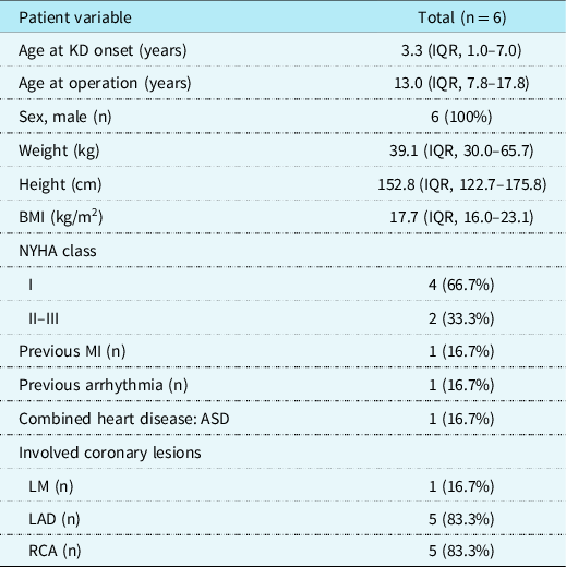
The study protocol was approved by the Institutional Ethics Committee/Review Board of Seoul National University Hospital (approval number 1909-009-1061), which exempted the requirement for patients’ informed consent because of the retrospective nature of the study. We analysed the clinical data of patients’ who underwent coronary artery bypass disease for Kawasaki disease-associated coronary artery lesions between March 2004 and March 2013. There were six patients (all male); their median age at the timing of coronary artery bypass grafting and presumed median age at the initial onset of Kawasaki disease were 13.0 years (interquartile range, 7.8–17.8 years) and 3.3 years (interquartile range, 1.0–7.0 years), respectively. The median interval from the initial onset of Kawasaki disease to operation was 7.0 years (interquartile range, 4.9–12.5 years). One of them underwent a coronary artery bypass grafting after the relapsed Kawasaki disease that occurred 6 years after first attack of Kawasaki disease. Most patients presented with multiple lesions including significant stenosis and aneurysmal change, while only two patients showed solitary lesions. The baseline characteristics of the pre-operative patients are summarised in Table 1. One patient had myocardial infarction symptoms such as exertional chest pain and dyspnoea on exertion, and another patient had ventricular tachyarrhythmia pre-operatively. In the patients diagnosed with myocardial infarction, troponin I increased to 16.1 ng/ml and CKMB to 1596 U/l and ST change and pathologic Q wave were observed in lead II, III, and augmented voltage vector of a foot on electrocardiogram.
Pre-operative characteristics of the patients.

BMI = body mass index; IQR = interquartile range; KD = Kawasaki disease; LAD = left anterior descending coronary artery; LM = left main coronary artery; MI = myocardial infarction; NYHA = New York Heart Association; RCA = right coronary artery.
Surgical technique
Our surgical strategy was conventional revascularisation under cardiopulmonary bypass. After a full median sternotomy, the left and/or right internal thoracic artery was harvested using a pedicled technique. Proximal dissection of the internal thoracic artery was moved to the inferior border of the subclavian vein, and the internal thoracic artery was then divided distally just proximal to the bifurcation. If needed, the saphenous vein was harvested from the proximal or distal portion of the leg. One case was performed using off-pump coronary artery bypass grafting in case that the patient was old enough, hemodynamically stable, and had a good ventricular function. In on-pump cases, cardiopulmonary bypass was established by standard aortic and both caval vein cannulation. After aortic cross-clamping and infusion of cardioplegia, the coronary arteries were revascularised in order beginning with the proximal part. Indications for operation should consider the patient’s age, performance status or exertional chest discomfort, history of myocardial infarction, and ventricular function. The optimal timing of surgical treatment for this disease was considered when the disease got into the chronic phase.
Echocardiographic and angiographic follow-up
In all cases, transthoracic echocardiography was performed before the surgery. All patients underwent echocardiography on average 6 days (interquartile range, 5–7 days) post-operatively before discharge. The last follow-up echocardiographic evaluation was performed 13.5 ± 6.6 years after surgery. We measured the left ventricular ejection fraction and regional wall motion abnormality using echocardiography. The size of the proximal coronary artery could also be evaluated through echocardiography, but there was a limitation in observing the distal coronary artery. All patients underwent coronary angiography or CT angiography to evaluate graft patency. The median interval from initial operation to the latest angiography was 11.6 years (interquartile range, 4.2–13.1 years). We measured the degree of occlusion at the graft body. We graded graft patency in the manner described by FitzGibbon et al: excellent graft for grade A, stenosis of 50% or less of vessel diameter for grade B, and stenosis of 75% or more or a totally occluded graft for grade O.Reference FitzGibbon, Kafka and Leach5 In this study, grades A and B were considered as patent and grade O was treated as occluded. Single-photon emission CT was performed to evaluate myocardial perfusion. All patients underwent a follow-up with an interval of six to twelve months. The follow-up was completely done in all patients, and the last follow-up observation was January 2019 in this study.
Statistical analysis
Statistical analysis was performed using IBM SPSS statistical software (version 23, IBM Inc., Armonk, New York, United States of America). Categorical variables were presented as numbers and percentages. Continuous variables were expressed as median and interquartile range or as proportions and differences between continuous variables were analysed using the Wilcoxon rank sum test. The significance of differences in categorical variables was assessed using the t-test or Fisher’s exact test. Kaplan–Meier methods were used to analyse the probability of survival and graft patency rates. A p-value of less than 0.05 was considered statistically significant.
Results
Pre-coronary artery bypass graft surgery status
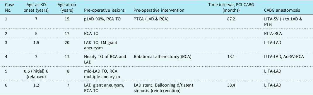
Before coronary artery bypass grafting, three patients needed pre-operative intervention. Of these, one patient underwent rotational atherectomy for right coronary artery total occlusion lesion at the age of 10 years in Japan, and one patient underwent percutaneous transluminal coronary angioplasty on the right coronary artery and left anterior descending artery lesions at the age of eight years. The other patient underwent stent insertion on the left anterior descending artery prior to coronary artery bypass grafting at the age of four years but needed reintervention on the left anterior descending artery due to in-stent stenosis at the age of five years. Table 2 shows the individual data of coronary artery lesions and coronary artery bypass grafting.
Individual data of coronary artery lesions and CABG.

Ao = aorta; CABG = coronary artery bypass grafting; KD = Kawasaki disease; LAD = left anterior descending coronary artery; LITA = left internal thoracic artery; LM = left main coronary artery; PCI = percutaneous coronary intervention; pLAD = proximal LAD; PLB = posterolateral branch; PTCA = percutaneous transluminal coronary angioplasty; RCA = right coronary artery; RITA = right internal thoracic artery; SV = saphenous vein; TO = Total occlusion.
Coronary artery bypass grafts
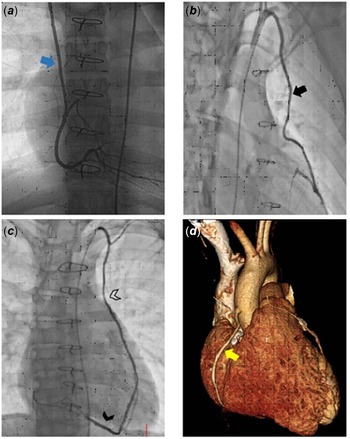
The most commonly used graft was the left internal thoracic artery (in 5 cases) followed by saphenous vein (2 cases) and right internal thoracic artery (1 case). Table 2 shows the operative details. The right internal thoracic artery was anastomosed to the right coronary artery (Fig 1a), and the left internal thoracic artery was anastomosed to the left anterior descending artery (Fig 1b). The saphenous vein was also harvested when the length of the harvested internal thoracic artery was insufficient. Saphenous vein grafts were used as an I-shaped composite graft anastomosed to the left internal thoracic artery (Fig 1c) and an aortocoronary bypass graft, respectively (Fig 1d). Four patients underwent single coronary artery bypass grafting, while two patients underwent multiple coronary artery bypass grafting. The concomitant procedure included atrial septal defect repair in one patient. Median cardiopulmonary bypass and aortic cross-clamping times were 128.0 minutes (interquartile range, 81.5–169.0 minutes) and 79.0 minutes (interquartile range, 45.5–96.5 minutes), respectively.
Grafts patency in coronary angiography or CT angiography. ( a ) Patent RITA graft to RCA (blue arrow). ( b ) Patent LITA graft to LAD (black arrow). ( c ) Patent LITA (empty arrowhead) to SV (full arrowhead) graft as I-shaped anastomosis. ( d ) SV was used as aorto-RCA bypass graft (yellow arrow). LAD, left anterior descending coronary artery; LITA, left internal thoracic artery; RCA, right coronary artery; RITA, right internal thoracic artery; SV, Saphenous vein.

Post-operative course
The time of assisted mechanical ventilation ranged from 5 to 22 hours (median, 8.5; interquartile range, 5.8–20.5 hours) post-operatively. The median time of intensive unit care was one day (interquartile range, 1.0–1.3 days), and the median duration of hospital stay was 7 days (interquartile range, 5.8–10.3 days). There were no post-operative complications or operative mortality. The median follow-up duration was 12.1 years (interquartile range, 9.5–13.1 years). Two patients who had shown definite exertional chest pain and dyspnoea and who had shown ventricular tachyarrhythmia pre-operatively did not show any coronary artery-related symptoms and signs post-operatively and neither did other patients during overall follow-up period.
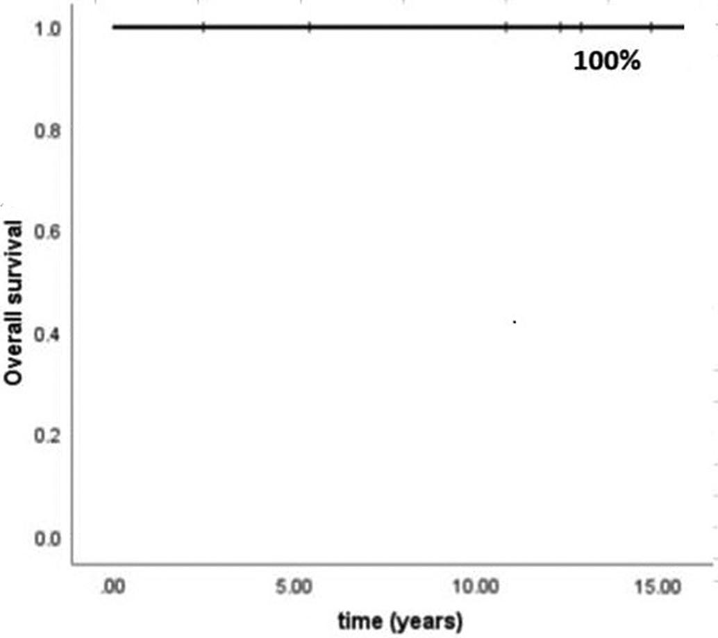
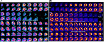
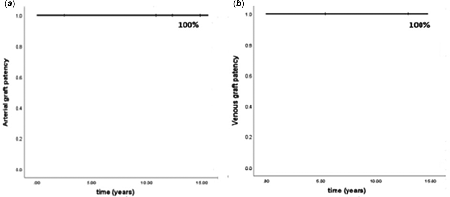
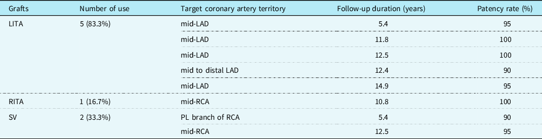
We have no late mortalities (Fig 2), and all patients were doing well without any obvious exercise restriction in their daily lives in the most recent follow-up. The New York Heart Association functional class at discharge improved compared to the pre-operative status (p < 0.05). Median left ventricular ejection fraction was preserved (64.5% pre-operatively vs. 63.2% post-operatively, p = 0.6). In all six patients, the pre-operative single-photon emission CT test showed a significant decrease in myocardial perfusion; the myocardial perfusion improved after coronary artery bypass grafting. For example, in the case where left internal thoracic artery was anastomosed to left anterior descending artery due to left anterior descending artery total occlusion, post-operative single-photon emission CT findings (Fig 3b) showed improved myocardial perfusion in the apico-mid anterior wall versus pre-operative single-photon emission CT (Fig 3a). All grafts including saphenous vein grafts stay patent in coronary angiography or CT angiography (Fig 4) and required no further interventions or re-operation after initial coronary artery bypass grafting (Table 3). We administered aspirin and checked the patient’s status by echocardiography or angiography every six months to one year at the outpatient clinic with a paediatrician.
Kaplan–Meier Curve for overall survival.

Comparison of SPECT on ( a ) pre-operative and ( b ) post-operative period. Post-operative SPECT findings showed the improved perfusion in the apico-mid anterior wall. SPECT, single-photon emission CT.

Long-term patency of arterial and venous graft. ( a ) All grafts were patent on arterial grafts. ( b ) Graft patency on venous grafts.

Follow-up according to grafts.

LAD = left anterior descending coronary artery; LITA = left internal thoracic artery; PL = branch, posterolateral branch; RCA = right coronary artery; RITA = right internal thoracic artery; SV = saphenous vein.
Discussion
There are several reports about clinical outcomes including graft patency after coronary artery bypass grafting in adult patients with myocardial ischemia. Goldman et al reported a patency rate of 95% for saphenous vein grafts and 99% for internal thoracic artery grafts at one week.Reference Goldman, Zadina and Moritz6 They found out if a saphenous vein or internal thoracic artery graft was patent in one week, then that graft patency had a 68% and 88% chance at 10 years, respectively. Shimokawa et al assessed saphenous vein graft patency at 11.8 ± 10.4 days after coronary artery bypass grafting using coronary angiography and similarly reported an early patency rate of 95.7%.Reference Shimokawa, Manabe and Sawada7 In another study, Puskas and colleagues reported a patency rate of 100% for internal thoracic artery grafts. According to their study, an angiographic follow-up showed an early graft patency rate of 88% for saphenous vein grafts and 95% for left internal thoracic artery grafts.Reference Puskas, Thourani and Marshall8
In contrast to adults, coronary artery bypass grafting using internal thoracic artery is technically demanding in children. According to reports on the long-term outcomes of surgical treatment, the internal thoracic artery long-term graft patency rate in young children was significantly lower.Reference Ochl9,Reference Takeuchi, Gomi, Okamura, Mori and Nagashima10 However, Kitamura et al recently reported that there was no age difference in graft patency among their paediatric cohort.Reference Tsuda, Kitamura and Kimura11 In infants or children, the internal thoracic artery is a thin-walled artery with a diameter of 1 mm or less. Therefore, coronary artery bypass grafting in children requires delicate and precise dissection and anastomosis, and cardiopulmonary bypass support with cardioplegic arrest is recommended as the best approach for exquisite procedures.Reference Tsuda, Kitamura and Kimura11,Reference Vida, Torregrossa and De Franceschi12 There were reports about using saphenous vein or right gastroepiploic artery as well as internal thoracic artery as graft in paediatric Kawasaki patients. Kitamura et al demonstrated that the internal thoracic artery graft implanted in young children could grow with somatic growth,Reference Kitamura, Seki and Kawachi13 while some other authors reported that the saphenous vein had poor growth potential in children and also had high incidence of obstructive and degenerative changes.Reference Kameda, Kitamura and Taniguchi14,Reference Chello, Mastroroberto, Perticone, Celi and Colonna15 Takeuchi et al reported a child with Kawasaki disease in which a right gastroepiploic artery graft was used.Reference Takeuchi, Gomi, Okamura, Mori and Nagashima10 The right gastroepiploic artery is widely used as a graft for revascularisation in adult coronary artery bypass grafting. However, the right gastroepiploic artery, the fourth branch of the abdominal aorta, is limited in use in small children and has poor patency because it carries a higher risk of flow competition than the internal thoracic artery in children with less developed body.Reference Ochl9 In the 25-year experience reported by Kitamura et al, patency of the internal thoracic artery and saphenous vein graft was 87% and 44%, respectively, in aged <20 years.Reference Kitamura, Tsuda and Kobayashi16
Unlike the systemic medium-sized artery, the internal wall of the internal thoracic artery has a well-developed elastic lamina layer with a very thin smooth muscle layer; inflammatory reactions are rare. Thus, internal thoracic artery can almost always be used as the focus for bypass graft in Kawasaki disease.Reference Marvroudis, Backer and Muster17,Reference Coskun, Coskun and El Arousy18 The effect of internal thoracic artery grafts on survival may depend on vascular endothelial function as well as graft longevity. However, endothelial function of the coronary artery vulnerable to inflammation could be impaired and depressed under Kawasaki disease.Reference McCrindle, McIntyre, Kim, Lin and Adeli19 Therefore, if we performed coronary arterial surgery when patients were in active phase of Kawasaki disease, we could not guarantee the patency of graft after coronary bypass surgery. We performed coronary artery bypass grafting surgery at a median of 7.0 years after they were diagnosed with Kawasaki disease, i.e., in the subacute or chronic phase of the disease. We can say that we were able to avoid an active phase of this disease in our cohort because the shortest intervals between the onset of Kawasaki disease and coronary arterial bypass surgery were 4.3 years. Presumably, we might explain our better graft patency versus other studies with this timing of coronary artery bypass grafting. Therefore, if patient’s clinical status permits, it would be reasonable to perform coronary arterial bypass surgery beyond the active phase. In addition to concerns about the effect of inflammation on graft patency in Kawasaki disease, an underdeveloped internal thoracic artery of small children sometimes has a flow capacity that depends on the severity of the anastomosed coronary artery stenosis.Reference Ochl9–Reference Vida, Torregrossa and De Franceschi12 Therefore, when considering sequential grafting or making a composite graft in young children, great care should be taken to avoid flow competition or possible technical errors.Reference Takeuchi, Gomi, Okamura, Mori and Nagashima10
We had three cases of percutaneous coronary intervention after Kawasaki disease prior to the coronary artery bypass grafting surgery. However, the patency of coronary artery did not maintain after percutaneous coronary intervention; thus, all of them required coronary artery bypass grafting in median 33.4 months after percutaneous intervention. There are several literatures that reported clinical results of percutaneous coronary intervention in children with Kawasaki disease. Muta et al reported that the rates of repeat-revascularisation in percutaneous coronary intervention group were higher than in surgery group.Reference Muta and Ishii20 Coronary artery stenotic lesions in children with Kawasaki disease are primarily due to severe calcification, unlike adult coronary artery disease, which is associated with atherosclerosis.Reference Kitamura and Tsuda2,Reference Jeong, Han and Lee4 Thus, the indication of coronary intervention in adult could not be applied as it is in children with Kawasaki disease. Moreover, the indications for intervention in Kawasaki patients have not been established, and the long-term result after intervention is still unclear.Reference Akagi21
We have some limitations in this study. This was a retrospective observational study with a small number of cases from a single centre. Statistical analyses for parameters that could affect some positive or negative outcomes were not possible. Therefore, our descriptive study does not represent the general children with Kawasaki disease requiring coronary artery bypass grafting. Further collection of multi-centre data would provide more reliable information.
In conclusion, our long-term results demonstrate acceptable results not only for the patency of the arterial graft but also for the saphenous vein graft. Coronary artery bypass graft surgery could be a good surgical option in patients with coronary lesions caused by Kawasaki disease in the subacute or chronic period in terms of graft patency and improvement in myocardial perfusion. We could anticipate better long-term clinical results, especially in graft patency and myocardial infarction-related symptoms or events, if we perform coronary artery bypass surgery at adequate timing during disease process in these patients.
Acknowledgements
None.
Financial support
This research received no specific grant from any funding agency, commercial or not-for-profit sectors.
Conflicts of interest
None.
Ethical standards
The authors assert that all procedures contributing to this work comply with the ethical standards of the relevant national guidelines on human experimentation and with the Helsinki Declaration of 1975, as revised in 2008, and has been approved by the institutional committees (the Institutional Ethics Committee/Review Board of Seoul National University Hospital).