Introduction
Endoscopic sinus surgery (ESS) has evolved from being a ‘cutting edge’ technique to now representing the standard surgical treatment for sinonasal pathology. One of the problems with the endoscope is that a lack of stereoscopic vision limits depth perception, which can only be perceived through motion and shading. Such limitations, along with the complexity of sinonasal anatomy, have led surgeons to search for advancements and refinements to improve the procedure, in order to optimise patient outcomes.
One of these has been the introduction of image guidance systems, which allow surgeons to continuously match intra-operative endoscopic findings to the patient's pre-operative computed tomography (CT) scan, with the use of specially designed instruments. These instruments, when placed within the sinonasal cavity, can be detected on a monitor.
The earliest of these systems were based on rigid servo arms and joints, making them difficult to prepare and manipulate during surgery.Reference Anon, Klimek, Mosges and Zinreich1 Today's interactive computer imaging systems have faster registration processes and are much less intrusive.Reference Govindaraj, Adappa and Kennedy2 Most studies have demonstrated the accuracy of such systems to be within 2 mm.Reference Anon3, Reference Labadie, Davis and Fitzpatrick4 In addition, the use of computer-assisted ESS has been found to enhance surgical efficiency and to facilitate trainees' acquisition of ESS skills.Reference Casiano and Numa5
Several papers have assessed the outcomes of computer-assisted ESS compared with non-assisted ESS. Many have found no significant difference in outcome measures.Reference Mueller and Caversaccio6, Reference Tabaee, Hsu, Shrime, Rickert and Close7 However, some have reported reductions in the incidence of major complications when computer assistance has been used.Reference Fried, Moharir, Shin, Taylor-Becker and Morrison8
The benefit of computer assistance during ESS is still undetermined. For this reason, we decided to assess our own experience of using image guidance during ESS for inflammatory sinus disease; we also undertook a meta-analysis to appraise the current evidence.
Materials and methods
Study design and population identification
Data were collected retrospectively from the records of all endoscopic sinus surgery cases performed between 30 June 2000 and 30 December 2003. Patients were only included if ESS was performed for inflammatory or infective sinus disease. Of the 380 cases operated upon during this period, 17 were excluded from the study due to incomplete data at the time of collection. Six cases of tumour excision and two cases of petrous apex cholesterol granuloma were also excluded. A total of 355 patients was included in the study.
Of these surgical cases, 333 involved use of the InstaTrak 3500+ surgical navigation system (GE Healthcare, Waukesha, Wisconsin, USA), while the other 22 cases were performed without computer guidance and prior to the computer-assisted ESS cases, as the guidance system was not available at the time. The advantages of the InstaTrak electromagnetic navigation system are the ease with which system registration and calibration are carried out (with the use of a headset) and the absence of the line-of-sight issues present with infrared systems. In our experience, issues relating to electromagnetic field disruption from metallic objects within the vicinity were easily addressed. Our department has previously published a comparison of the InstaTrak system versus an optical system (LandmarX; Medtronic-Xomed, Jacksonville, Florida, USA). Both were felt to be equally competent in terms of accuracy, but the InstaTrak was found to be more ‘user-friendly’;Reference Kherani, Javer, Woodham and Stevens9 therefore, we used this latter system.
Outcomes and data source
This study was performed at St Paul's Sinus Centre, Vancouver.
The primary outcomes of the study were complications and time to revision surgery (as an indicator of disease recurrence). The time to revision surgery was defined as the time period between the date of the index surgery and the date of the decision to undertake revision surgery. The date of this decision was used due to the long waiting list that some of our patients were placed on. Only patients who eventually underwent surgery were included.
The secondary outcomes of the study were the volume of blood loss during the index surgery and the duration of the index surgery.
Collated data included the duration of surgery, volume of blood loss, complications, and time to the decision to undertake revision surgery. Patient demographic information was also collected, including age, sex, and whether the index surgery was primary or revision. All the endoscopic sinus surgery procedures were performed by the same surgeon. All outcome results were reported as of 31 October 2010.
Technical aspects
Before surgery, patients underwent a standardised, 1-mm slice width, axial, sinus CT scan. The InstaTrak protocol was used for patients undergoing computer-assisted ESS. Lund–Mackay CT scores were calculated. Patients were categorised based on their total Lund–Mackay CT score as mild (score, 0–8), moderate (9–16) or severe (17–24). For computer-assisted ESS patients, the digital CT scan file was transferred to the InstaTrak navigation workstation.
After intubation, each patient was fitted with a headset which established a reference frame from which registration and calibration were performed. The computer-assisted ESS probes were in the form of straight and angled (45° and 90°) suction aspirators that were tracked on the system's monitor.
Following surgery, patients were discharged on the same day unless co-morbidities warranted an overnight stay.
Patients were seen six days post-operatively, at which time spacers were removed and endoscopic debridement performed. Patients were then seen four weeks later and further follow up was performed as needed by each individual.
Meta-analysis
A comprehensive literature review was performed using the Ovid Medline (1948 to present), PubMed and Ovid Embase databases. Search terms included ‘endoscopic sinus surgery’ and ‘computer-assisted surgery’. We limited our review to English language articles. We identified 126 articles on PubMed which fitted the search criteria, 82 articles on Medline and 3 articles on Embase.
For the purpose of the meta-analysis, two of the authors (VSS and DY) reviewed the abstracts of the articles and included only those studies that compared conventional ESS with computer-assisted ESS. Studies had to examine one of our two primary outcome measures – revision surgery rate or complication rate – in order to be included. We excluded studies that included tumour removal. If results were published several times, only the most recent article was included. Seven articles fulfilled these criteria.
Complications were categorised as major or minor. Major complications included major haemorrhage, orbital haematoma, cerebrospinal fluid (CSF) leakage, nasolacrimal injury, pneumocephalus, meningitis, stroke and carotid injury. Minor complications included minor haemorrhage requiring a return to hospital, hyposmia, headache, periorbital ecchymosis and periorbital emphysema.Reference Sunkaraneni and Nair10 Only those complications related to the surgery itself were included in the analysis.
Statistical analysis
Patients' baseline characteristics were summarised and compared for the two treatment groups. Continuous variables were compared using the Wilcoxon rank-sum test. For categorical variables, the chi-square or Fisher's exact test was applied.
Survival analysis was performed for the time to revision surgery. The cumulative probability of revision surgery was calculated for each group using the Kaplan–Meier method, and the two curves were compared using the log-rank test. The effect of treatment on the time to revision surgery was examined using Cox proportional hazards models.
To assess the validity of proportional hazard assumption in the Cox proportional hazard model, the cumulative residual was used with graphical displays and formal tests. In the presence of violation of the proportional hazard assumption, the piece-wise Cox proportional hazard model was performed in order to take account of the non-constant proportional hazards.
Constant hazard ratios were estimated for two time intervals: less than 1.5 years following index surgery and more than 1.5 years following index surgery. All models were adjusted for preselected confounding variables, age, gender, Lund–Mackay score and type of surgery.
Due to the low complication rate in the study, exact 95 per cent confidence intervals were calculated for complication rates. The Wilcoxon rank-sum test was used to analyse the two secondary outcomes: volume of blood loss during surgery and duration of surgery.
A separate meta-analysis was conducted for each of the outcomes of interest (i.e. revision surgery rate, and total, major and minor complication rates). The number of events and total number of patients per treatment arm were extracted from the seven studies selected from the literature plus our own study.
When no events were reported in one or both arms of a study, a treatment arm continuity correction was applied. The treatment arm continuity correction has been shown to out-perform the constant continuity correction method.Reference Sweeting, Sutton and Lambert11 Odds ratios and 95 per cent confidence intervals were estimated for each study.
A few studies reported no events in either treatment arm. There is ongoing debate in the literature over whether such studies should be included in meta-analyses.Reference Sweeting, Sutton and Lambert11, Reference Diamond, Bax and Kaul12 We opted to include them.
Cochran's Q test was used to evaluate heterogeneity across studies. The IReference Govindaraj, Adappa and Kennedy2 value was also calculated to describe the variation between studies which was due to heterogeneity rather than chance.Reference Higgins and Thompson13
The random effects model was used to calculate the odds ratios for the rates of complications and revision surgery, for non-assisted versus computer-assisted ESS groups. While there are no universally accepted criteria for choosing between a random effects model and a fixed effects model, the random effects model is more conservative compared with the fixed effects model and assumes greater heterogeneity between studies. Therefore, the random effects model was used in the current study in order to avoid overstating our findings.
Forest plots were generated to enable visualisation of the heterogeneity among the studies, and to show the overall odds ratios plus 95 per cent confidence intervals.
All reported p values are two-sided. P values of 0.05 or less were considered statistically significant. Analyses were conducted using the SAS (version 9.2) and R (version 2.12) software programs.
Results and analysis
Vancouver population
Our study population comprised 355 cases: 187 males (52.684 per cent) and 168 females (47.32 per cent). The mean patient age was 48.6 years (range, 14–83 years).
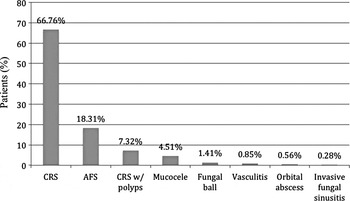
Indications for surgery are shown in Figure 1. There were 169 (47.9 per cent) primary cases and 184 (52.1 per cent) revision cases. Three hundred and thirty-three (93.8 per cent) cases involved computer-assisted ESS and 22 (6.2 per cent) involved non-assisted ESS.
Indications for endoscopic sinus surgery. CRS = chronic rhinosinusitis; AFS = allergic fungal sinusitis; w/= with

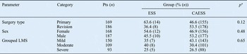
Comparison of patients' baseline characteristics in the two treatment groups revealed a mean patient age of 48.98 years in the computer-assisted ESS group and 42.64 years in the non-assisted ESS group; this difference was statistically significant. There was no statistically significant difference between the mean total Lund–Mackay CT scores for the computer-assisted ESS (11.11) and ESS (11.35) groups (Table I). There was no statistically significant difference between the groups as regards type of surgery (primary vs revision; p = 0.12), patient sex (p = 0.48) or stratified Lund–Mackay score (p = 0.65) (Table II).
Baseline continuous variables

*n = 22; †n = 333. ESS = non-assisted endoscopic sinus surgery; CAESS = computer-assisted ESS; SD = standard deviation; Med = median; IQR = interquartile range; LMS = Lund–Mackay score
Baseline categorical variables

*Two-sided. Pts = patients; ESS = non-assisted endoscopic sinus surgery; CAESS = computer-assisted ESS; LMS = Lund–Mackay score
Complications were divided into intra-operative and post-operative. Intra-operative complications included anterior and posterior ethmoid artery injury, skull base leak, orbital injury, and lacrimal injury. Post-operative complications included epiphora. Complications were further categorised on the basis of severity, as either minor or major.Reference Sunkaraneni and Nair10 Only one complication was noted in the computer-assisted ESS group (0.3 per cent; 95 per cent exact CI, 0.001–2 per cent), and no complications occurred in the ESS group (0 per cent; 95 per cent exact CI, 0–15 per cent). The single complication in the computer-assisted sinus surgery group was post-operative epiphora requiring dacryocystorhinostomy.
The median follow-up time was 7.43 years (range, 0.07–10.25 years). Eighty-eight patients underwent revision surgery during the follow-up time: 79 (23.7 per cent) in the computer-assisted ESS group and 9 (40.9 per cent) in the ESS group.
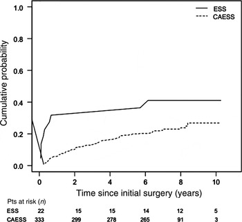
The cumulative probability plot for revision surgery (Figure 2) shows that the most significant difference in the rate of revision surgery between the two groups appeared in the early follow-up period. Log-rank testing indicated a statistically significant difference in the cumulative probability of revision surgery between the two groups (p = 0.04).
Cumulative probability of revision surgery for the non-assisted endoscopic sinus surgery (ESS) and computer-assisted endoscopic sinus surgery (CAESS) groups. P = 0.04 (log-rank test).

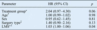
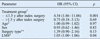
However, after utilising the Cox proportional hazard model with adjustment for preselected confounding variables, there was no statistically significant difference in the risk of recurrence between the non-assisted and computer-assisted ESS groups, over the whole follow-up period (hazard ratio, 2.04; 95 per cent CI, 0.97–4.30; p = 0.06) (see Table III). Due to the invalid proportional hazard assumption, further analysis was performed using a piece-wise Cox proportional hazard model.
Hazard ratio for revision sinus surgery

*Endosopic sinus surgery vs computer-assisted endoscopic sinus surgery. †Year. ‡Revision vs primary. **One unit. HR = hazard ratio; CI = confidence interval; LMS = Lund–Mackay score
This analysis revealed that, in the first 1.5 years following the index sinus surgical procedure, a hazard ratio of 4.54 (95 per cent CI, 1.86–11.06; p = 0.001) was noted for the risk of revision surgery, comparing the ESS and computer-assisted ESS groups. Thereafter, the hazard ratio for the risk of revision surgery was 0.75 (95 per cent CI, 0.18–3.13; p = 0.69) (see Table IV).
Hazard ratio for revision sinus surgery: piece-wise cox proportional hazard model*

* For 1.5-year interval. †Endoscopic sinus surgery vs computer-assisted endoscopic sinus surgery. ‡Year. **Revision vs primary. §One unit. HR = hazard ratio; CI = confidence interval; y = years; LMS = Lund–Mackay score
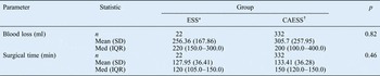
Analysis of secondary outcomes, namely duration of surgery and estimated blood loss, showed no statistically significant differences between the two groups (see Table V).
Secondary outcome measures

*n = 22; †n = 333. ESS = non-assisted endoscopic sinus surgery; CAESS = computer-assisted ESS; SD = standard deviation; IQR = interquartile range; min = minutes
Meta-analysis
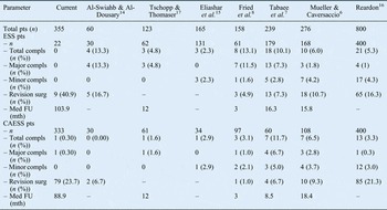
Meta-analysis was performed on eight studies, including our own. Data for each outcome were pooled and analysed separately (Table VI). Sample sizes ranged from 60 to 800 patients, with a total of 2176 ESS cases (1053 non-computer-assisted cases, 1123 computer-assisted cases). Of the seven studies obtained from the literature, six consisted of retrospective reviews that compared the outcomes for patients who had undergone non-assisted ESS versus patients with computer-assisted ESS.
Studies included in meta-analysis

Pts = patients; ESS = non-assisted endoscopic sinus surgery; compls = complications; surg = surgery; – = data not supplied; Med FU = median follow up; mth = months; CAESS = computer-assisted ESS
These six retrospective reviews presented level 3b evidence. One of the six studiesReference Tabaee, Hsu, Shrime, Rickert and Close7 collected data both retrospectively and prospectively (the prospective data referred to patient quality of life post-operatively); for the purpose of our meta-analysis, only the retrospective data were included. One of the seven studiesReference Tschopp and Thomaser17 was a prospective, non-randomised study and could be regarded as level 3b evidence. All seven studies utilised cases from a single institution.
Complications
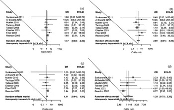
For total complications, the estimated overall odds ratio (ESS vs computer-assisted ESS) was 1.47 (95 per cent CI, 0.92–2.36; p = 0.11) (Figure 3a). Analysis of major complications generated an estimated overall odds ratio of 2.00 (95 per cent CI, 0.91–4.39; p = 0.09) (Figure 3b). For minor complications, the estimated overall odds ratio was 1.11 (95 per cent CI, 0.64–1.91; p = 0.71) (Figure 3c). This would suggest that the incidence of overall complications may be higher in the non-assisted ESS group than the computer-assisted ESS group, especially considering the higher proportion of major complications in patients undergoing ESS; however, neither of these results was statistically significant.
Forest plots showing random effects models of odds ratios (ORs) (conventional endoscopic sinus surgery vs computer-assisted endoscopic sinus surgery) for: (a) total complications; (b) major complications; (c) minor complications; and (d) revision surgery. CI = confidence interval; df = degrees of freedom

Revision
Six studies were included in the analysis of revision surgery. The Q statistic was 9.7 (p = 0.1) while the IReference Govindaraj, Adappa and Kennedy2 index was 48.5 per cent. This IReference Govindaraj, Adappa and Kennedy2 index value would be considered moderate, suggesting that there was wider variability between studies. The estimated overall odds ratio for revision surgery was 1.28 (95 per cent CI, 0.75–2.20; p = 0.37) (Figure 3d). These data suggest that there was a higher rate of revision surgery in patients undergoing non-assisted ESS compared with computer-assisted ESS, although this difference was not statistically significant.
Discussion
Development of image guidance systems
Today, ESS enjoys a reputation for great successReference Lee and Chiu18 and minimal risk. In contrast, an early, 1987 paper on ESS complicationsReference Stankiewicz19 reported a high incidence: major complications occurred in 8 per cent of cases. By the mid-1990s, May et al. Reference May, Levine, Mester and Schaitkin20 had reported much lower rates – 6.9 per cent minor and 0.85 per cent major complications – reflecting increased experience of ESS in the ENT community.
The perennial pursuit of the surgical fraternity is to develop techniques that achieve as high a success rate as possible with, ideally, a complication rate of zero. Despite the published, low complication rates, the significant morbidity and mortality associated with major complications warrants the adoption of any technique that can further reduce these risks.
Navigation systems were developed for this purpose in the 1980s and 1990s, having initially been pioneered in the field of neurosurgery. Since then, many surgical specialties have adopted the technology. Systems have evolved, from the early, rather cumbersome models developed by the Aachen group,Reference Anon, Klimek, Mosges and Zinreich1 to the electromagnetic or optically based, non-mechanically linked, frameless systems of today. All have a computer workstation, a tracking system and specially designed instruments that can be detected when placed within the surgical field. Software that allows rapid calculation of complex algorithms is pre-loaded onto the computer.
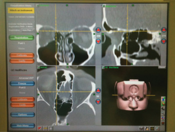
Data acquired from pre-operative CT scans are loaded onto the system, and images in all three planes are shown on the monitor. Positional data acquired in ‘real time’ from the instruments within the surgical field are incorporated into the three-dimensional data set from the CT images. The position is usually represented as a ‘cross-hair’ in all three CT images on the system monitor (Figure 4).
Screengrab showing GE InstaTrak image guidance system monitor display.

Indications
The American Academy of Otolaryngology – Head and Neck Surgery endorses computer-assisted ESS in selected sinus and skull base surgery cases, to assist the surgeon in clarifying complex anatomy. Examples of such cases include those involving: (1) revision sinus surgery; (2) distorted sinus anatomy of developmental, post-operative or traumatic origin; (3) extensive sino-nasal polyposis; (4) pathology involving the frontal, posterior ethmoid and/or sphenoid sinuses; (5) disease abutting the skull base, orbit, optic nerve or carotid artery; (6) CSF rhinorrhoea or conditions in which there is a skull base defect; and (7) benign and malignant sino-nasal neoplasms.21
A previous study showed that these guidelines have generally been adopted and are being followed.Reference Orlandi and Petersen22 We agree with these guidelines, although we also use image guidance routinely for all ESS procedures, as a matter of personal preference.
Complications
There is a perception that computer assistance might be helpful in minimising the incidence of complications. However, our own experience does not provide evidence for this, possibly because of the low numbers in our non-assisted ESS group. In our meta-analysis, only one studyReference Fried, Moharir, Shin, Taylor-Becker and Morrison8 provided evidence for fewer complications following computer-assisted ESS.
However, evidence indicates increased surgical accuracy, which suggests that, if used appropriately, complications arising from surgical error should occur less frequently.Reference Casiano and Numa5 Our meta-analysis revealed no significant difference in complication rates; however, despite the large numbers of patients, it remains uncertain whether this result represents actual reality or a type two error.
Indeed, Tabaee et al. Reference Tabaee, Hsu, Shrime, Rickert and Close7 calculated that, in order to demonstrate a 50 per cent improvement in the occurrence of a complication which itself has an incidence of 1 per cent, 6000 patients would be required in each study arm. Such a study is almost impossible to perform by a single surgeon or even a single institution. Multi-centre studies may help but will introduce further confounding variables.
Recurrence and revision
Compared with the other studies in our meta-analysis, our patient population was followed up for a much longer period of time. The most interesting finding was a significantly large hazard ratio for revision surgery requirements in the first 18 months following index surgery, comparing the two groups. Our analysis indicated that, during this period, the risk of disease recurrence in the non-assisted ESS group was over four times greater than that in the computer-assisted ESS group.
A number of anatomical factors contribute to the requirement for revision surgery.Reference Musy and Kountakis23 The majority of these relate to retained structures following incomplete surgery. A possible explanation for the large hazard ratio seen in our initial post-operative period is that computer-assisted ESS allows for a more complete clearance of cells and bony shelves, which would otherwise promote the persistence of disease and therefore the earlier development of recurrent symptoms. This benefit has been suggested previously.Reference Thong, Lee, Yeak and Siow24 None of the other studies analysed their revision surgery in chronological segments, so it was not possible to obtain stronger evidence for this parameter from our meta-analysis.
The meta-analysis did not demonstrate a significant difference in overall revision surgery rates. However, five of the six studies showed a greater revision surgery rate for ESS compared with computer-assisted ESS, although this difference was not significant. Only Reardon's studyReference Reardon16 demonstrated a higher rate of revision surgery for computer-assisted ESS (again, a non-significant difference). However, because this latter study included a large sample size, it had a significant impact on our analysis.
It is unclear why only Reardon's studyReference Reardon16 and our own found a higher rate of revision surgery in the computer-assisted ESS group, of the eight studies included in our meta-analysis. A possible explanation might be a difference in case mix.
Compared with the other studies, our population had a higher overall proportion of patients who went on to require revision surgery. There are two possible explanations.
Firstly, a very high proportion of patients referred to our centre had undergone previous sinus surgery performed elsewhere. Patients who require revision procedures are known to be much more likely to develop further residual disease.Reference Philpott, Dharamsi, Witheford and Javer25, Reference King, Caldarelli and Pigato26
Secondly, our study was performed very soon after the establishment of our hospital's Sinus Centre. A more recent paper from our CentreReference Philpott, Dharamsi, Witheford and Javer25 reported that, over a 10-year period, only 8 per cent of patients undergoing their first surgery at our Centre required revision surgery.
Use of image guidance amongst rhinologists
Two US national questionnaire surveys were reported in 2006.
Orlandi and PetersenReference Orlandi and Petersen22 distributed 1050 questionnaires but had a response rate of only 32.4 per cent. Of those who did respond, 81.9 per cent stated they used image guidance systems, but less than 10 per cent used such systems in more than 90 per cent of cases.
Hepworth et al. Reference Hepworth, Bucknor, Patel and Vaughan27 sent out 1100 questionnaires and gained a 61 per cent response. Seventy-three per cent of respondents used image guidance systems. Those who did not use them reported expense or lack of benefit as reasons for not doing so. Of those respondents who did use image guidance systems, 84 per cent stated that the most acceptable reason for doing so was revision surgery, closely followed by frontal sinus and sphenoid surgery. Eighty-five per cent of respondents report an increased level of confidence when using image guidance systems, although these were generally felt to prolong the surgical time.Reference Metson, Cosenza, Cunningham and Randolph28
In our experience, however, registration and calibration of the image guidance system occurs while the standard equipment trays are being prepared, and therefore adds negligible time to the procedure.
Training
It would seem reasonable to believe that the ability to identify intra-operative anatomy and compare it with CT findings in real time would be of benefit to trainee surgeons. However, the literature seems to be equivocal on this issue.
Casiano and Numa'sReference Casiano and Numa5 cadaver study of computer-assisted ESS for resident training showed that, even amongst inexperienced sinus surgeons, there was an improvement in surgical accuracy and efficiency.
A similar study from 200929 reported that, over a 2-day cadaver dissection course, trainees' confidence increased and their sinus dissection was demonstrably more complete. However, this result was independent of the use of an image guidance system.
Mueller et al. Reference Mueller and Caversaccio6 concluded that routine use of computer-assisted ESS should be discouraged as surgeons may become dependent upon it. Whilst there may be some merit to this argument, we believe that, supervised appropriately, computer-assisted ESS can in fact be a powerful learning tool for trainees.
Cost analysis
A cost analysis of computer-assisted ESS compared with non-assisted ESS was reported in 2001.Reference Gibbons, Gunn, Niwas and Sillers30 This study found that the use of image guidance systems was quantitatively more expensive, but that this was very different to ‘cost-effectiveness’, and that computer-assisted ESS provided many intangible benefits that might outweigh this added expense. Indeed, the authors believed that the true value of image guidance systems was inherently unquantifiable, as it was impossible to identify those patients in whom complications may have occurred had computer-assisted ESS not been used.
Meta-analyses such as ours may be helpful in assessing the financial aspects of image guidance systems. However, as indicated above, detecting a statistically significant change in financial parameters may require a prohibitive number of patients. Furthermore, the ethics of such a trial would be questionable.Reference Smith, Stewart, Orlandi, Setzen and Lanza31
However, it is certainly true that purchasing costs have come down, and are likely to continue to do so.
Patient satisfaction
One of the parameters that has been used to identify differences in surgical outcomes between computer-assisted and non-assisted ESS is subjective symptom score. A study at our CentreReference Javer and Genoway32 which used the 31-item Rhinosinusitis Outcome Measures Form questionnaire showed an improvement in quality of life in the early post-operative period when image guidance systems were used.
However, Tabaee et al. Reference Tabaee, Hsu, Shrime, Rickert and Close7 found no difference in patient satisfaction scores, based on the 20-item Sinonasal Outcome Test, more than 6 months after the date of surgery (mean patient follow up was 16.3 months). Following computer-assisted ESS, it would appear that any early gains in subjective benefits subsequently level out. This finding corresponds to the revision surgery requirements observed in our own study population.
Limitations of the Vancouver study
A significant limitation of the findings from our Centre is the small sample size in the non-assisted ESS group. A further limitation was that all the non-assisted ESS cases were performed before the computer-assisted ESS cases; however, due to the low numbers in the non-assisted group, there was only a short time period between the first non-assisted ESS cases and the first computer-assisted ESS cases. This was because the image guidance system was acquired quite soon after the senior surgeon's commencement at the St Paul's Sinus Centre. Nonetheless, it is important that this limitation is taken into account when considering the results.
• Image guidance systems have been used and developed for over a decade
• They may reduce complications and revision surgery rates following endoscopic sinus surgery
• Statistically significant differences are hard to demonstrate
• They are surgical tools, not substitutes for anatomical knowledge and surgical skill
For this reason, we felt it important to attempt to consolidate the findings in the literature by conducting a meta-analysis.
Conclusion
The results from our institution suggest that recurrence in the early to medium term after ESS for inflammatory sinus disease is significantly less frequent when an image guidance system is used. However, this result appeared to be unsupported by the meta-analysis, which showed no difference in the frequency of revision surgery following computer-assisted versus non-assisted ESS, although most studies showed a trend towards an overall reduction in revision surgery rates with computer-assisted ESS.
There was no significant difference in the incidence of post-operative complications; however, many of the meta-analysis studies showed some improvement in this outcome with computer-assisted ESS. On the face of it, this might suggest that computer-assisted ESS has no benefit over non-assisted ESS. However, the rarity of complications, particularly major complications, hinders the identification of real differences and increases the likelihood of a type two error.
We would emphasise what many authors have previously stated, that many advantages of image guidance systems are not easily assessed. As Gibbons et al. Reference Gibbons, Gunn, Niwas and Sillers30 have stated, ‘not all benefits…[of computer-assisted ESS]…are quantifiable’.Reference Gibbons, Gunn, Niwas and Sillers30 We believe that the intangible benefits of computer-assisted ESS outweigh the increased cost implication, if only by offering the surgeon added confidence and reassurance during surgery.
Nonetheless, it is also important to emphasise that computer assistance during ESS is no substitute for sound anatomical knowledge and surgical expertise.Reference Anon, Lipman, Oppenheim and Halt33 Like any other instrument used during the procedure, an image guidance system is simply a tool. Moreover, it is a tool that has the potential to malfunction during surgery. Surgeons should decide before surgery whether or not they have the appropriate training and experience to take on a particular case, without relying on the assistance of an image guidance system.
Image guidance systems are now in widespread use in North America.Reference Hepworth, Bucknor, Patel and Vaughan27 However, there is a reluctance to formally state that they represent the accepted ‘standard of care’, partly due to significant medico-legal implications. As more evidence is acquired, and as the use of image guidance systems becomes more widespread due to reduced purchase costs, this situation may eventually change.
Acknowledgement
We thank the secretarial staff at St Paul's Sinus Centre for their help with case note retrieval and administrative assistance.