Introduction
Post-auricular incisions are commonly performed for a range of otological procedures, including tympanoplasty and mastoid surgery. The popular post-auricular incision provides wide access and is advantageous in that the scar lies hidden from view during routine social interaction. However, the incision is often significantly longer than that of the main alternative, an endaural approach.
The sensory supply to the auricle and post-auricular skin is complex and variable,Reference Peuker and Filler1 with significant overlap in the distribution of cutaneous nerves. The size of the initial cutaneous sensory deficit from a post-auricular incision, and its subsequent potential for recovery, is therefore difficult to predict from the anatomy alone. Anecdotally, many patients seem to notice initial post-auricular numbness after their procedure, although for most it seems to be of significantly less concern at subsequent follow-up appointments.
The General Medical Council recommends that, as part of the process of gaining consent, surgeons should ‘…also tell patients about less serious side effects or complications if they occur frequently’.2 While most surgeons warn their patients of the risk of ear lobe numbness following parotidectomy, patients are often not warned of the potential for numbness following a post-auricular incision.
This study aimed to ascertain the frequency and severity of post-auricular numbness and its potential for recovery, so that appropriate information could be given to patients to secure informed consent.
Methods
Patient selection
The hospital operating theatre database was searched, by procedure code, to produce a list of patients who had undergone surgery potentially performed via a post-auricular approach between March and August 2008.
Data acquisition
Questionnaires were posted to these patients in May 2009. A second mailing was sent out in July to those who had not returned a completed questionnaire. Respondents' medical notes were then reviewed to record operative details, including the type of incision. In addition, any evidence of previous ear surgery or revision surgery between August 2008 and July 2009 was recorded.
Patients reported whether any area of the ear was numb, and if so whether the area affected was totally numb (i.e. anaesthetic) or partially numb (i.e. hypaesthetic). They were then asked to shade in, on lateral and oblique ear photographs, any areas in which they were experiencing such ongoing numbness. Respondents were also asked whether or not their numbness had increased, reduced or remained unchanged, in size and severity, over time. The impact on respondents' quality of life was ascertained by asking whether, on a day-to-day basis, they were unaware of the deficit, whether it caused them minor irritation, or whether they were constantly aware of it. Those whose sensory deficit had recovered were asked to shade the area previously affected, and to record how long it took this area to return to normal.
Respondents were excluded if (1) their procedure had included complex, extensive soft tissue resections or local flap reconstructions, or (2) their medical records were not available after three separate visits to the medical records department.
Results
General considerations
A total of 146 patients were sent questionnaires, of which 91 (62 per cent) responded after two requests.
Three respondents' notes were not found in the medical records department after three separate visits, and five respondents were excluded on review of their notes as they had undergone complex flap reconstructions.
Of the remaining 83 respondents, 61 (73 per cent) had received post-auricular incisions. Seventeen of these cases were revision procedures following previous surgery. Ten respondents had undergone further ear surgery between the procedure under consideration and their receipt of the questionnaire (one of whom had also had previous ear surgery). Therefore, 35 respondents had undergone primary operations via a post-auricular approach, and had undergone no other ear surgery at the time of completing the questionnaire.
Recovery of normal sensation and awareness of deficit
Primary surgery group
Table I shows results for this group. Of the 35 respondents in this group, 24 (69 per cent) reported experiencing initial numbness after their procedure, and one respondent could not remember whether he had experienced any transient numbness. Nine of the 35 respondents (26 per cent) reported continued numbness eight to 12 months after surgery. Of these nine, seven (78 per cent) reported hypaesthesia and two (22 per cent) anaesthesia. Six respondents (67 per cent) felt that the area involved had reduced in size, and seven (78 per cent) felt that the severity of the numbness had improved. One respondent was constantly aware of the numbness, six reported it as a minor irritation, and two felt it was not a hindrance during everyday activity.
Questionnaire: respondent data

Data represent respondent numbers (percentages) unless otherwise indicated. Mth = months; Hypaesth = hypaesthesia; Anaesth = anaesthesia; ↓= reduced; surg = surgery
Combined primary and revision surgery groups
Table I also shows combined results. Thirty-five of 51 respondents (69 per cent) reported initial numbness in the post-auricular area after surgery, and one could not remember clearly. Fifteen of these 51 respondents (29 per cent) had continued numbness eight to 12 months after surgery. Of the 15 with ongoing numbness, 11 (73 per cent) reported hypaesthesia and four (27 per cent) anaesthesia. Eight (53 per cent) felt that the area of numbness had reduced in size, and nine (60 per cent) that it had reduced in severity. Four respondents (27 per cent) were constantly aware of the numbness, seven (47 per cent) found it a minor irritation, and three (20 per cent) were not troubled by it at all; one respondent failed to answer.
Duration of recovery
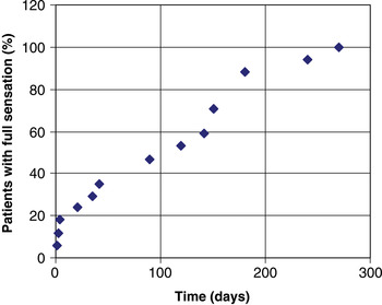
Eighty-eight per cent of respondents who made a subjective full sensory recovery within eight to 12 months post-operatively had done so by six months (see Figure 1), and 18 per cent had done so within four days.
Reported cumulative recovery of full sensation in patients making a complete recovery eight to 12 months post-operatively.

Cutaneous areas affected
Ongoing numbness
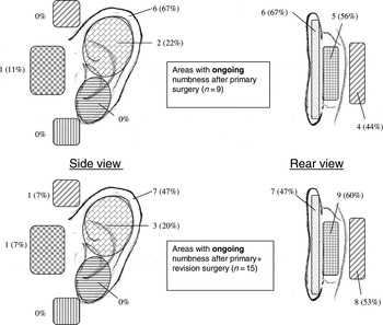
Of those respondents who had undergone primary surgery, the most common areas in which numbness continued beyond eight to 12 months post-operatively were the medial pinna (56 per cent of respondents reported ongoing numbness in this region), the area posterior to the post-auricular sulcus (44 per cent) and the helical rim (67 per cent; n = 9) (see Figure 2). A similar pattern was found after respondents receiving revision surgery (n = 15) were included in the analysis.
Diagrams showing the number of patients with ongoing numbness in specific anatomical locations, after eight to 12 months' recovery from primary surgery (upper diagrams) and after primary and revision surgery (lower diagrams; combined patient data).

Temporary numbness
In the 15 respondents who had undergone primary surgery and whose numbness had completely recovered, the areas previously affected were the helical rim in eight patients (53 per cent), the medial pinna in seven (47 per cent), the area posterior to the post-auricular sulcus in six (40 per cent) and the upper half of the lateral pinna in four (27 per cent) (see Figure 3). Again, a very similar pattern was found when revision cases were included in the analysis.
Diagrams showing the number of patients with temporary numbness in specific anatomical locations, after eight to 12 months' recovery from primary surgery (upper diagrams) and after primary and revision surgery (lower diagrams; combined patient data).

Discussion
Patients questioned after parotid surgery state that their ear numbness predisposes them to ear trauma in daily life. One study found that only 50 per cent of patients undergoing parotidectomy with confirmed great auricular nerve section went on to recover pinna sensation after one year.Reference Ryan and Fee3 There are several published studies examining the recovery of cutaneous sensation following parotidectomyReference Ryan and Fee4–Reference Brown and Ord6 and alveolar nerve section.Reference Robinson7 However, we are not aware of any, in the English language literature, which have assessed sensory deficit following post-auricular incision, and recovery of sensation over time.
Study design
Previous studies examining the recovery of cutaneous sensation at other anatomical sites have used objective measurement and multi-modality testing. However, it has been shown that patients' subjective questionnaire responses correlate well with calibrated anaesthesiometer testing.Reference Ryan and Fee4 In addition, the use of a questionnaire-based study enabled us to include a larger sample population without inconveniencing patients by requesting repeated attendance for objective testing.
Following inferior alveolar or great auricular nerve section, maximal recovery of light touch sensation has been shown to have occurred by eightReference Robinson7 or 12 months.Reference Ryan and Fee4 Another study found that, following great auricular nerve sacrifice, the majority of recovery of pin prick sensitivity had occurred by six months.Reference Porter and Wood5 Given that ours was a retrospective, questionnaire study, it was felt that patient responses might be too unreliable if the period of recollection was greater than a year. In addition, many combined approach tympanoplasty cases are re-opened for a ‘second look’ at one year, preventing a longer period of observation before surgery is repeated. As a result, we could not comment on further sensory recovery beyond eight to 12 months post-operatively.
It was difficult to achieve a high questionnaire completion rate, especially given the fact that a significant proportion of the patient group were members of the armed forces and were either posted overseas or had moved bases when the questionnaires were distributed. A second mailing was sent to maximise the return, and a final response rate of 62 per cent was achieved. While this is not a low figure by questionnaire standards, an even higher completion rate would clearly have been desirable.
Interestingly, while traditional teaching states that surveys with low response rates are unreliable due to probable sampling bias, recent studies in the non-medical literature have challenged this view, suggesting that the results from surveys with lower response rates are often no less accurate.Reference Keeter, Kennedy, Dimock, Best and Peyton8 Another author found no effect on study results after excluding data from subjects who initially refused to respond, or who required multiple prompts to respond,Reference Curtin, Presser and Singer9 suggesting that initial failure to respond does not necessarily produce sampling bias.
However, a higher response rate would have given us a greater degree of confidence in the accuracy of our results, and allowed us to perform more detailed subgroup analysis of the primary and revision surgery groups.
Results
The cutaneous supply of the auricle is complex, and varies between individuals. The auricle receives its cutaneous innervation from the lesser occipital nerve, great auricular nerve, auriculotemporal nerve and the auricular branch of the vagus nerve. The pattern of innervation varies between individuals, and there are frequently areas of dual, but not triple, innervation.Reference Peuker and Filler1
• This questionnaire study examined cutaneous sensory disturbance after otological post-auricular incision
• After primary surgery, initial numbness was present in 69 per cent of patients, persisting beyond eight months in 26 per cent
• Those with ongoing numbness mostly described: hypaesthesia (not anaesthesia), significant improvement with time, and little effect on daily activities
• Numbness mainly occurred on the helical rim, medial pinna, and posterior to the post-auricular sulcus
• Numbness after revision surgery showed less recovery over time
• Findings are relevant to the consenting process
Nerve injury sustained during a post-auricular surgical approach can encompass all degrees of severity, from neuropraxia to neurotmesis (nerve transection), in differing areas of the surgical field. When neurotmesis occurs, Wallerian degeneration of the distal segment follows, with degradation of axons and the surrounding myelin. This leaves empty endoneural tubes that can act as a scaffold for nerve regrowth from the proximal stump. Although reparative nerve growth along a graft can proceed as rapidly as 2–3 mm/day, it is often much slower in a wound scar, which is more difficult for the axons to breach. In such situations, growth may occur at a rate of approximately 0.25–1 mm/day in the human adult, and faster in children.Reference Lawrence, Kashyap, Achauer, Eriksson and Wilkins10 However, regenerating axons in vitro have been shown capable of traversing a gap as wide as 10 mm in a mesothelial chamber, so reinnervation from the proximal stump is certainly a possibility.Reference Lundborg and Lundborg11 Thus, post-auricular cutaneous nerves do theoretically have the ability to search out their distal separated segment, to allow more precise reinnervation.
Alternative mechanisms of sensation recovery may include activation of previously quiescent overlapping dermatomes, or active nerve in-growth by neighbouring nerves as reported elsewhere in the body.Reference Robinson7 However, studies blocking stump regeneration following inferior alveolar nerve section have suggested that surrounding nerves can only reduce the area of anaesthesia by 65 per cent, and that they are unable, alone, to provide complete recovery.Reference Robinson7 A similar study has not been performed in the region of the auricle.
Finally, the plasticity of the central nervous system may have a role in interpreting the new, disordered cutaneous signals following nerve regrowth, but this is likely to be a more significant factor in children than adults.Reference Florence, Jain, Pospichal, Beck, Sly and Kaas12 In view of the multiple cutaneous nerves in the auricular area, with overlapping distributions, it is likely that a combination of reinnervation methods occurs following a post-auricular incision.
Conclusion
These study findings emphasise the need for patients undergoing a post-auricular surgical approach to be informed of the likelihood of a cutaneous deficit, and its potential for improvement over time. Our findings provide data which will be useful in such discussions. Our results also raise the possibility that revision surgery may result in less complete sensory recovery, after eight to 12 months, which is relevant when obtaining consent from this patient group.
We now advise our patients that there is a 70 per cent chance of some numbness around the ear after their operation, but that in the majority of people this will fully recover within six months. Patients undergoing primary surgery have a 26 per cent chance of numbness continuing beyond eight months; however, in less than 3 per cent is such numbness troublesome in everyday life.