Introduction
The phylum Acanthocephala (spiny-headed worms) is a small group of endoparasites with approximately 1300 species, commonly occurring in the intestine of various fishes, amphibians, reptiles, birds and mammals (Amin, Reference Amin2013; Lisitsyna et al., Reference Lisitsyna, Kudlai, Cribb and Smit2019). In the past few decades, large numbers of new taxa of acanthocephalans have been reported worldwide (Pichelin and Cribb, Reference Pichelin and Cribb2001; Amin and Ha, Reference Amin and Ha2008), which contributed useful material for understanding the evolutionary history and reconstructing the classification of the phylum Acanthocephala. In contrast, our present genetic knowledge of Acanthocephala, especially the mitochondrial genomes, are still very limited. To date, only 17 acanthocephalan species with their mt-genomes are available in the NCBI database (Steinauer et al., Reference Steinauer, Nickol, Broughton and Ortí2005; Gazi et al., Reference Gazi, Sultana, Min, Park, García-Varela, Nadler and Park2012; Reference Gazi, Kim and Park2015; Reference Gazi, Kim, García-Varela, Park, Littlewood and Park2016; Pan and Nie, Reference Pan and Nie2013; Weber et al., Reference Weber, Wey-Fabrizius, Podsiadlowski, Witek, Schill, Sugár and Hankeln2013; Song et al., Reference Song, Zhang, Deng, Ding, Liao and Liu2016; Reference Song, Zhang, Gao, Cheng, Xie, Li and Wu2019; Pan and Jiang, Reference Pan and Jiang2018; Muhammad et al., Reference Muhammad, Ma, Khan, Li, Zhao, Ahmad and Zhu2019a, Reference Muhammad, Ma, Khan, Wu, Zhu and Lib, Reference Muhammad, Khan, Li, Zhao, Ullah, Zhu and Ma2020a, Reference Muhammad, Li, Zhao, Bannai, Mohammad, Khan and Mab). The current classifications of Acanthocephala are established mainly based on morphological characters and ecological traits (Amin, Reference Amin2013). The systematic status of many genera or families of Acanthocephala remains controversial.
The family Centrorhynchidae currently including approximately 120 species are placed in three genera, Centrorhynchus Lühe, (1911), Sphaerirostris Golvan (Reference Golvan1956) and Neolacunisoma Amin and Canaris (Reference Amin and Canaris1997) (Amin, Reference Amin2013). However, the phylogenetic relationships of these three genera have not been solved. Golvan (Reference Golvan1956) established the subgenus Sphaerirostris in the genus Centrorhynchus. Later, Golvan (Reference Golvan1969) raised the subgenus Sphaerirostris to full genus rank. This proposal was accepted by most of the subsequent studies, i.e. Dimitrova et al. (Reference Dimitrova, Murai and Genov1995, Reference Dimitrova, Georgiev and Genov1997), Amin and Canaris (Reference Amin and Canaris1997), Amin et al. (Reference Amin, Heckmann, Halajian and Eslami2010), and Amin (Reference Amin2013). The genus Sphaerirostris can be distinguished from Centrorhynchus mainly based on the shape and size of trunk and testes, the pattern of the lacunar system and the morphology and armature of proboscis. However, due to the paucity of genetic data of the Centrorhynchidae, there was no phylogenetic study using molecular data to clarify the relationships of the two genera before 2020. Recently, Muhammad et al. (Reference Muhammad, Khan, Li, Zhao, Ullah, Zhu and Ma2020a, Reference Muhammad, Li, Zhao, Bannai, Mohammad, Khan and Mab) provided a phylogenetic analysis of Acanthocephala using mitogenomic data that displayed S. picae nested in some species of Centrorhynchus, but there was only one species of Sphaerirostris included in the phylogeny. However, a more rigorous phylogeny including more species from Sphaerirostris and Centrorhynchus with their mitogenomics are required to solve the phylogenetic relationships of these two genera.
In the present study, the nearly complete mitochondrial (mt) genome of S. lanceoides was sequenced and annotated for the first time, based on specimens collected from the Indian pond heron Ardeola grayii (Sykes) (Ciconiiformes: Ardeidae) in Pakistan. Moreover, in order to clarify the phylogenetic relationships of Centrorhynchus and Sphaerirostris, and to test the systematic position of S. lanceoides in the Centrorhynchidae, the phylogenetic analyses were performed using Bayesian inference (BI) and maximum likelihood (ML) methods based on concatenated nucleotide sequences of 12 protein-coding genes (PCGs), rRNAs and tRNAs genes.
Materials and methods
Parasites collection
The specimens of S. lanceoides (Petrochenko, 1949) were isolated from the intestine of the Indian pond heron Ardeola grayii (Sykes) (Ciconiiformes: Ardeidae) in the district Swabi (34°07′07.23″ N, 72°36′32.38″ E), Khyber Pakhtunkhwa Province, Pakistan, and then stored in 75% ethanol at −20°C for molecular studies. The specimens were identified as the cystacanths of S. lanceoides using morphological characters and genetic data (cox1 sequences) according to Sato et al. (Reference Sato, Suzuki, Uni and Kamiya2005, Reference Sato, Suzuki and Aoki2006), Torracca et al. (Reference Torracca, Macchioni, Masetti, Gabrielli, Guardone, Prati and Magi2010), and Kang and Li (Reference Kang and Li2018).
Molecular procedures
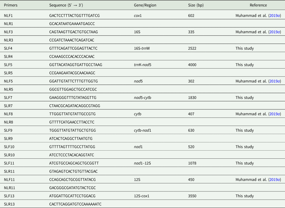
The genomic DNA was extracted from the specimens using sodium dodecyl sulfate/proteinase K solution and column purification (Wizard® SV Genomic DNA Kit, Promega, Madison, USA) following the protocol (Gasser et al., Reference Gasser, Hu, Chilton, Campbell, Jex, Otranto, Cafarchia, Beveridge and Zhu2006). Five short fragments (cox1, rrnL, nad5, cytb and rrnS) of the mt genome of S. lanceoides were amplified using a primer sets of NLF1/NLR1, NLF3/NLR3, NLF5/NLR5, NLF8/NLR8 and NLF11/NLR11 (Muhammad et al., Reference Muhammad, Ma, Khan, Li, Zhao, Ahmad and Zhu2019a). For amplification of the remaining sequences of the S. lanceoides mt genome, seven pairs of primers were designed from the amplified short fragments. The detailed information of primers was shown in Table 1. Polymerase chain reactions (PCR) were organized in a 50 μL reaction mixture, containing 2 μL DNA template, 1.5 μL of each primer, 22.5 μL dd H2O and 22.5 μL PrimeSTAR Max DNA polymerase (Takara, Dalian, China). Amplification was carried out under the following conditions: initial denaturation at 98°C for 3 min followed by 30 cycles at 96°C for 10 s, 45–60°C for 15 s, 65°C for 1 min/kb, and final extension for 10 min at 68°C. The obtained PCR products were sequenced at Genewiz company (Beijing, China) using primer walking strategy Table 2.
PCR primers used for the amplification of nearly complete mt genome of S. lanceoides

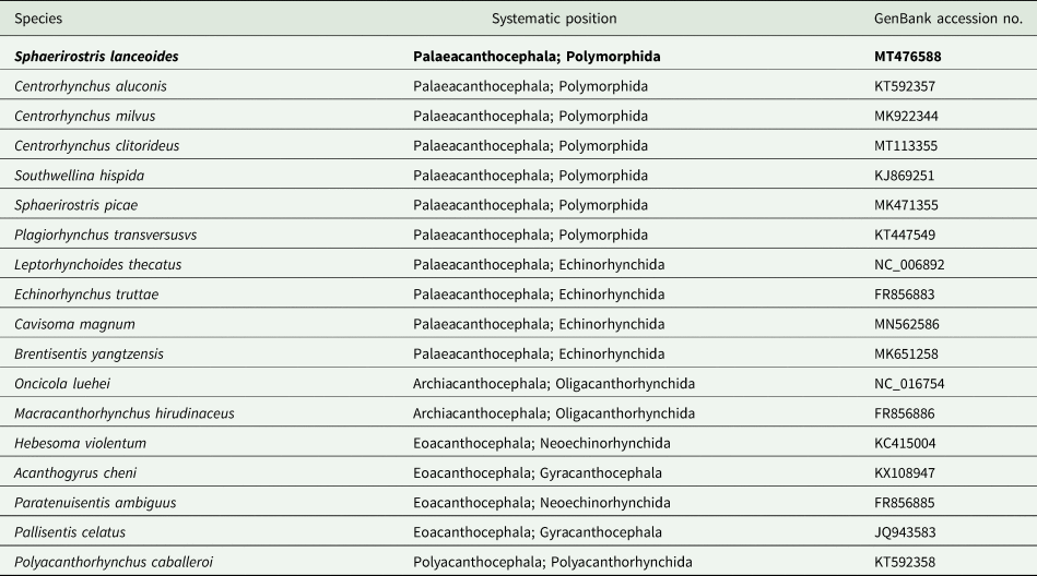
The species, taxonomy and GenBank accession numbers for acanthocephalan used in the phylogenetic analysis in this study

Newly sequenced species is shown in bold.
The chromatograms were examined using Chromas v.1.62. The complete mt genome sequences of S. lanceoides were assembled manually using the program DNAstar v7.1 (Burland, Reference Burland2000). The MEGA7 (Kumar et al., Reference Kumar, Stecher and Tamura2016) was used to analyse the nucleotide composition and codon usage. The boundaries of two ribosomal RNA genes (rrnL and rrnS) and 12 PCGs were identified by comparison with S. picae (MK471355) and Centrorhynchus aluconis (KT592357). The sequences alignment was performed using MAFFT 7.130. The 12 PCGs amino acid sequences were obtained with the help of MEGA7, while invertebrate mt code was chosen. The transfer RNA genes (tRNAs) were identified using ARWEN (Laslett and Canback, Reference Laslett and Canback2008) and MITOS web server (Bernt et al., Reference Bernt, Donath, Juhling, Externbrink, Florentz, Fritzsch, Putz, Middendorf and Stadler2013) or by alignment with other centrorhynchid species. The 12 PCGs codon usage and relative synonymous codon usage (RSCU) were determined using PhyloSuite v1.1.15 (Zhang et al., Reference Zhang, Gao, Jakovlić, Zou, Zhang, Li and Wang2020). RSCU figure was drawn by ggplot2 plugin (Wickham, Reference Wickham2016).
Phylogenetic analysis
Phylogenetic analyses were conducted using the newly sequenced S. lanceoides and 17 other acanthocephalans mitogenome based on 12 PCGs, two rRNAs and 22 tRNAs sequences. Two species of Bdelloidea Rotaria rotatoria (NC013568.1) and Philodina citrina (FR856884.1) were chosen as the out-groups according to the previous studies (García-Varela et al., Reference García-Varela, Cummings, Perez-Ponce de Leon, Gardner and Laclette2002; García-Varela and Nadler, Reference García-Varela and Nadler2006; Gazi et al., Reference Gazi, Sultana, Min, Park, García-Varela, Nadler and Park2012, Reference Gazi, Kim and Park2015, Reference Gazi, Kim, García-Varela, Park, Littlewood and Park2016). The nucleotide sequences of 12 PCGs, 22 tRNAs and two rRNAs of all included mitogenomes were collected from GenBank files, using PhyloSuite (Zhang et al., Reference Zhang, Gao, Jakovlić, Zou, Zhang, Li and Wang2020). Phylogenetic analyses were operated using the codon-based alignment of nucleotide sequences of PCGs + Q-INS-i strategy of alignment for tRNAs and rRNAs. The nucleotide sequences of each gene were aligned in batches using MAFFT Ver 7.122, poorly aligned regions were excluded using Gblocks 0.91b (Talavera and Castresana, Reference Talavera and Castresana2007).
The sequences were finally concatenated into a single alignment used to generate nexus files in PhyloSuite. Phylogenetic analyses were conducted using BI and ML methods. Based on the Akaike's information criterion, as implemented in ModelFinder (Kalyaanamoorthy et al., Reference Kalyaanamoorthy, Minh, Wong, Von Haeseler and Jermiin2017), GTR + F + I + G4 and GTR + F + R5 were selected as best-fitting models for nucleotide evolution for the BI and ML analyses, respectively. Phylogenetic analysis was done based on the ML method using IQ-Tree (Nguyen et al., Reference Nguyen, Schmidt, Von Haeseler and Minh2015) by carrying out ultrafast bootstraps (Minh et al., Reference Minh, Nguyen and Von Haeseler2013) with 5000 replicates. Phylogenies based on BI were constructed using MrBayes 3.2.6 (Ronquist et al., Reference Ronquist, Teslenko, Van der Mark, Ayres, Darling, Höhna, Larget, Liu, Suchard and Huelsenbeck2012) with default setting and BI analysis was run for 3 × 10⁶ metropolis-coupled Markov chain Monte Carlo generations and sampling a tree with every 1000 generations. The first 25% (750) trees were treated as ‘burn-in’. Finally, the phylograms were viewed and annotated by iTOL (Letunic and Bork, Reference Letunic and Bork2016) and Adobe Illustrator® with the help of PhyloSuite generated dataset files.
Results and discussion
Identification of S. lanceoides
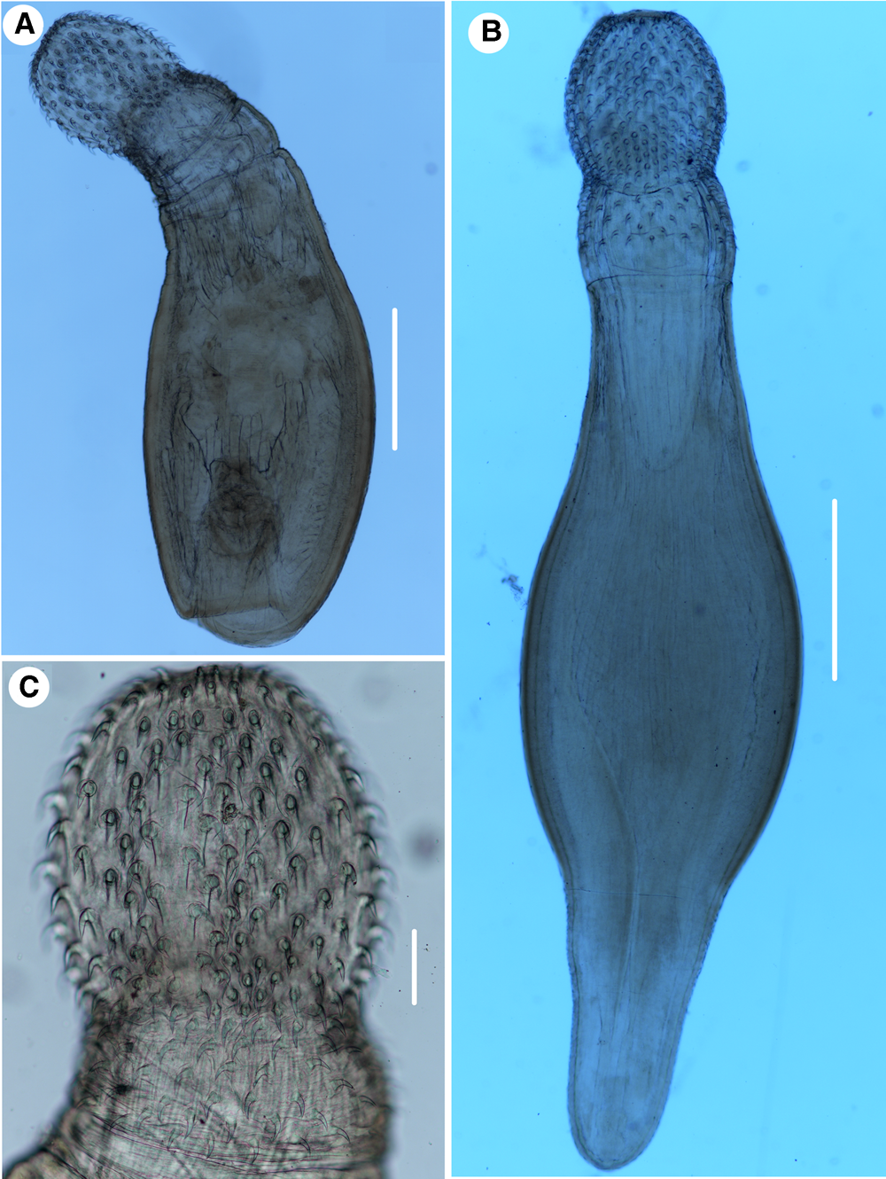
Small, whitish worms, trunk spindle-shaped, 3.07 mm long (posterior end retracted), with maximum width 0.84 mm. Proboscis is divided into two parts separated by constriction; anterior proboscis nearly spherical, 0.48 × 0.42 mm2, with 34 longitudinal rows of 11 hooks each. Posterior proboscis cylindrical, 0.18 × 0.41 mm2, with 30 longitudinal rows of 4 hooks each. Proboscis receptacle and lemnisci not clear. Uterus 0.10 × 0.06 mm2; vagina 0.17–0.10 mm. Eggs absent.
The morphology and morphometrics of our specimens are almost identical with the descriptions of the cystacanths of S. lanceoides by Sato et al. (Reference Sato, Suzuki, Uni and Kamiya2005, Reference Sato, Suzuki and Aoki2006), Torracca et al. (Reference Torracca, Macchioni, Masetti, Gabrielli, Guardone, Prati and Magi2010) and Kang and Li (Reference Kang and Li2018), including the size and morphology of the trunk and proboscis, the number of the longitudinal rows of proboscis hooks and the hooks per longitudinal row (Fig. 1). Moreover, pairwise comparison of the cox1 sequences of our specimens with that of S. lanceoides (MG931942.1) available in GenBank provided by Kang and Li (Reference Kang and Li2018), displayed 98.21% of nucleotide similarity. The morphological and genetic evidence both supported that our present specimens collected from A. grayii in Pakistan to be the species S. lanceoides.
Photomicrographs of cystacanths of Sphaerirostris lanceoides. A, sample collected from A. grayii (Ciconiiformes: Ardeidae) in Pakistan (posterior end of trunk retracted); B, sample collected from Bufo gargarizans (Amphibia: Anura) in China; C, proboscis of sample collected from Pakistan. Scale bars: A,B = 0.5 mm, C = 0.1 mm. B, anterior part of female; C, region of testes; D, posterior part of male; E, posterior end of male (arrow indicated gonopore); F, posterior end of female (arrow indicated gonopore). B, bursa; C, cement glands; G, gonopore; L, lemnisci; N, neck; R, proboscis receptacle; T, testes.

General characterization of S. lanceoides mt genome
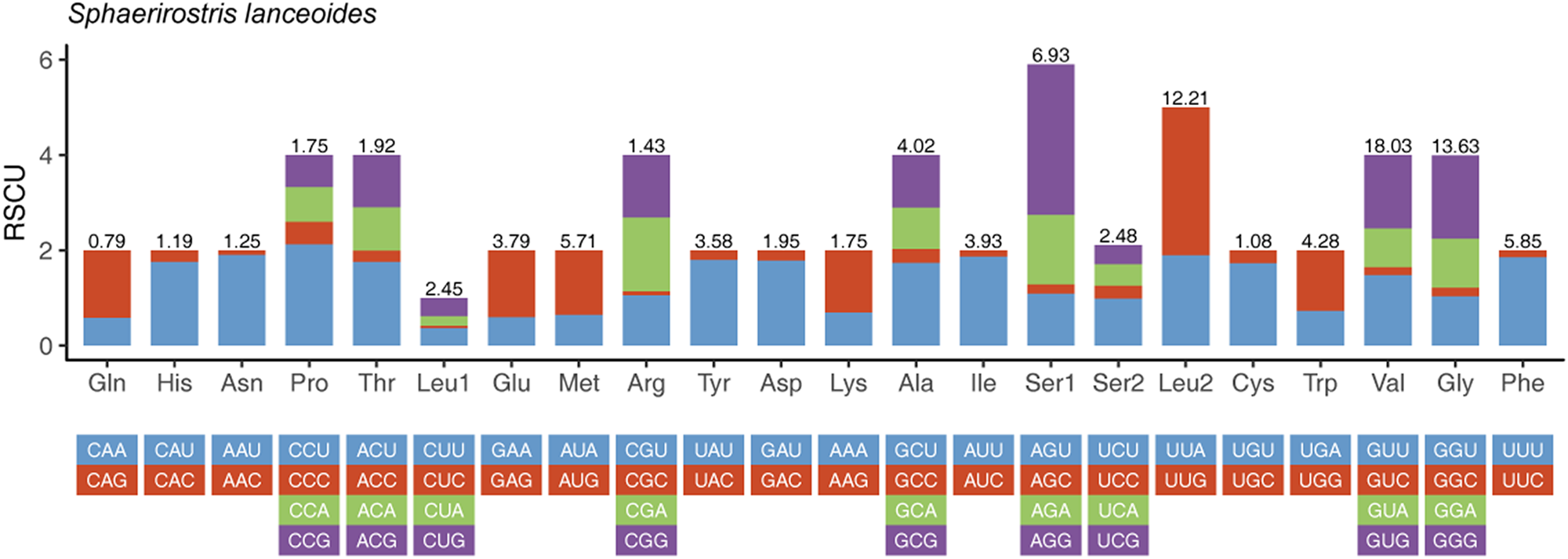
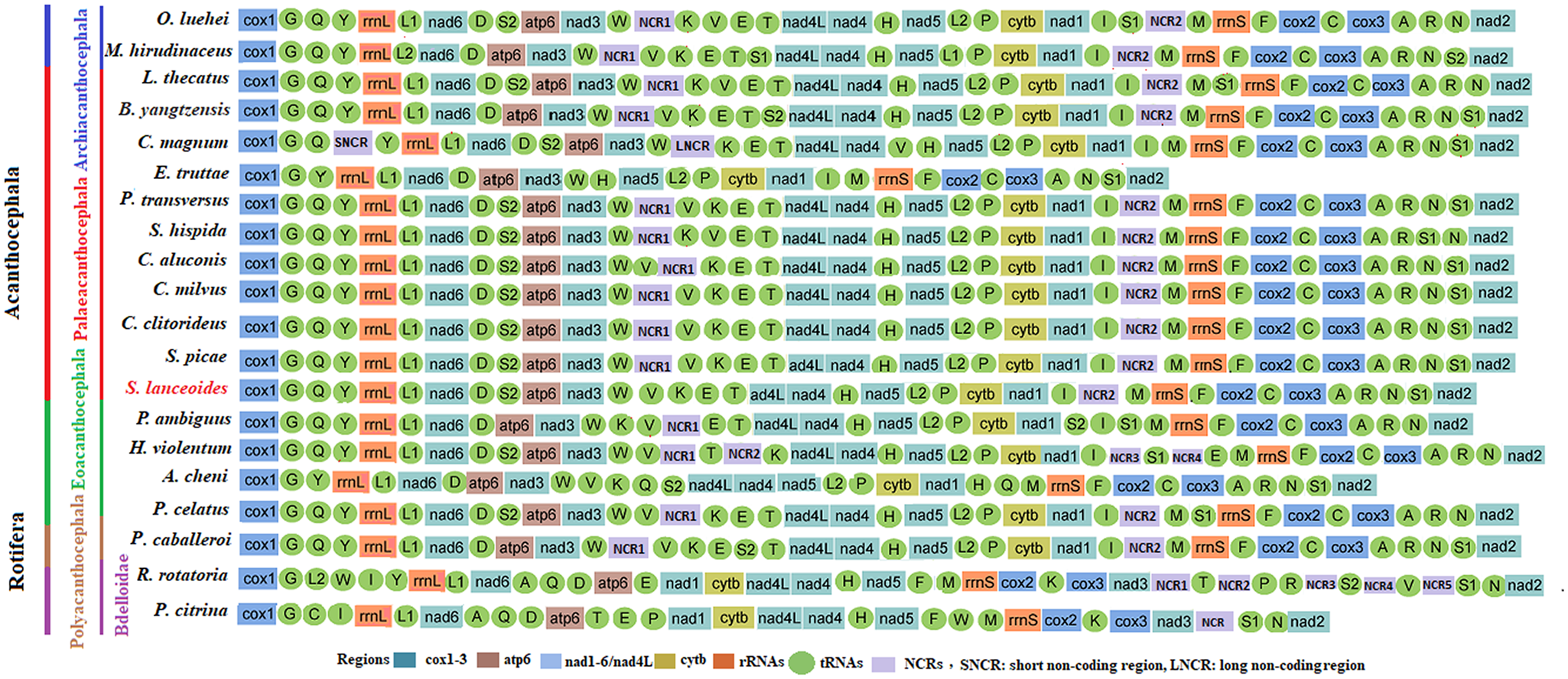
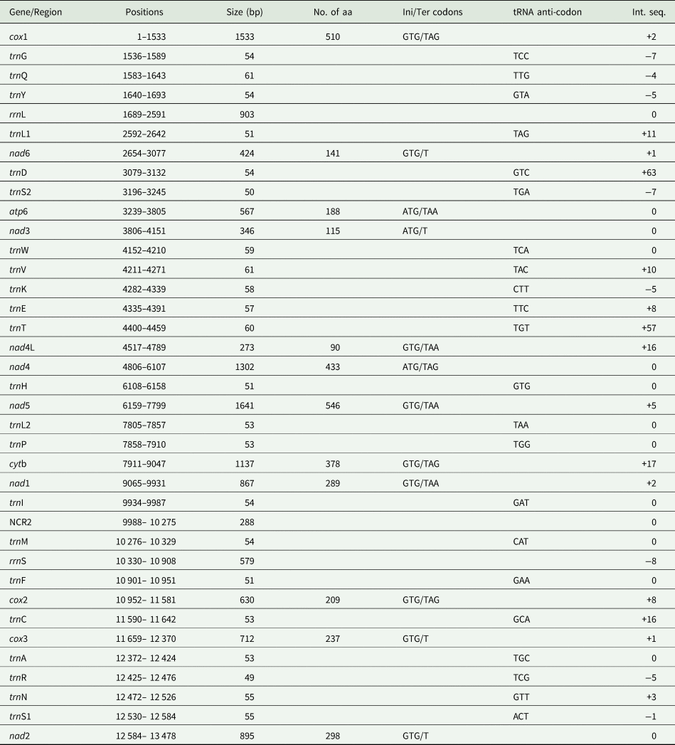
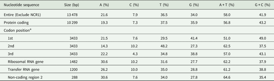
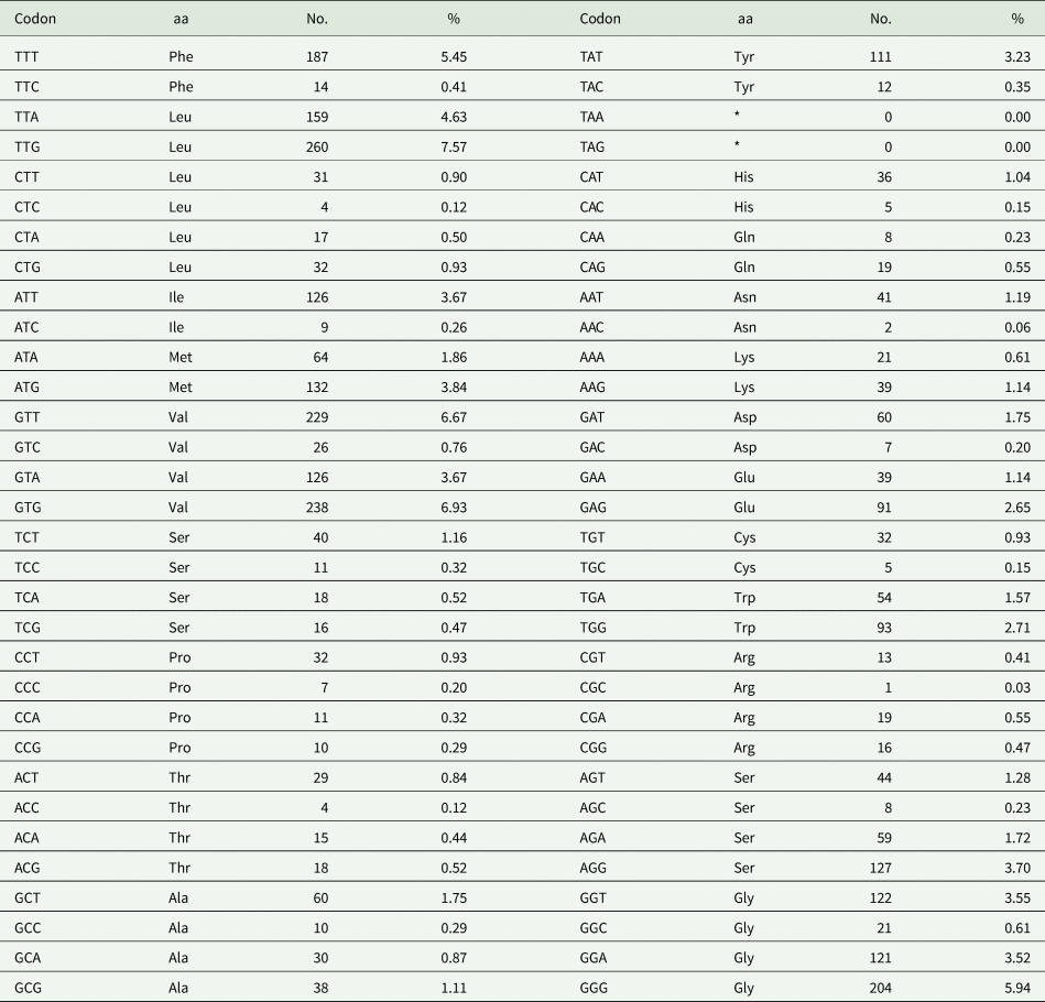
The nearly complete mt genome (excluding NCR1) of S. lanceoides was 13 478 bp in length, containing 12 PCGs (missing atp8) (cox1–3, atp6, nad1–6, nad4L and cytb), 22 tRNAs and two rRNA (rrnL or 16S and rrnS or 12S) (Table 3). The 12 PCGs were 10 302 bp in length, excluding termination codons, which encoded 3434 amino acids (Table 4). The component and usages of codons in the S. lanceoides mt genome were shown in Fig. 2 and Table 5. GTG and ATG were used as start codons in 12 PCGs of mt genome of S. lanceoides, whereas TAA, TAG and T were used as termination codons (Table 3). The gene arrangement of all 36 genes and NCRs (Fig. 3) was the same as that in the mitogenome of S. picae (Muhammad et al., Reference Muhammad, Ma, Khan, Li, Zhao, Ahmad and Zhu2019a).
Relative Synonymous Codon Usage (RSCU) of 12 PCGs of S. lanceoides, on the x-axis the codon families are labelled. Amino acid usage in percentage is represented by values on the top of each bar.

Linearized comparison of the mitochondrial gene arrangement for 18 acanthocephalan species and two rotifer species. Genome and Gene size are not to the scale. All genes are encoded from left to right in the same direction. The tRNAs are labelled by single-letter code for the corresponding amino acid. The red colour indicates the newly complete mtDNA sequenced species in this study.

Organization of the nearly complete mitochondrial genome of Sphaerirostris lanceoides

NCR, non-coding region; bp, base pair; aa, amino acid; Ini/Ter, initial/terminal codons; Int. seq, intergenic sequences.
Nucleotide composition of the mitochondrial genome of Sphaerirostris lanceoides

a Termination codons are excluded.
Genetic code and codon usage for the 12 mitochondrial protein-coding genes of Sphaerirostris lanceoides

No., number of copies; *, stop (termination) codon; aa, amino acid.
Phylogenetic analyses
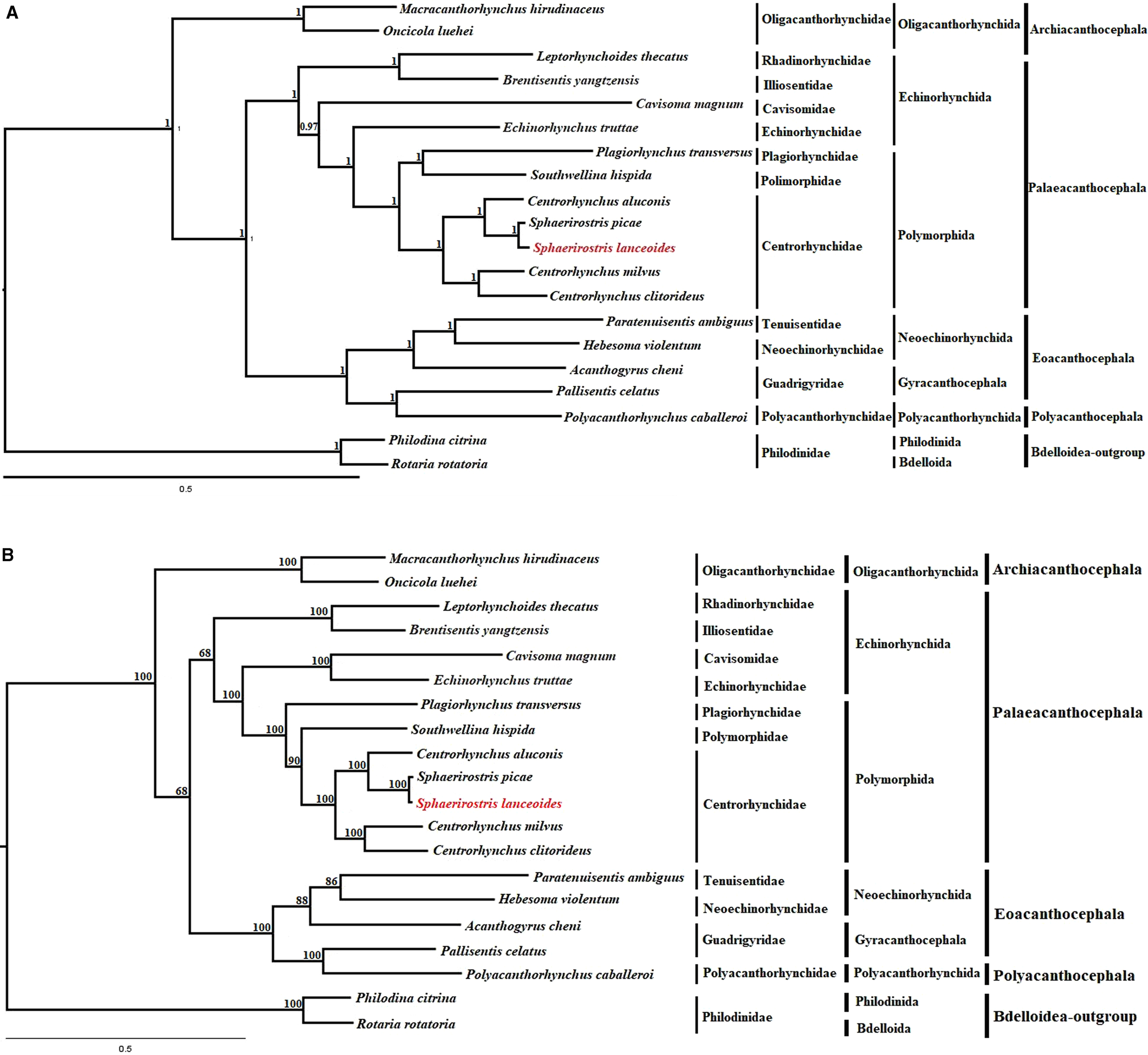
The phylogenetic trees were built using BI and ML methods. The phylogenetic tree using BI analysis showed nearly identical topology to the ML tree (Fig. 4a and b). Both phylogenetic trees showed that the class Archiacanthocephala, including representatives O. luehei and M. herudinaceus (Oligacanthorynchida: Oligacanthorynchidae), was a monophyletic group, which represented a basal clade of Acanthocephala. The present results agreed well with the previous molecular phylogenetic studies (Near et al., Reference Near, Garey and Nadler1998; García-Varela et al., Reference García-Varela, Perez-Ponce de Leon, De la Torre, Cummings, Sarma and Laclette2000; Near, Reference Near2002; García-Varela and Nadler, Reference García-Varela and Nadler2005; Verweyen et al., Reference Verweyen, Klimpel and Palm2011; Weber et al., Reference Weber, Wey-Fabrizius, Podsiadlowski, Witek, Schill, Sugár and Hankeln2013; Gazi et al., Reference Gazi, Kim and Park2015). Our phylogenetic results also revealed that the representative of the class Polyacanthocephala (P. caballeroi) nested within the class Eoacanthocephala, which seems to suggest that the members of the class Polyacanthocephala should be considered as members of the class Eoacanthocephala (Fig. 4a and b). The interrelationships among the four classes of Acanthocephala revealed by the present phylogenetic analysis are consistent with the previous phylogenomic studies (Gazi et al., Reference Gazi, Kim, García-Varela, Park, Littlewood and Park2016; Muhammad et al., Reference Muhammad, Ma, Khan, Li, Zhao, Ahmad and Zhu2019a, Reference Muhammad, Ma, Khan, Wu, Zhu and Lib, Reference Muhammad, Khan, Li, Zhao, Ullah, Zhu and Ma2020a, Reference Muhammad, Li, Zhao, Bannai, Mohammad, Khan and Mab; Song et al., Reference Song, Zhang, Gao, Cheng, Xie, Li and Wu2019).
(A) Phylogenetic analysis of Acanthocephala using Bayesian inference based on concatenated amino acid sequences of 12 protein-coding genes. Philodina citrina (FR856884.1) and Rotaria rotatoria (GQ304898.1) (Bdelloidea: Rotifera) treated as out-groups. (B) Phylogeny of Acanthocephala based on available mitochondrial genome data. The phylogram was constructed based on maximum likelihood (ML) methods using concatenated nucleotide sequences of protein-coding genes, rRNAs and tRNAs of 18 acanthocephalan mitogenomes. The newly sequenced species is indicated by red colour. Rotaria rotatoria (GQ304898.1) and Philodina citrina (FR856884.1) (Bdelloidea: Rotifera) was used as outgroup.

In Palaeacanthocephala, the order Polymorphida with only 7 representatives was supported to be a monophyletic clade in the phylogenetic tree, in contrast, the order Echinorhynchida was displayed to be a paraphyletic group. The phylogenetic results agreed well with the recent phylogenetic study (Muhammad et al., Reference Muhammad, Khan, Li, Zhao, Ullah, Zhu and Ma2020a, Reference Muhammad, Li, Zhao, Bannai, Mohammad, Khan and Mab) and some previous molecular studies based on some nuclear and mitochondrial genetic markers i.e. SSU/LSU + SSU/SSU + LSU + cox1 (García-Varela et al., Reference García-Varela, Cummings, Perez-Ponce de Leon, Gardner and Laclette2002; García-Varela and Nadler, Reference García-Varela and Nadler2005, Reference García-Varela and Nadler2006; García-Varela and González-Oliver, Reference García-Varela and González-Oliver2008; Verweyen et al., Reference Verweyen, Klimpel and Palm2011). In our phylogenetic trees, the species S. lanceoides grouped together with S. picae, and they both nested in the representatives of the genus Centrorhynchus in the family Centrorhynchidae with strong nodal support (BPP = 1). The results indicated that the validity of the genus Sphaerirostris seems to be challenged. It seems that these morphological characters for differentiating the genera Sphaerirostris and Centrorhynchus, i.e. the shape and size of trunk and testes, the pattern of the lacunar system and the morphology and armature of proboscis, have restricted taxonomic value of generic importance. However, we do not make any immediate change on the systematic status of Sphaerirostris, because only a few species of Sphaerirostris were included in the present phylogenetic analyses. A more rigorous phylogenetic study with broader representatives of the family Centrorhynchidae based on the nuclear and mt genomic data is required to further clarify the systematic status of Sphaerirostris in the future.
Concluding remarks
In the present study, the characterization of the nearly complete mt genome of S. lanceoides was reported for the first time based on specimens collected from the Indian pond heron (Ardeola grayii, Ardeidae) in Pakistan. The nearly complete mt genome (excluding NCR1) of S. lanceoides is 13 478 bp in length. Phylogenetic analyses using concatenated nucleotide sequences of 12 PCGs, rRNAs and tRNAs further confirmed the monophyly of the order Polymorphida and the paraphyly of the order Echinorhynchida in the class Palaeacanthocephala. Our phylogenetic results also challenged the validity of the genus Sphaerirostris (Polymorphida: Centrorhynchidae) and showed the sister relationship between S. lanceoides and S. picae.
Financial support
This study was supported by the National Natural Science Foundation of China (Grant nos. 31872197 and 31702225), the International Science and Technology Cooperation Project of Gansu Provincial Key Research and Development Program (Grant No. 17JR7WA031), the Elite Program of Chinese Academy of Agricultural Sciences, and the Agricultural Science and Technology Innovation Program (ASTIP) (Grant No. CAAS-ASTIP-2016-LVRI-03) and the Strategic Priority Research Program of the Chinese Academy of Sciences (XDB26000000).
Conflict of interest
The authors declare that there are no conflicts of interest.
Ethical standards
The collection of parasite specimens from the Indian pond heron (Ardeola grayii) was approved by the Animal Ethics Committee of the University of Swabi, Pakistan. All animals were handled in strict accordance with good animal practice according to the Animal Ethics Procedures and Guidelines of Pakistan.