Implications
Incubation of chicken eggs has a duration of about 21 days. The first hatched chicks are 30 to 48 h ahead of the last ones. In addition, before the start of rearing on the farm, chicks are sometimes deprived of feed and water for 24 to 48 h. In the present study, it was aimed to investigate the effects of hatching time and to combine this with the often seen practice in the poultry industry: delay in feed access after hatch. The interaction and underlying mechanisms are not only an interesting scientific topic, but also provide useful insight for animal husbandry.
Introduction
Early feeding after hatch improves the initiation of growth in neonatal chicks (Bigot et al., Reference Bigot, Mignon-Grasteau, Picard and Tesseraud2003a), and this growth promoting effect lasts until marketing (Noy and David, Reference Noy and David1999). However, it is not rare that newly hatched chicks remain without feed for up to 48 h before they are placed on farms due to a series of operations, such as sexing, vaccination and transportation. Furthermore, there is also an inevitable spread of hatch of 30 to 48 h for late v. early hatchers. In other words, the early hatchers will undergo even longer feed deprivation due to the spread of hatch. Hence, in practical situations, a complex combination of spread of hatch and delayed feed access is present.
Several studies have paid close attention to the spread of hatch. Previous study indicated that the spread of hatch resulted in chicks of different qualities (Tona et al., Reference Tona, Bamelis, De Ketelaere, Bruggeman, Moraes, Buyse, Onagbesan and Decuypere2003a). Careghi et al. (Reference Careghi, Tona, Onagbesan, Buyse, Decuypere and Bruggeman2005) studied the interaction of spread of hatch and feed delay, and introduced the concept of biological age (BA: age counted from the precise time of hatch), which is more relevant than the chronological age (CA: age counted from end of hatch of the whole batch) for comparison between early, middle and late hatching chicks. Interestingly, they found that the spread of hatch had effects on relative growth rate and plasma triiodothyronine (T3) levels up to BA day 7. A recent study demonstrated that the spread of hatch not only affected the post hatch growth (CA was employed in this study), but also had interaction with hatching systems (van de Ven et al., Reference Van de Ven, Van Wagenberg, Debonne, Decuypere, Kemp and Van den Brand2011).
Early access to feed is of crucial importance. Delayed feed access and early feed restriction will cause detrimental effects on juvenile performance. Layer chicks with 48 h delayed feed access had a lower BW and decreased serum glucose, total protein and triglycerides concentration up to 56 days (Gaglo-Disse et al., Reference Gaglo-Disse, Tona, Aliou, Debonne, Aklikokou, Gbeassor and Decuypere2010). Newly hatched broiler chicks subjected to 14 days of feed restriction (feed provided on alternate days) had lower BW and lateral gastrocnemius weight accompanied by reduced serum T3 levels and changes of mRNA expression for growth-related genes in muscle (Li et al., Reference Li, Yuan, Yang, Ni, Xia, Barth, Grossmann and Zhao2007).
Despite the notable progress, as yet, it is still elusive how the spread of hatch and early feed deprivation interact and what the effects are on chicken post hatch performance and metabolism. Therefore the aim of the present study was to investigate the effects of hatching time, combined with a delay in feed access of 48 h, on chicken development, especially on the post hatch performance up to day 5 and underlying causal physiological mechanisms.
Material and methods
Incubation and hatch
A total of 1200 eggs produced by a commercial flock of Ross 308 breeders were obtained from a local hatchery (Belgabroed, Merksplas, Belgium). All eggs were set in a forced-draft incubator at specific dry bulb temperature of 37.6°C and wet bulb temperature of 29°C. The eggs were turned every hour until day 18 of incubation, when the eggs were candled and those with evidence of living embryos were transferred from turning trays to hatching baskets. Between 480 and 524 h of incubation, the eggs were checked every 2 h, and hatched chicks were sexed, marked with leg tags and weighed individually. Only female chicks were employed in this study. Newly hatched female chicks were evenly divided into immediate feed access and 48 h feed delay groups forthwith after removing from the incubator.
Early, middle and late hatchers and spread of hatch
After the total hatching period, a hatching curve of all the female chicks, approaching normal distribution, was obtained. Three hatching groups were defined according to the standard deviation (σ) and average hatching time (µ) of the hatching curve: early hatchers (µ−2σ~µ−1σ), middle hatchers (around µ but with µ−1σ~µ+1σ as limits), and late hatchers (µ+1σ~µ+2σ). The duration of spread of hatch was calculated as the confidence interval of the hatching curve, namely spread of hatch=4×σ.
According to the hatching time and feeding status of chicks, six experimental groups were established: namely early hatcher access to feed immediately (EI), early hatcher 48 h feed delay (ED), middle hatcher access to feed immediately (MI), middle hatcher 48 h feed delay (MD), late hatcher access to feed immediately (LI) and late hatcher 48 h feed delay (LD).
Feed intake and BW
The chicks (n=45 per group) were raised at standard conditions of light and temperature. A broiler corn-wheat-soybean-based starter diet (AVEVE, Merksem, Belgium) was provided ad libitum immediately when they were removed from the incubator or ad libitum after the 48 h feed delay process based on BA. Chicks had always free access to water. For daily feed intake study, 25 chicks from each group were further divided into three pens (n=8, 8 and 9). Feed intake and BW were recorded daily exactly at their BA up to 5 days post hatch. These BWs together with the hatch weight were used to calculate relative growth rate or BW loss. Relative growth=100×(W t5−W t0)/W t0; Weight loss=100×(W t0−W t2)/W t0 where W t0 is the weight at hatch, W t2 the weight at BA day 2; W t5 the weight at BA day 5.
Sampling
Ten chicks per hatching time were sampled at BA day 0 (immediately after being removed from the incubator; early, middle and late time of hatch was estimated based on experience of the hatching process, and verified by the hatching curve after the termination of complete hatching process). On BA day 2 (before the feed-delay group had access to feed) and BA day 5, 10 chicks from each group were sacrificed. At each sampling time, blood samples from the vena jugularis, residual yolk sac (BA day 0 and 2), breast muscle, liver and hatching muscle (BA day 0 and 2) were collected. Blood was centrifuged (3000 r.p.m., 15 min, 4°C) to obtain plasma which was stored at −20°C for further analysis. Liver and breast muscle were weighed to calculate the proportional organ weight (proportional organ weight (%)=100×organ weight/BW). The absolute weight of residual yolk sacs (at hatch and BA day 2) was recorded for yolk utilization comparison. The present experiment was approved by the Ethical Commission for Experimental Use of Animal of the KU Leuven.
Radioimmunoassay (RIA) of plasma T3
Plasma T3 concentrations were measured by RIA as described by Darras et al. (Reference Darras, Vanderpooten, Huybrechts, Berghman, Dewil, Decuypere and Kühn1991). The antisera for T3 was purchased from Byk Belga (Brussels, Belgium).
Plasma glucose determination
Plasma glucose concentration was determined by a commercial kit (no. 298-65701; WAKO Pure Chemical Industries Ltd., Osaka, Japan). The assay was carried out according to manufacturer’s protocol.
Glycogen determination
A protocol based on the method of Dreiling et al. (Reference Dreiling, Brown, Casale and Kelly1987) was employed to determine the glycogen concentration in the hatching muscle or liver. In short, tissue was homogenized in 7% HClO4 (1 µl/mg tissue). Homogenates were centrifuged at 4°C at 14 000×g until a clear supernatant was obtained. The supernatant was washed with 1 ml of petroleum ether and stored at −20°C. An iodine colour reagent (0.39 ml of an iodine solution (0.104 g I2, 1.04 g KI2 in 4 ml of MQ water)+30 ml of 10% CaCl2) was added to standards or tissue extracts in a microtiter plate. After mixing and a reacting period of 10 min, the absorbance was measured at 450 nm (Victor 1420 Multilabel counter; PerkinElmer, MA, USA). Tissue glycogen concentration was calculated using a standard curve prepared with rabbit liver glycogen (Sigma, Bornem, Belgium).
Statistical analysis
All statistical analyses were performed with SAS version 9.2 (SAS Institute Inc., Cary, NC, USA). Comparisons between groups were performed using one-way ANOVA or a general linear model, with access to feed (yes/no) and time of hatching (early, middle and late) and their interaction as factors. The level of significance was set at P<0.05. When the means of the general linear model were statistically different, means were compared using least squares means with Tukey’s adjustment for multiple comparisons. All data are shown as average±s.e.m.
Results
The hatching time and spread of hatch
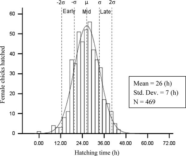
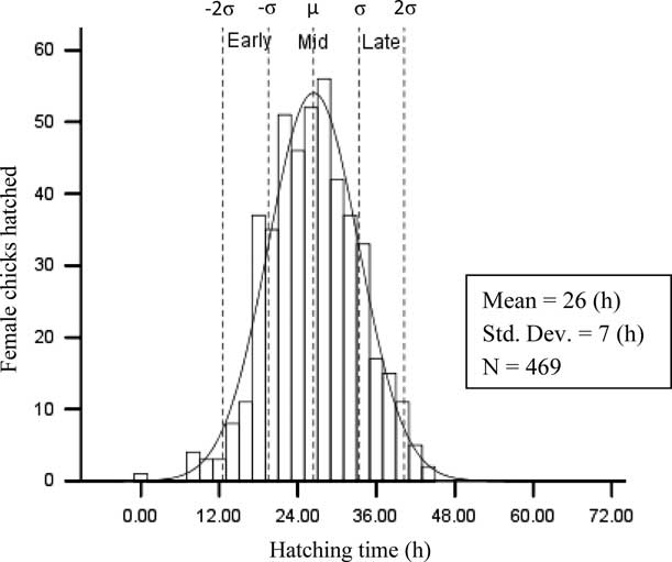
Based on the hatching time of all the female chicks, a hatching curve approaching normal distribution was obtained (Figure 1). The σ of the hatching curve of female chicks was 6 h 55 min. The hatching time (early, middle and late) of female chicks was divided by the µ and σ. The complete hatching period of female chicks lasted 44 h, but the spread of hatch of female chicks was 27 h 40 min (4×σ).
Hatching curve, early, middle and late hatchers and spread of hatch of female chicks (n=469). Distance between two close dotted lines is σ.
Daily feed intake
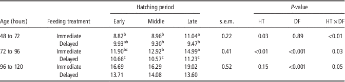
The interaction of feed delay and hatching time on daily feed intake was significant on BA day 3 (BA 48 to 72 h, P=0.01) and BA day 4 (BA 72 to 96 h, P=0.03), and almost significant on day 5 (BA 96 to 120 h, P=0.05; Table 1). Furthermore, spread of hatch had a significant effect on feed intake on BA day 3 (P=0.03) and day 4 (P=0.01), but not on day 5. LI chicks had markedly higher daily feed intake than the EI and MI chicks on day 3 and day 4 (P<0.01). At BA day 5 feed intake of LI chicks was also 14% and 17% higher than that of the EI (P=0.15) and MI (P=0.07) groups, although these differences were not significant. After 48 h delay in feed access, the feed intake of the three hatch groups was similar. The effect of feeding treatment was significant on day 4 (P<0.001) and day 5 (P<0.001), but not on day 3.
Effects of hatching time and delay in feed intake on daily feed intake from BA 48 h up to BA 120 h
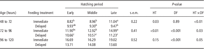
BA=biological age; HT=hatching; DF=delay in feed intake; Immediate=fed immediately; Delayed=delay in feed intake.
a–cValues within the same age with different superscripts differ significantly at P<0.05.
BW and relative growth rate
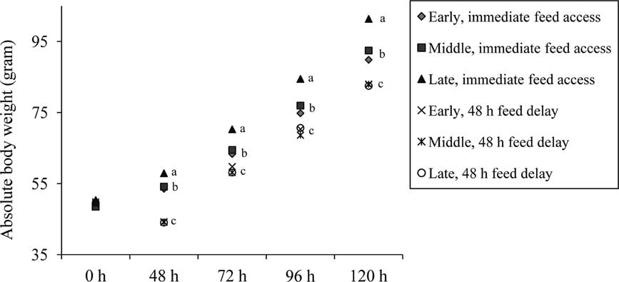
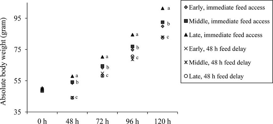
The three hatch groups had similar BW at hatch (Figure 2). The effect of hatching time, delay in feed access and their interaction were all significant from BA day 2 up until day 5 (P<0.01). When chicks were fed immediately after hatch, the BW of late hatchers was higher than their early and middle counterparts from day 2 up to day 5 (P<0.01). Moreover, after the 48 h fasting, the weight loss of each hatch group was not different, and the BW among the feed delay chicks was equal up to BA day 5 (data not shown).
Effect of hatching time and feeding treatments (immediate access to feed and 48 h delay in feed access) on absolute BW up to biological age day 5 (n=25). Data sharing no common letter are different between hatching times within the same feeding treatment (P<0.05).
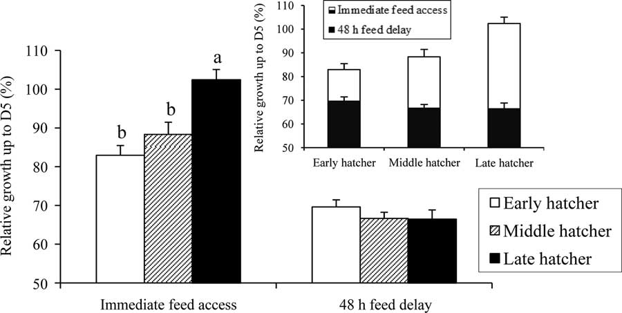
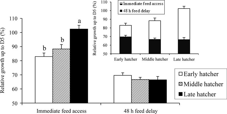
The effect of hatching time and feeding treatment on the relative growth rate up to day 5 was significant (P<0.01; P<0.001), and they showed a pronounced interaction (P<0.001, Figure 3). More specifically, the LI chicks had greater relative growth rate compared with the EI and MI chicks (P<0.01), which were similar in this respect. When feed access was delayed, there was no difference in relative growth rate between the three hatching groups.
Effect of hatching time and feeding treatments (immediate access to feed and 48 h delay in feed access) on relative growth up to biological age day 5 (n=25). Data sharing no common letter are different between hatching times within the same feeding treatment (P<0.05). HT=hatching time; DF=delay in feed intake.
Proportional breast muscle and liver weights
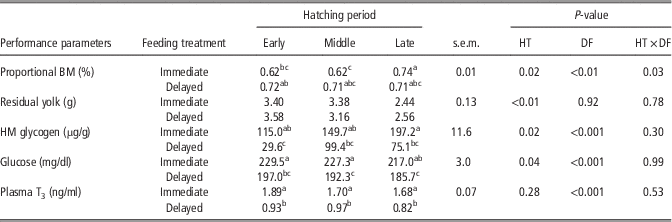
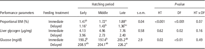
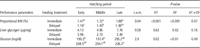
The hatching time did not affect the proportional breast muscle weight at hatch (data not shown). However, hatching time affected the proportional breast muscle on BA day 2 (P=0.02, Table 2) and day 5 (P<0.001, Table 3). The effect of the feeding treatment was significant on BA day 2 (P<0.01) and day 5 (P<0.001). Moreover, the interaction of hatching time and delay in feed intake was significant on BA day 2 (P=0.03, Table 2), and almost significant on day 5 (P=0.07, Table 3). When chicks were fed immediately after hatch, the late hatchers had a higher proportional breast muscle weight on BA day 2 compared with their early (P=0.03) and middle (P<0.01) hatched counterparts, however this parameter did not differ between the fasted chicks (Table 2).
Effects of hatching time and delay in feed intake on proportional breast muscle weight, residual yolk, hatching muscle glycogen content, plasma glucose and T3 levels on BA day 2
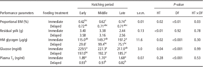
BA=biological age; HT=hatching; DF=delay in feed intake; BM=breast muscle; Immediate=fed immediately; Delayed=delay in feed intake; HM=hatching muscle; Glucose=plasma glucose.
a–cValues within the same parameter with different superscripts differ significantly at P<0.05.
Effects of hatching time and delay in feed intake on proportional breast muscle weight, liver glycogen content and plasma glucose level on BA day 5
BA=biological age; HT=hatching; DF=delay in feed intake; BM=breast muscle; Immediate=fed immediately; Delayed=delay in feed intake; Glucose=plasma glucose.
a–cValues within the same parameter with different superscripts differ significantly at P<0.05.
The spread of hatch did not influence the proportional liver weight at hatch. No effect of hatching time and feeding treatment or interactions were observed on BA day 2 and 5 (data not shown).
Absolute residual yolk weights
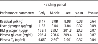
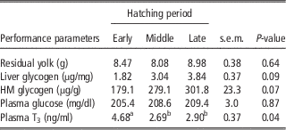
There was no effect of spread of hatch on the residual yolk weight at hatch (Table 4). However, the hatching time significantly affected the yolk weight on post hatch day 2 (P<0.01, Table 2): the late hatchers had lower yolk sac weights than their early and middle counterparts. The feeding treatment had no effect on the residual yolk weight on BA day 2, nor was there an interaction with the hatching time.
Effect of hatching time on residual yolk weight, liver glycogen content, hatching muscle glycogen content, plasma glucose and T3 levels at hatch (BA day 0)
BA=biological age; HM=hatching muscle.
a,bValues within the same parameter with different superscripts differ significantly at P<0.05.
Liver glycogen content
At hatch, even though the effect of spread of hatch on the liver glycogen content was not statistically significant (P=0.09, Table 4), the liver glycogen content of the late hatchers was 111% higher than that of the early hatchers, and the middle hatchers had an intermediate value. The liver glycogen of LI chicks was 25% lower than that of MI and 18% lower than EI on BA day 2, but they were not statistically different (data not shown). In the chicks subjected to 48 h of feed delay, on the other hand, the liver glycogen level was below the detection limit on BA day 2. The previous starvation significantly decreased the liver glycogen content on BA day 5 (P=0.02). No effect of hatching time, nor the interaction with the feeding treatment on the liver glycogen content was observed on BA day 5 (Table 3).
Hatching muscle glycogen contents
At hatch, the effect of spread of hatch on hatching muscle glycogen content was almost statistically significant (P=0.07, Table 4). The glycogen content in the hatching muscle was 69% higher for late hatchers than that of the early hatchers, and the middle hatchers had an intermediate value. The hatching muscle glycogen content was decreased at BA day 2 compared with that at hatch (Table 2). Delayed feed intake resulted in a lower hatching muscle glycogen content (P<0.001). The effect of the hatching time was also significant on BA day 2 (P=0.02): the late hatchers had higher hatching muscle glycogen content than the early hatchers (P=0.02), and the middle hatchers had an intermediate value. On BA day 2, no interaction between spread of hatch and feeding treatment was seen on this parameter.
Plasma glucose levels
The plasma glucose levels of newly hatched chicks were not affected by the hatching time (Table 4). The 48 h starvation period dramatically decreased the blood glucose level on BA day 2 (P<0.001). Moreover, the effect of spread of hatch was significant (P=0.04): the plasma glucose level was higher in early hatchers than the late hatchers (P=0.05), and the middle hatchers had an intermediate value (Table 2). No interaction of hatching time and feeding treatment on the blood glucose level was observed on BA day 2.
On BA day 5, the blood glucose level was significantly higher in the chicks fed with a delay than their immediately fed counterparts (P<0.01). The spread of hatch significantly influenced the blood glucose on BA day 5 (P=0.02): the late hatchers had a higher level than the early hatchers (P=0.03), and the middle hatchers had an intermediate value (Table 3). There was no interaction between hatching time and feeding treatments on blood glucose level on BA day 5.
Plasma T3 levels
The plasma T3 levels at hatch were significantly higher in the early hatchers than the middle and late hatchers (P=0.02, 0.04, Table 4). However, on BA day 2, the spread of hatch had no effect on the blood T3 levels. The feed delay decreased the plasma T3 levels on day 2 compared with chicks that had access to feed immediately (P<0.001, Table 2). No interaction of hatching time and feeding treatment was observed on BA day 2. The effect of hatching time, feeding treatment and their interaction on T3 level were not significant on BA day 5 (data not shown).
Discussion
Spread of hatch
The spread of hatch is a factor that potentially contributes to the different chick qualities and physiological traits in one batch of hatched chicks (Decuypere et al., Reference Decuypere, Tona, Bruggeman and Bamelis2001; Careghi et al., Reference Careghi, Tona, Onagbesan, Buyse, Decuypere and Bruggeman2005). Therefore, the effect of spread of hatch on post hatch performance and its potential mechanisms are of interest and importance.
Hatching time is known to be influenced by factors such as parental age, egg storage time and conditions, and incubation conditions (temperature, humidity, air velocity, etc.) and gender (Tona et al., Reference Tona, Malheiros, Bamelis, Careghi, Moraes, Onagbesan, Decuypere and Bruggeman2003b; Careghi et al., Reference Careghi, Tona, Onagbesan, Buyse, Decuypere and Bruggeman2005; Decuypere and Bruggeman, Reference Decuypere and Bruggeman2007). To limit the number of affecting factors, eggs from the same young flock, that were laid on the same day were chosen, with very short storage period (1 to 2 days) before incubation and were incubated in one incubator but switched eggs location in the incubator daily. After hatch, only female chicks were employed in the present study. Hence, genetic and nutritional variation between eggs might be two major factors that affected the spread of hatch in this experiment. Additionally, the standard deviation was used as a criterion to partition the different hatching time, which resulted in a quantifiable and repeatable design to perform experiments investigating the spread of hatch.
The present study showed that the spread of hatch did not affect the hatch weight, which is consistent with observation of Careghi et al. (Reference Careghi, Tona, Onagbesan, Buyse, Decuypere and Bruggeman2005) and van de Ven et al. (Reference Van de Ven, Van Wagenberg, Debonne, Decuypere, Kemp and Van den Brand2011). In addition, no difference in residual yolk weight was found at hatch, which agrees with Careghi et al. (Reference Careghi, Tona, Onagbesan, Buyse, Decuypere and Bruggeman2005). van de Ven et al. (Reference Van de Ven, Van Wagenberg, Debonne, Decuypere, Kemp and Van den Brand2011), on the other hand, observed a lower residual yolk weight at hatch for the late hatchers, whereas in the present study this was only seen on BA day 2.
The process of hatching is dependent on the proper development of the supporting musculature, especially the hatching muscle (Musculus complexus), and the available energy storage (Decuypere et al., Reference Decuypere, Dewil and Kuhn1990). The higherliver and hatching muscle glycogen at hatch of the late hatchers suggests that the late hatchers stored more energy during incubation, and/or consumed less which brought about the later hatching of these birds. As embryonic metabolism and the hatching time are known to be determined by regulators of thyroid hormone availability and action (Beck et al., Reference Beck, Roelens, Maervoet, Schepens and Darras2005; Van Herck et al., Reference Van Herck, Geysens, Delbaere and Darras2013), differences concerning these factors between eggs of one batch might also have induced differences in the spread of hatch.
It is well known that thyroid hormones affect the time of hatching and hence the length of incubation and play a crucial role during chicken post hatch development (Decuypere et al., Reference Decuypere, Dewil and Kuhn1990). In accordance with the study of van de Ven et al. (Reference Van de Ven, Van Wagenberg, Debonne, Decuypere, Kemp and Van den Brand2011), the early hatchers had higher levels of plasma T3 in the present study, which also agrees with findings of Decuypere et al. (Reference Decuypere, Dewil and Kuhn1990) showing that treatment of embryos with T3 on embryonic day 19 advanced hatching time. As it was reported that T3 could decrease the liver and skeletal muscle glycogen (Potenza et al., Reference Potenza, Via and Yanagisawa2009; Ribeiro et al., Reference Ribeiro, Teixeira, Aparecido da Silva, Dalia, Júnior, Bertolini, Rostom de Mello and Luciano2012), through glycogen synthase kinase 3 β (GSK 3 β) (Kuzman et al., Reference Kuzman, Gerdes, Kobayashi and Liang2005), the higher plasma T3 level of early hatchers at hatch is consistent with the lower glycogen in liver and hatching muscle.
When chicks had access to feed immediately after hatch, the late hatchers grew faster than chicks that hatched earlier. This improved growth performance of the late chicks, when measured on their BA, is in agreement with Careghi et al. (Reference Careghi, Tona, Onagbesan, Buyse, Decuypere and Bruggeman2005). However, it is notable that when the CA was used to measure the BW in former investigations, no difference or even a decreased growth performance was observed for the late hatchers (Careghi et al., Reference Careghi, Tona, Onagbesan, Buyse, Decuypere and Bruggeman2005; van de Ven et al., Reference Van de Ven, Van Wagenberg, Debonne, Decuypere, Kemp and Van den Brand2011). Interestingly, despite the higher BW of the late hatchers on BA day 2 and 5, the proportion of the breast muscle to BW of the late chicks was still higher than the other two fed hatchers.
It is worth noting that the augmented metabolic rate caused by T3 is normally accompanied by an increased feed intake (Kong et al., Reference Kong, Martin, Smith, Gardiner, Connoley, Stephens, Dhillo, Ghatei, Small and Bloom2004). In the present study, however, the early hatched chicks were characterized by higher plasma T3 levels yet had lower feed intake at the beginning of their post hatch life, indicating that factors other than T3 are involved in manipulating the energy intake.
The different hatching time did not affect the blood glucose level at hatch, which disagrees with van de Ven et al. (Reference Van de Ven, Van Wagenberg, Debonne, Decuypere, Kemp and Van den Brand2011) who observed a higher plasma glucose for the late hatchers compared to their earlier hatched counterparts, suggesting that more glucose was available for the late hatchers. In our study, as the late hatchers started their post hatch life with a higher hepatic glycogen reserve and had equal (or even somewhat lower) levels of hepatic glycogen on day 2, they consumed more glucose than their early counterparts, as also implicated by the higher feed intake but a lower plasma glucose concentration on BA day 2. The available glucose on these first two post hatch days might have stimulated the utilization of yolk lipids, as the intermediate of the TCA cycle is then used for lipolysis (Bergman and Kon, Reference Bergman and Kon1964; Ørngreen et al., Reference Ørngreen, Jeppesen, Andersen, Taivassalo, Hauerslev, Preisler, Haller, Van Hall and Vissing2009), resulting in a lower residual yolk of the late hatched chicks on day 2. This glucose and yolk consumption hypothesis was also true for the fasted chicks. Consequently, all these factors together with the higher feed intake and increased yolk utilization more than probably caused the higher BW (from day 2 onwards) of the fed late hatchers.
Delay in feed access
In practice, the first feed of chicks is commonly delayed due to the logistics of commercial production (Kornasio et al., Reference Kornasio, Halevy, Kedar and Uni2011). The 48 h of feed delay resulted in retarded early growth which is in agreement with reports from Bigot et al. (Reference Bigot, Mignon-Grasteau, Picard and Tesseraud2003a) and Careghi et al. (Reference Careghi, Tona, Onagbesan, Buyse, Decuypere and Bruggeman2005). According to Noy and Sklan (Reference Noy and Sklan2001), feed deprivation results in a slower yolk consumption in the newly hatched chicks, which is caused by a poor stimulation of the development of the gastrointestinal tract. Nevertheless, in this experiment no effect of fasting was observed on yolk sac utilization, which is analogous to some previous observations (Careghi et al., Reference Careghi, Tona, Onagbesan, Buyse, Decuypere and Bruggeman2005; Gaglo-Disse et al., Reference Gaglo-Disse, Tona, Aliou, Debonne, Aklikokou, Gbeassor and Decuypere2010).
Interestingly, when these chicks had access to feed, the feed intake was lower than their immediately fed counterparts, which implies that delayed feed access had impaired the feed intake which potentially further aggravated the growth retardation. In addition, the effects of hatching time on feed intake and relative growth observed in immediately fed chicks were eliminated by the 48 h feed delay. The reduction in relative growth rate caused by the delayed feed access was larger in the late hatchers than the other two hatching time groups (Figure 3, the smaller figure), which indicated that the 48 h feed delay had more impact on the late hatchers. Indeed, the advantage of the late hatchers, whether caused by nutrient availability or genetic potential, disappeared due to the 48 h feed delay. In the same line, Bigot et al. (Reference Bigot, Mignon-Grasteau, Picard and Tesseraud2003a) found that post hatch starvation reduced interfamily variation in BW that is expressed in chicks given immediate access to feed, which may mask the expression of the genetic potential. Surprisingly, the increased yolk consumption of late hatchers from hatch to day 2 compared with their earlier counterparts, was also observed for the feed deprived chicks, however, without any effect on BW (loss).
In accordance with previous studies (Decuypere and Kühn, Reference Decuypere and Kühn1984; Careghi et al., Reference Careghi, Tona, Onagbesan, Buyse, Decuypere and Bruggeman2005), decreased plasma T3 levels were observed immediately after the feed delay, which might be an adaptation in order to reduce oxygen consumption and metabolism.
Very similar to a former report (Decuypere and Kühn, Reference Decuypere and Kühn1984), the blood glucose level of previous starved chicks was significantly higher than their immediately fed counterparts after refeeding (BA day 5), although their feed intake was markedly lower at that time.
The proportional breast muscle weight on day 2 was higher when chicks were starved for the first 48 h of their life, while it was proportionally lower on day 5 compared with their fed counterparts. As the intestines are barely developing during the early post hatch starvation, the breast muscle takes a good proportion of the BW, while on day 5, when the starved chicks had access to feed, their intestines were most probably developing fast, which resulted in a proportionally lower breast muscle weight compared with their fed counterparts. The retardation in muscle development has been shown by several authors. Bigot et al. (Reference Bigot, Taouis, Picard and Tesseraud2003b) concluded that muscle ribosomal S6 kinase 1 (S6K1), a key element in the control of protein synthesis, is activated only when food is available, without an altered improvement on the response of the S6K1 pathway after post hatch starvation. Additionally, another study suggested that early nutritional status affected the percentage of muscle fiber type and changed the mRNA expression for the growth-related genes in muscle (Li et al., Reference Li, Yuan, Yang, Ni, Xia, Barth, Grossmann and Zhao2007). Interestingly, Kornasio et al. (Reference Kornasio, Halevy, Kedar and Uni2011) reported that delayed feed access (36 h post hatch) declined the number of the breast muscle cells and myofiber diameter in post hatch chicks.
In conclusion, when chicks were immediately fed after hatch, the spread of hatch influenced post hatch performance: chicks hatched later benefitted from a better appetite and faster growth at least until day 5. In this experimental set-up, the spread of hatch was due to their genetic potential, or the availability/consumption of less nutrients during embryonic development. The different T3 levels at hatch may have contributed to the distinct post hatch performance, although other factors might be involved in the differential feed intake, which is under further investigation in our lab. Delayed feed intake diminished the ‘non-uniformity’ caused by the different hatching time, but induced growth retardation compared with the chicks immediately fed after hatch, which was more pronounced for the late hatchers.
Acknowledgements
The authors would like to thank the technical staff Andre Respen, Marcel Samain, Inge Vaesen and Daniel Vermeulen of the Division of Livestock-Nutrition-Quality of the KU Leuven. This research was funded by the ‘Fonds Wetenschappelijk Onderzoek – Vlaanderen’ (FWO G.0620.11N). Dr Nadia Everaert is a post-doctoral fellow of FWO.