Introduction
Synthetic biology initially focused on the use of molecular biology tools to engineer bacterial hosts, but has also made considerable progress in the engineering of mammalian cells with potential therapeutic applications.[Reference Ho and Chen1,Reference Esensten, Bluestone and Lim2] The strategies of synthetic biology have been used to engineer immune cells to treat cancer using chimeric antigen receptors (CARs). CARs are synthetic receptors that can improve the cancer-targeting capabilities of immune cells by equipping them to recognize tumor-associated antigens. CAR-engineered autologous T cells have shown remarkable success in treating hematologic malignancies, but have had disappointing clinical results in targeting solid tumor due to the immunosuppressive tumor microenvironment (TME). Additionally, CARs targeting tumor-associated antigens that are also expressed on normal tissue may result in “on-target, off-tumor” toxicity and have caused significant safety concerns.[Reference Sadelain, Brentjens and Rivière3] These unresolved issues of CAR-related treatments necessitate better CAR design. A number of tools and approaches can program the therapeutic CAR-modified immune cells to improve persistence as well as safety in cancer treatment.
Genetic engineering and synthetic biology strategies have been developed to improve CAR-T cell proliferation, persistence, cytotoxic function, and safety in vivo. For example, T cells can be engineered to overexpress cytokine transgenes such as IL-12 and IL-15 that work to promote T-cell proliferation and cytotoxic functions.[Reference Koneru, Purdon, Spriggs, Koneru and Brentjens4,Reference Hurton, Singh, Najjar, Switzer, Mi, Maiti, Olivares, Rabinovich, Huls, Forget, Datar, Kebriaei, Lee, Champlin and Cooper5] Control systems through the addition of a small molecule or biologic circuit to the CAR design have been shown to increase the safety of CAR-T therapy. One strategy involves the use of a split CAR system containing drug-inducible heterodimerization domains that remain inactive but can be turned on by the addition of a heterodimering drug.[Reference Wu, Roybal, Puchner, Onuffer and Lim6] T cells have also been engineered to express an inducible suicide gene caspase 9 (iCAS9) as a safety switch which responded to a chemical inducer of dimerization (CID) and resulted in dose-dependent levels of apoptosis.[Reference Straathof, Pule, Yotnada, Dotti, Vanin, Brenner, Heslop, Spencer and Rooney7,Reference Diaconu, Ballard, Zhang, Chen, West, Dott and Savoldo8] This suicide switch is a tool for physicians to regulate treatment when complications including “on-target, off-tumor” toxicity or cytokine release syndrome occur.[Reference Bonifant, Jackson, Brentjens and Curran9]
Among the candidates of immune cells to express CARs for the cellular immunotherapy, the well-characterized cytotoxic natural killer (NK) cell line, NK-92 cell line, is a promising one due to its lack of immunogenicity and availability as an “off-the-shelf” product.[Reference Rezvani and Rouce10–Reference Zhang, Oberoi, Oelsner, Waldmann, Linder, Tonn and Wels12] The standardized NK92 cell line is thus an ideal target for a synthetic biology approach which equips cells with CARs or other engineered structures for improved antitumor efficacy.
NK cells typically rely on interleukin (IL)-2 for growth and cytotoxic function, so during animal studies or clinical trials, they are often co-administered with exogenous IL-2, which may cause acute toxicity in high doses.[Reference Rezvani and Rouce10] Instead, lentiviral vectors encoding IL-15 can transduce NK92 cells to stably express the cytokine and sustain continued proliferation and cytotoxic capabilities without exogenous IL-2.[Reference Zhang, Sun, Wei, Zhang and Tian13,Reference Sahm, Schönfeld and Wels14] Moreover, the receptor subunit IL-15Rα can form a stable complex with IL-15 on cell surfaces and enables autocrine stimulation of NK cells by membrane-bound IL-15 in cis presentation.[Reference Imamura, Shook, Kamiya, Shimasaki, Chai, Coustan-Smith, Imai and Campana15] This membrane-bound IL-15/IL-15Rα complex has shown to contribute to the long survival of CD8 memory T cells and the persistence of T cells engineered to co-express CAR and the complex in a tethered form.[Reference Hurton, Singh, Najjar, Switzer, Mi, Maiti, Olivares, Rabinovich, Huls, Forget, Datar, Kebriaei, Lee, Champlin and Cooper5,Reference Sato, Patel, Waldmann and Tagaya16] We hypothesized that NK92 cells engineered to co-express CAR and the membrane-bound IL-15/IL-15Rα complex would also survive and proliferate without exogenous IL-2.
For both preclinical animal studies and clinical trials using NK92 cells to treat cancer, the cells are typically irradiated prior to infusion for safety concerns to prevent engrafting cells from in vivo tumorigenicity.[Reference Rezvani and Rouce10,Reference Suck, Odendahl, Nowakowska, Seidl, Wels, Klingemann and Tonn11] As a result, the infused NK92 cells experience limited expansion without long-term persistence. For NK92 cells engineered to express membrane-bound IL-15/IL-15Rα complex, irradiation prior to infusion would greatly defeat the purpose of the complex to improve NK92 proliferation and persistence in vivo. The iCAS9 suicide gene, when transduced into the CAR-NK cells with membrane-bound IL-15/IL-15Rα complex, serves as a safety mechanism other than irradiation without affecting the proliferation and persistence of infused NK92 cells.
Mesothelin is a cell-surface molecule overexpressed in many carcinomas and is an attractive target for CAR-engineered immune therapies.[Reference Hassan, Bera and Pastan17] A third-generation anti-mesothelin CAR consisting of a single-chain variable fragment domain targeting mesothelin and two co-stimulatory domains, CD28 and CD137 (4-1BB), transduced into T cells has shown to eradicate large, established tumors overexpressing mesothelin antigen.[Reference Carpenito, Milone, Hassan, Simonet, Lakhal, Suhoski, Varela-Rohena, Haines, Heitjan, Albelda, Carroll, Riley, Pastan and June18]
Therefore, we utilized synthetic biology technology and engineered NK92 cells to express anti-mesothelin CAR and membrane-bound IL-15/IL-15Rα complex for specific killing and better proliferation in an ovarian cancer mouse model. We further transduced the engineered CAR-NK cells with the iCAS9 suicide gene as a safety switch to eliminate infused CAR-NK cells in case of adverse side effects or post-treatment, in order to mitigate the risk of tumorigenicity from the infused cells.
Materials and methods
Cell culture
SKOV3 (ATCC HTB-77) tumor cell lines were maintained in a 5% CO2 environment in RPMI 1640 (Gibco) media supplemented with 10% FBS, 1% pen-strep, and 2 mM L-glutamine. NK92 cells (Dr. Jihane Khalife, Children's Hospital Los Angeles; ATCC CRL-2407) were maintained in MEM-α (Gibco) supplemented with 10% FBS, 10% horse serum, 1% NEAA, 1% pen-strep, 1% sodium pyruvate, 0.1 mM 2-β mercaptoethanol, 0.2 mM myo-inositol, and 2.5 µM folic acid. Mesothelin+ SKOV3 (SKOV.meso) cells were generated by transducing SKOV3 cells with lentivirus containing mesothelin cDNA and sorting mesothelin+ cells with fluorescence-activated cell sorting.
Virus production
The CAR constructs consisted of ss1, the murine-derived scFv anti-mesothelin antigen-binding domain; CD8 hinge and CD28 transmembrane regions; and CD28, 4-1BB, and CD3ζ cytoplasmic regions in the retroviral MP71 vector. The membrane-bound IL-15/IL-15Rα complex was included in one of the CAR constructs following a 2A linker. The iCAS9 suicide switch with GFP marker, pMSCV-F-del Casp9.IRES.GFP, was purchased from Addgene. The plasmid for mesothelin consisted of human mesothelin cDNA cloned into a lentiviral FUW backbone. These plasmids were used to transfect HEK 293T cells in 30 mL plates using CaCl2 precipitation methods. Fresh media (high glucose DMEM supplemented with 10% FBS and 1% pen-strep) was plated onto the cells 4 h after initial transfection. Supernatants were harvested and filtered (0.45 µm) 48 h later and used fresh.
Transduction of NK92 and SKOV3 cells
NK92 cells were transduced with anti-mesothelin CAR and iCAS9 retroviral vectors. Non-tissue culture-treated 12-well plates were coated overnight with 25 µg RetroNectin per well (Clontech Laboratories, Mountain View, CA). Retroviral vectors were subsequently spin-loaded onto the plates by centrifuging at 2000 × g for 2 h at 32 °C. NK92 cells were resuspended at a concentration of 5 × 10⁵/mL with fresh media complete with 200 U/mL human IL-2 and added to the vector-coated plates. The plates were centrifuged at 600 × g for 30 min at 32 °C and incubated overnight at 37 °C and 5% CO2. CAR+ or GFP+ NK92 cells were sorted using fluorescence-activated cell sorting.
SKOV3 cells were similarly transduced to express human mesothelin via a lentiviral vector. One million cells were added to 2 mL of fresh viral supernatant and centrifuged at 1050 × g for 90 min at room temperature and resuspended in fresh media. Mesothelin+ cells were sorted using fluorescence-activated cell sorting.
CAR detection on NK cell surface
After transduction and cell sorting, 1 × 10⁵ NK92 cells were incubated with recombinant human mesothelin-Fc chimera (R&D Systems, Minneapolis, MN) at a volume ratio of 1:50 (2 µg/mL) in PBS at 4 °C for 30 min and rinsed with PBS. The cells were subsequently incubated with PE-labeled goat anti-human Fc (Jackson ImmunoResearch) at a volume ratio of 1:150 in PBS at 4 °C for 10 min, rinsed, and read using flow cytometry. Non-transduced NK92 cells served as a negative control.
Cytokine release assay
The 1 × 10⁵ NK cells per well were coincubated with target cells in 96-well round-bottom plates at a 1:1 ratio for 6 h at 37 °C. One microgram Brefeldin-A (Sigma, St. Louis, MO) was added to each well to prevent protein transport. At the end of the incubation, cells were permeabilized using the CytoFix/CytoPerm kit (BD Biosciences, San Jose, CA) and stained for CD45 and interferon-γ (IFN-γ) using Pacific Blue-conjugated anti-human CD45 (Biolegend, San Diego, CA) and PE-conjugated anti-human IFN-γ (Biolegend). NK cells without target cells were used as a negative control. Results were read using flow cytometry. The data were determined in triplicate and presented as the mean ± SEM.
Cytotoxicity assay
The 2 × 10⁴ target cells were labeled with 5 µM carboxyfluorescein succinimidyl ester (CFSE, Life Technologies, Waltham, MA) as previously described[Reference Han, Bryson, Zhao, Cinay, Li, Guo, Siriwon and Wang19] and coincubated with NK cells at various ratios in 96-well round-bottom plates for 24 h at 37 °C. The cells were then incubated in 7-AAD (Life Technologies) in PBS (1:1000 dilution) for 10 min at room temperature and analyzed via flow cytometry. Percentages of killed cells were calculated as [CFSE+7-AAD+ cells/(CFSE+7-AAD− + CFSE+7-AAD+)] cells, with live/dead gates based on control wells of target cells only to account for spontaneous cell death. The cytotoxicity was determined in triplicate and presented as the mean ± SEM.
Chemical inducer of dimerization sensitivity assay
CID AP20187 (B/B Homodimerizer, Takara, Mountain View, CA) was diluted in complete NK cell media at a concentration of 10 nM. The 5 × 10⁴ NK cells were incubated in media or 10 nM CID for 24 h and analyzed for viability. Number of viable cells were counted under a light microscope via trypan blue exclusion and percentage of viable cells was calculated using AnnexinV/7-AAD staining (BD Biosciences) per the manufacturer's instructions.
Xenograft tumor model
All animal experiments were conducted according to the animal protocol approved by the University of Southern California Institutional Animal Care and Use Committee (IACUC). Eight-week-old female NOD.Cg-PrkdcscidIL2Rγ tm1Wj1/SZ (NSG) mice (Jackson Laboratories) were inoculated subcutaneously with 3.5 × 10⁶ SKOV.meso cells, and treatment began when tumors grew to 50–70 mm3. The mice were injected intravenously through the tail vein with 10 × 106 NK cells twice a week, three times total. Tumor growth and body weight of the remaining mice were recorded until the end of the experiment. The tumor length and width were measured with a fine caliper three times a week, and tumor volume was calculated as ½ × (length) × (width)2. The survival endpoint was determined to be when the tumor reached 1000 mm3 or became ulcerated.
Ex vivo NK cell staining
One week after the final NK cell injection, a subset of mice were sacrificed and organs were harvested. Each tissue sample was made into a single-cell suspension through a 70 µm strainer, washed with PBS, centrifuged, and resuspended in 3 mL TAC buffer for 10 min at room temperature. The samples were then washed and resuspended with PBS. Fluorescent-conjugated antibodies (Biolegend) were used to stain the samples in 100 µL PBS at a 1:100 volume ratio for 15 min at 4 °C in a 96-well plate. Samples were analyzed using flow cytometry in triplicate and presented as the mean ± SEM.
Statistical analysis
Statistical analysis was performed in Microsoft Excel and GraphPad Prism 6. One-way analysis of variance with Tukey's multiple comparison was used to assess the data between different groups. P values of <0.05 were considered significant and denoted as follows: not significant (NS) P > 0.05; *P < 0.05; **P < 0.01; ***P < 0.001.
Results and discussion
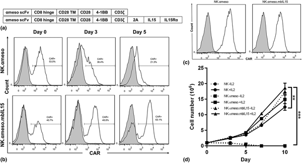
The addition of a membrane-bound cytokine to the CAR construct is one method to program immune cells to be more therapeutically effective. Anti-mesothelin CARs with or without membrane-bound IL-15/IL-15Rα complex were transduced efficiently in NK92 cells. Retroviral vectors as described in “Materials and methods” section were used to transduce NK92 cells with a third-generation anti-mesothelin CAR, either without (NK.αmeso) or with (NK.αmeso.mbIL15) membrane-bound IL-15/IL-15Rα complex as shown in the schematic representation in Fig. 1(a). Genetically engineering CAR-NK cells with membrane-bound IL-15/IL-15Rα complex allowed CAR-NK cells to expand in the absence of exogenous IL-2 in vitro. Unsorted transduced NK.αmeso and NK.αmeso.mbIL15 cells were cultured in complete media without IL-2 supplementation over 10 days. NK.αmeso cells were mostly dead by day 5, and the CAR-expressing population of the live cells also decreased over time, while NK.αmeso.mbIL15 increased the purity of the CAR-expressing population over time in the absence of IL-2 [Fig. 1(b)]. Although CAR+ cells ultimately were sorted using flow cytometry, this demonstrates that starving unsorted cells of IL-2 can serve to enrich the transduced population. After selection for transduced NK92 cells via fluorescence-activated cell sorting, the cells displayed high CAR expression [Fig. 1(c)]. Non-transduced NK92 cells were used as a negative control. Previous research indicates that the membrane-bound IL-15/IL-15Rα complex acts in cis instead of trans, behaving as an autocrine stimulation by binding to the β and γ IL-15R subunits expressed on the transduced cells instead of those on surrounding immune cells.[Reference Imamura, Shook, Kamiya, Shimasaki, Chai, Coustan-Smith, Imai and Campana15] This data supports that finding, as CAR+ populations selectively proliferated, not the entire cell population—cells were stimulated by the membrane-bound IL-15/IL-15Rα complex only if the cells already expressed that transgene. It was further demonstrated that NK.αmeso.mbIL15 cells could survive and proliferate in the absence of exogenous IL-2 by observing the expansion over 10 days of sorted CAR-NK cells in IL-2-free media [Fig. 1(d)]. All cells expanded well in media with IL-2 supplementation, but only the NK.αmeso.mbIL15 cells expanded without IL-2—non-transduced NK92 cells and NK.αmeso cells died by day 5 in media without IL-2 (P < 0.001). NK.αmeso.mbIL15 cells also expanded significantly more than non-transduced NK92 cells (P < 0.01).
(a) Schematic representation of CAR constructs. (b) CAR expression over time in the absence of exogenous IL-2. (c) CAR expression of sorted cell populations. (d) Expansion of NK cells with or without exogenous IL-2.

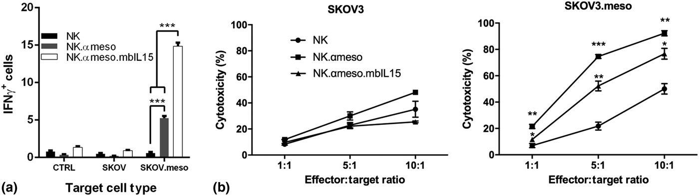
CAR-NK cells with membrane-bound IL-15/IL-15Rα complex secreted cytokines and exhibited cytotoxicity to mesothelin-expressing target cells. In vitro functionality of the CAR-NK cell lines was assessed by performing IFN-γ release and cytotoxicity assays. NK cells incubated without target cells or with SKOV3 cells served as a negative control, as they do not express mesothelin, while SKOV.meso cells transduced to overexpress mesothelin were used as the desired target cells. In the IFN-γ assay, flow cytometry was used to analyze the IFN-γ + NK cell populations after coincubation with each of the target cell lines [Fig. 2(a)]. None of the NK cell lines reacted to SKOV3 cells, indicating specificity toward mesothelin. Non-transduced NK92 cells did not release IFN-γ in response to SKOV.meso cells, demonstrating the necessity of an anti-mesothelin CAR to generate an immune response. NK.αmeso cells had a significantly larger IFN-γ + population when coincubated with SKOV.meso cells when compared with non-transduced NK cells (P < 0.001), but NK.αmeso.mbIL15 cells had significantly higher percentages of IFN-γ + cells after exposure to SKOV.meso cells compared with both non-transduced (P < 0.001) and NK.αmeso cells (P < 0.001). These data are in accordance with previous work showing that IL-15 aids in greater production of IFN-γ in NK cells.[Reference Tagaya, Bamford, DeFilippis and Waldmann20–Reference Mah and Cooper22] Cytotoxicity assays were performed using the above-mentioned target and effector cells in different ratios. NK.αmeso.mbIL15 cells killed lower percentages of SKOV.meso target cells compared with NK.αmeso cells, perhaps due to slightly lower CAR expression on the cell surface. Both CAR-NK cell lines were significantly more cytotoxic to SKOV.meso cells compared with non-transduced NK92 cells at all effector-to-target ratios [Fig. 2(b); significance stars are comparing non-transduced NK cells to NK.αmeso and non-transduced NK cells to NK.αmeso.mbIL15].
(a) Interferon-γ secretion by NK cells coincubated with mesothelin+ or mesothelin− target cells. (b) Cytotoxicity of NK cells coincubated with mesothelin+ or mesothelin− target cells. Significance stars compare non-transduced NK cells to NK.αmeso and non-transduced NK cells to NK.αmeso.mbIL15 (*P < 0.05, **P < 0.01, ***P < 0.001).

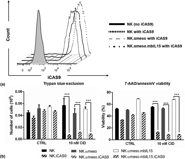
The iCAS9 suicide gene harnesses technology from genetic engineering and small molecule drugs. This kill switch was efficiently transduced in NK92 cells (NK92 without CAR, NK.αmeso, and NK.αmeso.mbIL15) using the commercially available retroviral vector described in “Materials and methods” section. After fluorescence-activated cell sorting, all cell lines showed similar high expression levels of iCAS9 [Fig. 3(a); NK92 at 98.5% iCAS9+, NK.αmeso at 97.8%, and NK.αmeso.mbIL15 at 98.3%]. Uniform transgene expression across all groups is important, as higher iCAS9 expression leads to greater sensitivity to the CID AP20187 used when triggering apoptosis.[Reference Diaconu, Ballard, Zhang, Chen, West, Dott and Savoldo8]
(a) iCAS9 expression of sorted NK cell lines. (b) Viability of NK cells, analyzed by both trypan blue staining and 7-AAD/AnnexinV flow cytometry, after incubation in either plain media or 10 nM CID for 24 h.

NK92 cells expressing iCAS9 were highly sensitive to low concentrations of the CID AP20187, an otherwise non-toxic small molecule dimerizer. To test the capability of the suicide transgene to induce apoptosis in cells after exposure to the AP20187, viable cell populations were observed using either trypan blue exclusion or AnnexinV/7-AAD staining. Six cell lines were tested—non-transduced NK92, NK.αmeso, and NK.αmeso.mbIL15 cells, each either with or without iCAS9 transgene. None of the cells without iCAS9 displayed any sensitivity to the CID after 24 h coincubation, while cells expressing iCAS9 showed markedly decreased viability after CID exposure (P < 0.001) in both trypan blue exclusion and AnnexinV/7-AAD staining [Fig. 3(b)]. These experiments demonstrate that, if CAR-NK therapy needs to be stopped, either due to unwanted toxicity or simply at the end of treatment, CID can be administered and quickly trigger apoptosis only in cells expressing the iCAS9 transgene. However, a small percentage of cells remained viable after CID treatment, likely due to iCAS9− NK cells which remained in the sorted population or which gradually lost iCAS9 expression over time. This emphasizes the importance of complete iCAS9 expression if the cells are to be used in a clinical setting; these issues can be addressed by obtaining the therapeutic cell product from a single cell clone and carefully monitoring for transgene downregulation or silencing prior to treatment.
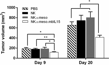
To evaluate the antitumor efficacy of NK.αmeso.mbIL15 cells in vivo, 10 million cells were injected intravenously into NSG mice with established SKOV.meso tumors twice a week for 2 weeks. The data demonstrate that NK.αmeso.mbIL15 cells slowed tumor growth and exhibited better tumor growth control than NK.αmeso cells (Fig. 4). The treatment with NK.αmeso cells did not show better antitumor efficacy than that of NK92 or PBS only. This was unexpected given the cytokine secretion and cytotoxicity of NK.αmeso cells against target cells in vitro, but immune cell exhaustion and lack of persistence in the TME is not faithfully replicated in in vitro conditions.
Tumor volumes of treated mice.

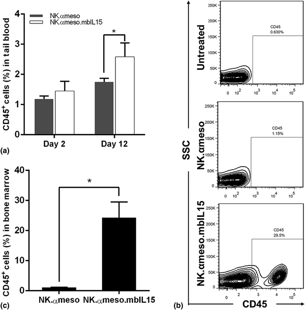
Infused NK.αmeso.mbIL15 cells proliferated more in vivo compared with NK.αmeso cells. Tumor-bearing mice were separated into two groups receiving NK.αmeso.mbIL15 cells and NK.αmeso treatment, respectively, on days 0, 5, and 9. The mice were tail bled three times a week following the first NK injection. By day 12, the group receiving NK.αmeso.mbIL15 cells had significantly higher NK cell populations in tail blood [Fig. 5(a)]. One week after the last injection of NK cells, the mice were euthanized, and organs were harvested and analyzed for NK cell populations. Interestingly, the group receiving NK.αmeso.mbIL15 cells treatment exhibited significantly higher populations of NK cells homing to the bone marrow, while NK.αmeso cells had few NK cells in the bone marrow. Representative flow cytometry plots are shown in Fig. 5(b), and quantitative data are displayed in Fig. 5(c). NK92 cells used for adoptive cell therapy has not been reported in the literature to be able to home to bone marrow, making NK92 cells expressing membrane-bound IL-15/IL-15Rα a potential therapy for malignancies which heavily metastasize to the bone marrow.[Reference Boissel, Betancur-Boissel, Lu, Krause, Van Etten, Wels and Klingemann23]
(a) Percentage of NK cells in mouse peripheral blood over time. (b) Representative FACS plots of NK cells in mouse bone marrow 7 days after the final NK injection. (c) Percentage of NK cells in mouse bone marrow.

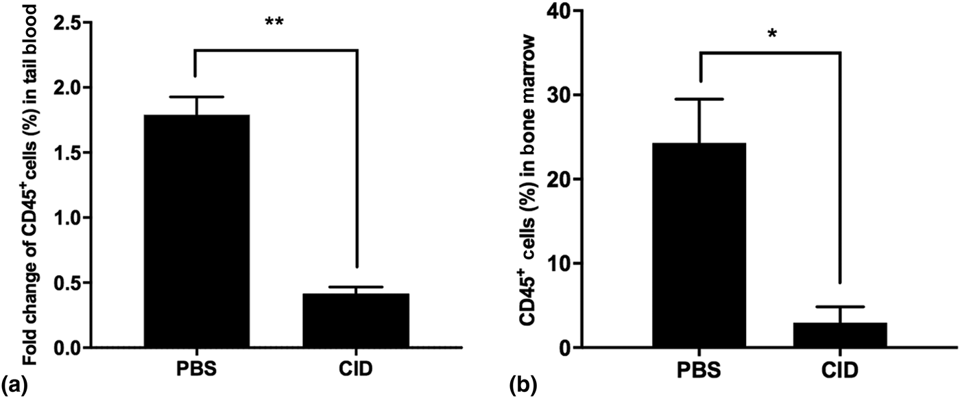
AP20187 injected into the mice induced apoptosis of NK cells expressing the iCAS9 transgene. Tumor-bearing mice received three injections of 10 million NK.αmeso.mbIL15 cells as described above on days 0, 5, and 9. The mice were tail bled on days 2, 7, 12, and 16 and were analyzed for NK cell populations in tail blood. One group of mice received 50 ug CID encapsulated in a previously reported drug-delivery liposomal nanoparticle[Reference Liu, Fang, Kim, Wong and Wang24] through intravenous injection, and another group of mice received PBS-only injections. The fold changes of the percentage of CD45+ cells in tail blood on day 16 over the percentage of CD45+ cells in tail blood on day 12 were calculated for each mouse and averaged for each group. Mice without any CID treatment had nearly doubled NK cell populations in tail blood while mice receiving CID treatment had less than half of the NK cell populations remaining [Fig. 6(a)]. On day 16, the mice were euthanized, and their organs were analyzed for NK cell populations. Mice treated with CID had minimal NK cell populations in the bone marrow, while mice without CID treatment retained high levels of NK cell populations that homed to the bone marrow [Fig. 6(b)]. Overall, the addition of an engineered cytokine and suicide switch allows for the optimization of biologic functions in CAR-NK cells.
(a) Fold-change of NK cells in mouse peripheral blood comparing before and after CID or PBS administration. (b) Percentages of NK cells in mouse bone marrow after CID or PBS administration.

Conclusions
CAR-engineered immune cells have been successful in treating a variety of hematologic cancers, but that measure of clinical success in treating solid tumors is lacking. CARs are a clinically relevant application of synthetic biology, and their modular design offers the potential to target a variety of diseases simply by switching out the antigen-binding domain. CARs can be further improved by genetically modifying human immune cells with additional engineered receptors. This research presents two more synthetic molecules in addition to the anti-mesothelin CAR: a membrane-bound cytokine, IL-15/IL-15Rα, and a suicide switch, iCAS9, for immune stimulation and safety mechanisms. Instead of autologous T cells, which are used in most CAR studies, the NK92 cell line has proven effective in executing CAR-mediated antitumor functions in preclinical settings and provides a renewable, off-the-shelf source of engineered cells. A homogenous, allogenic cellular product has the potential for more precise modulation of biologic outputs than autologous cells, which vary widely in function from patient to patient. There are many more applications of synthetic biology for CARs beyond the scope of this research letter, including logic gates[Reference Kloss, Condomines, Cartellieri, Bachmann and Sadelain25,Reference Zah, Lin, Silva-Benedict, Jensen and Chen26] and synthetic notch receptors.[Reference Roybal, Williams, Morsut, Rupp, Kolinko, Choe, Walker, McNally and Lim27] CARs require fine-tuning through molecular engineering to have the precision and persistence needed to eradicate solid tumors, and additional artificial receptors modulating the immune environment and cell response are key to more effective CARs in the future.
Acknowledgments
This work was supported by National Institutes of Health grants (R01AI068978, R01CA170820, R01EB017206 and P01CA132681) and a translational acceleration grant from the Joint Center for Translational Medicine.