Introduction
Experiencing a traumatic event can result in the development of a post-traumatic stress disorder (PTSD; Breslau et al. Reference Breslau, Kessler, Chilcoat, Schultz, Davis and Andreski1998). PTSD is characterized by re-experiencing the traumatic event, avoiding and numbing symptoms, and hyperarousal symptoms (APA, 2013). The amygdala is a brain structure involved in emotional processing, and is particularly associated with fear (LeDoux, Reference Ledoux2000). Heightened amygdala responsivity in general was hypothesized to be an important neurobiological characteristic of PTSD (Shin et al. Reference Shin, Rauch and Pitman2006). However, this hypothesis is mainly based on specific findings regarding the brain response to trauma/fear-related stimuli. An increased amygdala response to trauma-related stimuli (e.g. Shin et al. Reference Shin, Kosslyn, Mcnally, Alpert, Thompson, Rauch, Macklin and Pitman1997; Reference Shin, Orr, Carson, Rauch, Macklin, Lasko, Peters, Metzger, Dougherty, Cannistraro, Alpert, Fischman and Pitman2004) or fearful faces (e.g. Rauch et al. Reference Rauch, Whalen, Shin, Mcinerney, Macklin, Lasko, Orr and Pitman2000; Shin et al. Reference Shin, Wright, Cannistraro, Wedig, Mcmullin, Martis, Macklin, Lasko, Cavanagh, Krangel, Orr, Pitman, Whalen and Rauch2005; Bryant et al. Reference Bryant, Kemp, Felmingham, Liddell, Olivieri, Peduto, Gordon and Williams2008) was often, but not always (e.g. Bremner et al. Reference Bremner, Narayan, Staib, Southwick, Mcglashan and Charney1999a ,Reference Bremner, Staib, Kaloupek, Southwick, Soufer and Charney b ; Lanius et al. Reference Lanius, Williamson, Densmore, Boksman, Gupta, Neufeld, Gati and Menon2001; Britton et al. Reference Britton, Phan, Taylor, Fig and Liberzon2005), observed in PTSD patients, using different imaging techniques. Trauma reminders elicit strong fear responses in PTSD patients, mediated by the amygdala (Rauch et al. Reference Rauch, Shin and Phelps2006). Fearful faces may represent indicators of danger, and provoke a larger amygdala response than emotional pictures (Hariri et al. Reference Hariri, Tessitore, Mattay, Fera and Weinberger2002). Three studies investigated the response to trauma-unrelated emotional pictures (Phan et al. Reference Phan, Britton, Taylor, Fig and Liberzon2006; Brohawn et al. Reference Brohawn, Offringa, Pfaff, Hughes and Shin2010; Brunetti et al. Reference Brunetti, Sepede, Mingoia, Catani, Ferretti, Merla, Del Gratta, Romani and Babiloni2010), but study samples were small and results opposing: a hyper- and hyporesponsive amygdala to negative stimuli was observed. To test the hypothesis that the amygdala of PTSD patients is hyperresponsive in general, a thorough investigation of trauma-unrelated emotional processing in PTSD is required.
Other areas involved in emotional processing are the hippocampus, the insula (Phillips et al. Reference Phillips, Drevets, Rauch and Lane2003a ), and the anterior cingulate cortex (ACC), in particular the dorsal ACC (dACC) and subgenual ACC (sgACC) (Beckmann et al. Reference Beckmann, Johansen-Berg and Rushworth2009). Structural MRI studies have implicated the hippocampus (Gilbertson et al. Reference Gilbertson, Shenton, Ciszewski, Kasai, Lasko, Orr and Pitman2002; Bremner et al. Reference Bremner, Vythilingam, Vermetten, Southwick, Mcglashan, Nazeer, Khan, Vaccarino, Soufer, Garg, Ng, Staib, Duncan and Charney2003; Karl et al. Reference Karl, Schaefer, Malta, Dörfel, Rohleder and Werner2006), insula (Herringa et al. Reference Herringa, Phillips, Almeida, Insana and Germain2012), and ACC (Dickie et al. Reference Dickie, Brunet, Akerib and Armony2013) in PTSD. PTSD was associated with hippocampal hyperactivity (Werner et al. Reference Werner, Meindl, Engel, Rosner, Riedel, Reiser and Fast2009; St. Jacques et al. Reference St. Jacques, Botzung, Miles and Rubin2011) as well as decreased hippocampal activity (Bremner et al. Reference Bremner, Vythilingam, Vermetten, Southwick, Mcglashan, Nazeer, Khan, Vaccarino, Soufer, Garg, Ng, Staib, Duncan and Charney2003; Milad et al. Reference Milad, Pitman, Ellis, Gold, Shin, Lasko, Zeidan, Handwerger, Orr and Rauch2009). Increased insula was demonstrated in PTSD patients (Etkin & Wager, Reference Etkin and Wager2007; Fonzo et al. Reference Fonzo, Simmons, Thorp, Norman, Paulus and Stein2010; Aupperle et al. Reference Aupperle, Allard, Grimes, Simmons, Flagan, Behrooznia, Cissell, Twamley, Thorp, Norman, Paulus and Stein2012). Furthermore, increased dACC activity in PTSD was found in several studies (Bryant et al. Reference Bryant, Felmingham, Kemp, Barton, Peduto, Rennie, Gordon and Williams2005; Felmingham et al. Reference Felmingham, Williams, Kemp, Rennie, Gordon and Bryant2009; Milad et al. Reference Milad, Pitman, Ellis, Gold, Shin, Lasko, Zeidan, Handwerger, Orr and Rauch2009; Rougemont-Bucking et al. Reference Rougemont-Bucking, Linnman, Zeffiro, Zeidan, Lebron-Milad, Rodriguez-Romaguera, Rauch, Pitman and Milad2011; Shin et al. Reference Shin, Bush, Milad, Lasko, Brohawn, Hughes, Macklin, Gold, Karpf, Orr, Rauch and Pitman2011). In contrast, the sgACC is particularly associated with major depressive disorder (MDD) (Drevets et al. Reference Drevets, Price, Simpson, Todd, Reich, Vannier and Raichle1997; Mayberg et al. Reference Mayberg, Liotti, Brannan, Mcginnis, Mahurin, Jerabek, Silva, Tekell, Martin, Lancaster and Fox1999). As the hippocampus, insula, dACC and sgACC are implicated in both emotional processing and PTSD, they should also be examined in a thorough study on emotional processing in PTSD.
Exposure to traumatic events and prolonged (uncontrollable) stress is common during military deployment. Consequently, PTSD rates are higher in war veterans than in non-deployed military personnel (Magruder et al. Reference Magruder, Frueh, Knapp, Davis, Hamner, Martin, Gold and Arana2005). In one study, war veterans demonstrated increased amygdala and insula activity at 1.5 months post-deployment compared with a non-deployed military control group (van Wingen et al. Reference Van Wingen, Geuze, Vermetten and Fernandez2011). This indicates that prolonged exposure to stress and trauma may cause altered reactivity of the amygdala and insula in the absence of PTSD, and is important to consider when investigating the neurobiology of PTSD. To differentiate the effects of long-term stress and trauma exposure (during deployment) from PTSD-related alterations, a PTSD group should be compared with two control groups, i.e. one group with stress/trauma exposure, and one healthy group (Patel et al. Reference Patel, Spreng, Shin and Girard2012).
Here we study trauma-unrelated emotional processing in male war veterans with PTSD, male war veterans without PTSD (combat controls; CC) and healthy non-military men (healthy controls; HC). With functional magnetic resonance imaging (fMRI), the response to emotional neutral, negative and positive pictures from the International Affective Pictures System (IAPS; Lang et al. Reference Lang, Bradley and Cuthbert1997) was investigated. We hypothesize an increased amygdala and dACC response to negative pictures, and a decreased hippocampal response. Positive pictures were included to test the hypothesis that emotional numbing symptoms in PTSD result in a decreased amygdala response to positive stimuli (Phan et al. Reference Phan, Britton, Taylor, Fig and Liberzon2006). Co-morbid MDD is common in PTSD, and altered emotional processing has been observed in MDD (Phillips et al. Reference Phillips, Drevets, Rauch and Lane2003b ). In addition, two fMRI studies presenting fear/trauma-related stimuli observed differences between PTSD patients with and without MDD in the amygdala and ACC (Kemp et al. Reference Kemp, Felmingham, Das, Hughes, Peduto, Bryant and Williams2007; Lanius et al. Reference Lanius, Frewen, Girotti, Neufeld, Stevens and Densmore2007). Therefore, in this study we will also distinguish PTSD with and without MDD.
Methods
Participants
A total of 31 war veterans with PTSD, 28 war veterans without a current psychiatric illness (CC) and 25 non-military men without a current psychiatric illness (HC) participated in this study. All veterans had been deployed at least once. Most common military deployments included missions to Lebanon, the Balkans, Iraq and Afghanistan. All veterans were exposed to typical war-zone stressors such as enemy fire, armed combat, and seeing seriously injured fellow soldiers and civilians. PTSD patients were recruited from one of four outpatient clinics of the military mental health care, the Ministry of Defence, The Netherlands. PTSD patients met DSM-IV criteria for PTSD as confirmed by the Clinician Administered PTSD Scale (CAPS; Blake et al. Reference Blake, Weathers, Nagy, Kaloupek, Klauminzer and Charney1990). The Structured Clinical Interview for DSM-IV Axis I Disorders (SCID I; First et al. Reference First, Spitzer, Gibbon and Williams1997) was used to examine (co-morbid) disorders. All PTSD patients were medication naive, except for one who used benzodiazepines occasionally up to 2 weeks before the MRI scan. After the MRI scan, the dissociative state symptom scale – short form (DSS; Stiglmayr et al. Reference Stiglmayr, Schimke, Wagner, Braakmann, Schweiger, Sipos, Fydrich, Schmahl, Ebner-Priemer, Kleindienst, Bischkopf, Auckenthaler and Kienast2010) was used to examine the presence of dissociative symptoms during the MRI scan.
Participants received monetary compensation for participation. All participants gave written informed consent after having received complete written and verbal explanation of the study, in accordance with procedures approved by the University Medical Center Utrecht ethics committee.
Ethical standards
The authors assert that all procedures contributing to this work comply with the ethical standards of the relevant national and institutional committees on human experimentation and with the Helsinki Declaration of 1975, as revised in 2008.
Task
In this study, 96 pictures from the IAPS were presented to the participants. The standardized IAPS pictures directly elicit general emotional experience and response (Lang et al. Reference Lang, Bradley and Cuthbert1997), whereas the commonly used trauma-related stimuli or fearful faces specifically induce fear. The pictures were categorized as neutral, positive or negative based on the validated ratings of the IAPS pictures. The valence of all pictures range from 1.50 to 8.39. Mean valence and arousal levels (mean±s.d.) in our study were 5.01 ± 0.31 and 2.76 ± 0.49 for the neutral pictures, 7.58 ± 0.42 and 5.64 ± 0.82 for the positive pictures and 2.28 ± 0.28 and 5.81 ± 1.01 for the negative pictures (Lang et al. Reference Lang, Bradley and Cuthbert1997). To investigate general emotional processing, pictures comprising a war-related content were excluded.
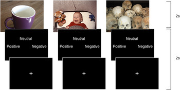
As shown in Fig. 1 (see also van Buuren et al. Reference Van Buuren, Vink, Rapcencu and Kahn2011), participants were instructed to view each picture for 2 s and to rate it as neutral, positive or negative by pressing a button when the evaluation screen was shown (maximum of 2 s). After each response, a fixation-cross appeared for the remaining trial duration. All subjects performed the task with their right hand. The task comprised four blocks of 96 s in which 24 pictures, eight of each condition, were presented in pseudo-randomized order. Each block was followed by a baseline block in which a fixation-cross was shown in the middle of the screen (32 s). Group differences in behavioural performance were analysed (Supplementary Information, S1).
Experimental task. Neutral (an example displayed left), positive (middle) and negative (right) emotional pictures from the international affective picture system (IAPS; Lang et al. Reference Lang, Bradley and Cuthbert1997) were presented to the participant for 2 s. The picture was followed by an evaluation screen, which was shown until the participant has responded (maximum 2 s). The fixation-cross appeared for the remaining trial time. This figure has been adapted with kind permission from van Buuren et al. (Reference Van Buuren, Vink, Rapcencu and Kahn2011).

Image acquisition
Functional images were acquired using a 3.0 T whole-body magnetic resonance imaging scanner (Philips Medical System, The Netherlands). Three hundred and twenty-two functional images were acquired during the task using a two-dimensional echo planar imaging-sensitivity encoding (EPI-SENSE) sequence with the following parameters: voxel size 4 mm isotropic; repetition time (TR) = 1600 ms; echo time (TE) = 23 ms; flip angle = 72.5°; 64 × 51 matrix, 4 × 4 mm in-plane resolution, 4 mm slice thickness; field of view (FOV) 256 × 204 mm; 30-slice volume; SENS-factor R = 2.4 (anterior–posterior). A T1-weighted image (200 slices, repetition time = 10 ms, echo time = 3.8 ms, flip angle = 8°, field of view = 240 × 240 × 160 mm, matrix of 304 × 299) was used for within-subject registration purposes.
Data analyses
First-level analysis
The fMRI data were pre-processed and analysed with SPM 5 (http://www.fil.ion.ucl.ac.uk/spm). Volumes were slice-time corrected to the middle slice and realigned to the first acquired volume. The data were spatially normalized to the Montreal Neurological Institute (MNI) T1-weighted template. The data were smoothed using an 8 mm full-width-at-half-maximum (FWHM) Gaussian kernel.
A general linear model regression analysis was used to model the fMRI data. Based on the stimuli and responses of the participants, three conditions were created when the participant's response was congruent with the neutral, positive or negative IAPS rating. Incongruent trials were not included in the analyses. The onsets and duration (2 s) of these three conditions were modelled as factors in the design matrix of the regression model. The six realignment parameters were included in the design matrix as regressors of no interest to correct for head movement. A high-pass filter with a cut-off frequency of 0.0058 Hz was applied to the data to correct for low-frequency scanner drifts.
For each participant three first-level contrasts were created: (1) the neutral pictures compared with rest (neutral>rest; baseline contrast), (2) the negative pictures compared with the neutral pictures (negative>neutral; negative contrast), and (3) the positive pictures compared with the neutral pictures (positive>neutral; positive contrast). The negative and positive contrasts were created this way to measure the effect of valence correcting for attentional and visual processes.
Group analyses
To test if the emotion network was activated by the task, a whole brain one-sample analysis was performed for all groups separately for both the negative and positive contrast.
Because of our specific interest in the amygdala, hippocampus, insula, dACC and sgACC, region of interest (ROI) analyses were performed for these areas on all three contrasts. Mean activation levels were extracted from predefined ROIs for both hemispheres separately. The amygdala, insula, dACC and sgACC ROIs were defined based on the WFU Pick atlas. For the dACC and sgACC Brodmann's areas (BA) were used (dACC: BA 32, sgACC: BA 25). The hippocampus was defined using the LONI Probabilistic Brain Atlas with a probability threshold of 80%. A 3 (group) × 5 (ROI) × 2 (hemisphere; left, right) repeated-measures general linear model (GLM) analysis was performed for the baseline, negative and positive contrast (multivariate test, Wilks’ lambda p < 0.05). Post-hoc tests were performed separately for each ROI when the group × ROI (or group × ROI by hemisphere) interaction was significant. Multiple testing was corrected for with Bonferroni.
To investigate group differences outside the ROIs, whole brain group analyses were performed for the negative and positive contrasts. The resulting maps were tested for significance at a cluster-defining threshold of p < 0.005, and a p < 0.05 family-wise error-corrected critical cluster size was calculated separately for each contrast. The cluster sizes were determined using SPM and a script (CorrClusTh.m; http://www2.warwick.ac.uk/fac/sci/statistics/staff/academic-research/nichols/scripts/spm), which uses estimated smoothness (estimated FWHM: 8 mm) and Random Field Theory to find these corrected thresholds. When a group effect was observed, post-hoc t tests masked with the main group effect were performed. Finally, a whole brain analysis on the baseline contrast was performed to confirm the absence of baseline group differences.
Psychophysiological interaction analyses
Changes in functional coupling between a voxel of interest (VOI) and all other brain areas during negative pictures compared to neutral pictures were assessed with psychophysiological interaction (PPI) analysis (Friston et al. Reference Friston, Buechel, Fink, Morris, Rolls and Dolan1997; Gitelman et al. Reference Gitelman, Penny, Ashburner and Friston2003). The centres of both the left and right amygdala ROI were used as VOI. A 4 mm-radius sphere around the centre of the VOIs was used as the seed region for the PPI analyses. For each individual, the eigenvariate of the BOLD signal within the seed region was calculated and adjusted for mean activation during negative v. neutral pictures and head motion. The interaction between activity within the seed region and the psychophysiological factor (PPI regressor) was calculated and activity that was (positively or negatively) related to each interaction was investigated.
For each group, one-sample t tests were performed to investigate areas that show increased (positive PPI) or decreased (negative PPI) coupling with the left and right amygdala, respectively. Then, a one-way ANOVA with group as a factor was performed to investigate group differences for functional coupling. The resulting maps were tested for significance at a cluster-defining threshold of p < 0.005, and a p < 0.05 family-wise error-corrected critical cluster size calculated separately for the left and right amygdala. Post-hoc tests were performed when the groups differed significantly.
Depression
To investigate the effect of co-morbid major depressive disorder (MDD) in our PTSD sample, the PTSD group was divided into a group with current MDD (PTSD+MDD) and a group without current MDD (PTSD–MDD) group. The same ROI analyses were performed to examine differences between PTSD with and without MDD.
Results
Demographics
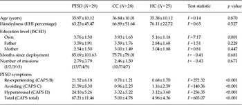
Demographics are presented in Table 1. Two PTSD patients were removed from the analysis because of excessive head movement (>3 mm). A total of 29 PTSD patients, 28 CC and 25 HC were included in the analyses. All PTSD patients fulfilled the DSM-IV criteria for PTSD as confirmed with a CAPS score ⩾45. Sixteen patients had a current co-morbid MDD, six a co-morbid anxiety disorder and two a co-morbid pain disorder according to DSM-IV criteria. The two control groups did not meet the criteria for PTSD as confirmed with a CAPS score ⩽15, and had no current psychiatric disorder. Age and handedness [according to the Edinburgh Handedness Inventory; EHI (Oldfield, Reference Oldfield1971)] were comparable for the three groups. Education level of the subject (but not of parents) differed significantly between the groups (F 2,79 = 7.17, p = 0.001). The HC group had a higher education level than the PTSD group (p = 0.001) and CC group (p = 0.003). The PTSD and CC groups did not differ. Year and country of deployment, number of military missions and time since deployment were comparable for the CC and the PTSD groups. None of the participants showed evidence for dissociation during the MRI scan according to the DSS.
Demographics of the PTSD, CC and HC group

Data are presented as means ± s.d.
PTSD, Post-traumatic stress disorder patients; CC, combat controls; HC, healthy non-military controls; EHI, Edinburgh Handedness Inventory (Oldfield, Reference Oldfield1971); ISCED, International Standard Classification of Education (Schneider, Reference Schneider2013); CAPS, Clinician Administered PTSD Scale (Blake et al. Reference Blake, Weathers, Nagy, Kaloupek, Klauminzer and Charney1990).
Task activation
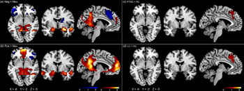
In all three groups brain areas involved in emotion regulation, i.e. amygdala, middle temporal gyrus, medial superior frontal gyrus and hippocampus, were significantly activated or deactivated during the negative and positive contrasts (Fig. 2, displayed for HC; Supplementary Table S2).
Whole brain activation during (a) the negative contrast (negative v. neutral pictures) and (b) the positive contrast (positive v. neutral pictures) is displayed for the non-military healthy control (HC) group in the left panel. Results of the whole brain group analyses for the PTSD patients (PTSD), combat controls (CC) and non-military healthy controls (HC) are displayed in the right panel. Only post-hoc t tests with significant differences between groups are displayed; clusters showing increased activation in (c) PTSD compared with HC and (d) CC compared with HC. Brain regions in warm colours show increased activation; brain regions in cool colours are deactivated in this contrast. Significant activated or deactivated clusters are displayed on a standardized brain (MRIcron). Significance is based on a cluster-defining threshold of p < 0.005, and a p < 0.05 family-wise error-corrected critical cluster size calculated separately for each contrast. Left=left.

ROI results
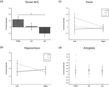
A significant group × ROI by hemisphere interaction was observed for the negative contrast (F 8,152 = 1.99, p = 0.05). Post-hoc tests within the ROIs showed a significant group difference in the dACC (F 2,79 = 4.10, p = 0.02; Fig. 3 a). The PTSD group showed increased dACC activation compared with the HC group (p = 0.02). The CC group did not differ from the PTSD (p = 0.43) or the HC (p = 0.49) groups. A significant group × hemisphere interaction was observed in the hippocampus (F 2,79 = 6.07, p < 0.01) and insula (F 2,79 = 4.73, p = 0.01). PTSD patients showed more left than right hippocampal and insula activation, whereas the CC and HC groups showed comparable left and right activation (Fig. 3 b,c). No group differences for the negative contrast were observed in the amygdala (F 2,79 = 0.11, p = 0.90; Fig. 3 d) or the sgACC (F 2,79 = 0.78, p = 0.46). Analyses of the positive and baseline contrast revealed no significant group differences in any of the ROIs [no group × ROI (by hemisphere) interactions; all F values <1].
Region of interest (ROI) analysis of the (a) dorsal anterior cingulate cortex (dACC), (b) hippocampus, (c) insula, and (d) amygdala in PTSD patients (PTSD; left), combat controls (CC; middle) and non-military healthy controls (HC; right) for the negative contrast (negative>neutral pictures). (a) Bars represent group mean bilateral dACC activation levels (i.e. parameter estimates). Error bars indicate the s.e. and ** means p < 0.005. (b) The group mean of left and right hippocampus activation levels are presented. (c) The group mean of left and right insula activation levels are displayed. (d) The scatterplot shows the amygdala activation level for each subject.

Whole brain results
The HC group showed a significant deactivation of the left medial superior frontal gyrus for the negative contrast. When compared with the HC group, both veterans with and without PTSD showed significantly more activation in this area, as they did not show this deactivation (Fig. 2, Supplementary Table S3). The PTSD group also showed significantly more activation compared to the HC group in the right ACC and supplementary motor area. The whole brain analysis did not reveal differences between the PTSD and CC groups. Finally, the positive and baseline contrast yielded no significant group differences.
Functional connectivity results
No significant whole brain group differences in functional connectivity of the left and right amygdala were observed. However, analysis per group showed that the PTSD group had increased functional coupling between right amygdala and left medial superior frontal gyrus, bilateral supplementary motor area, left medial frontal gyrus, and the right inferior frontal gyrus during negative v. neutral pictures (Supplementary Table S4).
Depression
The PTSD group was divided into a PTSD+MDD group (N = 16) and a PTSD–MDD group (N = 13). Total PTSD severity was higher in the PTSD+MDD group (t 27 = −3.02, p = 0.01; Supplementary Table S5). The two groups did not differ in age (t 27 = −1.28, p = 0.21), re-experiencing symptoms (t 27 = −0.34, p = 0.74), or hyperarousal symptoms (t 27 = −0.66, p = 0.52). Avoiding and numbing symptoms were more severe in the PTSD+MDD group (t 27 = −3.57, p < 0.01).
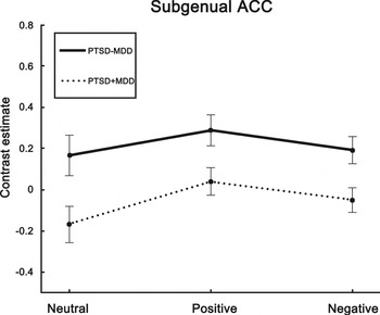
The PTSD+MDD group showed a decreased sgACC response to neutral pictures (baseline) compared to the PTSD–MDD group (t 1,27 = 4.65, p = 0.04), therefore, analyses for the negative and positive pictures were performed v. rest instead of neutral pictures. The PTSD+MDD group showed a decreased sgACC response to negative pictures (t 1,27 = 7.35, p = 0.01) and positive pictures (t 1,27 = 6.09, p = 0.02; Fig. 4) v. rest. The difference in total PTSD severity between the PTSD+MDD and PTSD–MDD group could not account for the sgACC differences, because no significant correlation within the whole PTSD group was observed between PTSD severity and sgACC activity to neutral (r = −0.20, p = 0.30), negative (r = −0.33, p = 0.08) and positive (r = −0.26, p = 0.17) pictures v. rest. No group differences were observed in the other ROIs.
Region of interest (ROI) analysis of the subgenual anterior cingulate cortex (sgACC) in PTSD patients with major depressive disorder (MDD; PTSD+MDD, dotted line) and without MDD (PTSD–MDD; solid line) during the viewing of neutral, positive and negative pictures. The dots represent group mean activation levels (i.e. parameter estimates), and error bars indicate the s.e. The sgACC response is significantly different between the two groups for all pictures. Significance is based on a cluster-defining threshold of p < 0.005, and a p < 0.05 family-wise error-corrected critical cluster size calculated separately for each contrast.

Discussion
This fMRI study investigated the response to trauma-unrelated emotional neutral, negative, and positive pictures in war veterans with PTSD, war veterans without PTSD (CC) and non-military HC. Our findings do not support the hypothesis that heightened amygdala activation in PTSD extends to trauma-unrelated negative emotional stimuli. Instead, PTSD patients showed increased dACC activation during the viewing of negative v. neutral pictures compared with the HC group. Furthermore, PTSD patients showed asymmetric activation in the hippocampus and insula (more left than right), which was not observed in the control groups. Deactivation of the medial superior frontal gyrus was only observed in the HC group and not in the veterans with and without PTSD. No group differences were observed in ROI or whole brain activation to the neutral or positive contrasts. Last, decreased sgACC activation to all stimuli was observed in PTSD patients with MDD compared to PTSD patients without MDD.
No evidence for heightened amygdala activation in our PTSD sample was observed. The type of stimuli we used or the population that was studied could account for these findings. First, heightened amygdala activity has consistently been demonstrated in studies where trauma-related stimuli or fearful faces were presented (Shin et al. Reference Shin, Kosslyn, Mcnally, Alpert, Thompson, Rauch, Macklin and Pitman1997, Reference Shin, Orr, Carson, Rauch, Macklin, Lasko, Peters, Metzger, Dougherty, Cannistraro, Alpert, Fischman and Pitman2004, Reference Shin, Wright, Cannistraro, Wedig, Mcmullin, Martis, Macklin, Lasko, Cavanagh, Krangel, Orr, Pitman, Whalen and Rauch2005; Rauch et al. Reference Rauch, Whalen, Shin, Mcinerney, Macklin, Lasko, Orr and Pitman2000; Bryant et al. Reference Bryant, Kemp, Felmingham, Liddell, Olivieri, Peduto, Gordon and Williams2008). Trauma-related stimuli induce strong fear responses in PTSD mediated by the amygdala (Rauch et al. Reference Rauch, Shin and Phelps2006), and fearful faces represent indicators of danger and provoke a larger amygdala response than general emotional pictures (Hariri et al. Reference Hariri, Tessitore, Mattay, Fera and Weinberger2002). Patients with an anxiety disorder (including PTSD) respond more strongly to fearful stimuli than controls (Etkin & Wager, Reference Etkin and Wager2007). This would explain why studies presenting fear-evoking stimuli did observe heightened amygdala activation in PTSD patients, whereas we did not. We also presented positive stimuli to test the hypothesis of Phan et al. (Reference Phan, Britton, Taylor, Fig and Liberzon2006) that the absence of amygdala hyperresponsivity was due to emotional numbing, but our findings did not support this hypothesis.
Second, it has previously been suggested that increased amygdala activation could only be detected when PTSD patients were compared to a HC group (Patel et al. Reference Patel, Spreng, Shin and Girard2012). Indeed, Simmons et al. (Reference Simmons, Matthews, Strigo, Baker, Donovan, Motezadi, Stein and Paulus2011) showed increased amygdala activation in all veterans regardless of PTSD status to faces v. shapes. Increased amygdala activation was observed in PTSD patients compared to combat and healthy controls specifically for the contrast fearful v. happy faces. Here, we included a large group of PTSD patients and compared them with both trauma and healthy control groups, thus this cannot explain the absence of differences. Like Phan et al (Reference Phan, Britton, Taylor, Fig and Liberzon2006), who also did not observe an increased amygdala response to IAPS pictures in PTSD patients, we only studied males. The two studies that presented IAPS pictures and observed increased amygdala activity in PTSD patients included predominantly females (Brohawn et al. Reference Brohawn, Offringa, Pfaff, Hughes and Shin2010; Brunetti et al. Reference Brunetti, Sepede, Mingoia, Catani, Ferretti, Merla, Del Gratta, Romani and Babiloni2010). This could indicate an effect of sex on the amygdala response to (trauma-unrelated) emotional stimuli in PTSD, which has been suggested to explain the higher prevalence of PTSD in women (Hamann, Reference Hamann2005).
Aside from studies presenting IAPS pictures, two studies on trauma-unrelated stressful or negative emotional imagery were performed in PTSD patients. No group differences were observed in the amygdala, but PTSD patients showed increased ACC activation (Lanius et al. Reference Lanius, Williamson, Hopper, Densmore, Boksman, Gupta, Neufeld, Gati and Menon2003; Gold et al. Reference Gold, Shin, Orr, Carson, Rauch, Macklin, Lasko, Metzger, Dougherty, Alpert, Fischman and Pitman2011). In contrast, two studies that used non-emotional tasks, i.e. an auditory oddball (Bryant et al. Reference Bryant, Felmingham, Kemp, Barton, Peduto, Rennie, Gordon and Williams2005) and trauma-unrelated memory recognition task (Whalley et al. Reference Whalley, Rugg, Smith, Dolan and Brewin2009), did see an increased amygdala response in the PTSD group. Taken together, the fMRI response to trauma-unrelated stimuli has previously been studied in PTSD patients, but findings were mixed and studies were limited by small sample sizes. Our study is the largest fMRI study to our knowledge and our findings strongly suggest that increased amygdala activity in PTSD does not extend to non-fear evoking negative stimuli in males. Potentially, trauma-unrelated stimuli could evoke stronger amygdala responses in female PTSD patients.
The dACC was found to be hyperresponsive to negative v. neutral pictures in the PTSD patients compared with the HC group. Increased dACC activation in PTSD has previously been demonstrated in several studies during cognitive tasks with emotional components, such as an emotional face-matching task and an attention bias task with threat stimuli (Fonzo et al. Reference Fonzo, Simmons, Thorp, Norman, Paulus and Stein2010; Fani et al. Reference Fani, Jovanovic, Ely, Bradley, Gutman, Tone and Ressler2012), or during general cognitive tasks (Bryant et al. Reference Bryant, Felmingham, Kemp, Barton, Peduto, Rennie, Gordon and Williams2005; Shin et al. Reference Shin, Bush, Milad, Lasko, Brohawn, Hughes, Macklin, Gold, Karpf, Orr, Rauch and Pitman2011). The dACC is thought to play a role in the regulation of both cognitive and emotional processing (Phan et al. Reference Phan, Wager, Taylor and Liberzon2002; Etkin et al. Reference Etkin, Egner and Kalisch2011). Increased dACC activity was associated with increased attention to negative emotions (Etkin et al. Reference Etkin, Egner and Kalisch2011), and reappraisal or rating of emotional stimuli v. passive viewing (Ochsner et al. Reference Ochsner, Bunge, Gross and Gabrieli2002; Taylor et al. Reference Taylor, Phan, Decker and Liberzon2003; Hutcherson et al. Reference Hutcherson, Goldin, Ochsner, Gabrieli, Feldman Barrett and Gross2005). The increased dACC activation we observed during presentation of negative v. neutral pictures in PTSD is therefore thought to reflect an attentional bias to negative stimuli, previously demonstrated as attentional bias to threat stimuli (Fani et al. Reference Fani, Jovanovic, Ely, Bradley, Gutman, Tone and Ressler2012). This is in accordance with cognitive models that emphasize the importance of attentional bias for PTSD (Ehlers & Clark, Reference Ehlers and Clark2000). The attentional bias is specific for negative stimuli, as a difference was only observed for the negative contrast and not for the positive contrast. Moreover, we demonstrate that an increased dACC response in PTSD is not specific to trauma- or fear-related stimuli, as is the amygdala, but it implicates an attentional bias that generalizes to trauma-unrelated negative stimuli. Unlike the amygdala response which is implicated in various psychiatric disorders, the dACC has specifically been associated with PTSD (Etkin & Wager, Reference Etkin and Wager2007); our findings highlight the importance of the (d)ACC in PTSD.
Increased activation in the left compared to the right hippocampus and insula was observed in PTSD patients, but not in the two control groups, during the negative contrast. The hippocampus and insula, specifically in the left hemisphere, have often been implicated in PTSD, both structurally and functionally. Reduced volume of particularly the left hippocampus has been found in PTSD (Karl et al. Reference Karl, Schaefer, Malta, Dörfel, Rohleder and Werner2006). Furthermore, increased left hippocampal activation is observed during the administration of pain stimuli (Geuze et al. Reference Geuze, Westenberg, Jochims, de Kloet, Bohus, Vermetten and Schmahl2007), and the encoding of negative words (Thomaes et al. Reference Thomaes, Dorrepaal, Draijer, de Ruiter, Elzinga, van Balkom, Smoor, Smit and Veltman2009, Reference Thomaes, Dorrepaal, Draijer, de Ruiter, Elzinga, Sjoerds, van Balkom, Smit and Veltman2013). Activation in the left parahippocampal region differed between PTSD patients and controls during presentation of masked emotional stimuli (Sakamoto et al. Reference Sakamoto, Fukuda, Okuaki, Rogers, Kasai, Machida, Shirouzu, Yamasue, Akiyama and Kato2005), and positive emotional processing (Jatzko et al. Reference Jatzko, Schmitt, Demirakca, Weimer and Braus2006). Activation in left insula during anticipation of positive or negative images differentiated PTSD patients who respond to treatment v. non-responders (Simmons et al. Reference Simmons, Norman, Spadoni and Strigo2013). Other studies showed reduced bilateral insula activation to anticipation of negative or positive pictures after treatment (Aupperle et al. Reference Aupperle, Allard, Grimes, Simmons, Flagan, Thorp, Norman, Paulus and Stein2013), and bilateral increased insula activation to pain stimuli (Geuze et al. Reference Geuze, Westenberg, Jochims, de Kloet, Bohus, Vermetten and Schmahl2007). The asymmetry observed in the current study has not been demonstrated in previous functional imaging studies. A potential explanation is that previous studies analysed the left and right hemisphere separately, or that the left and right hippocampus or insula were analysed as one ROI. Our findings suggest that future functional imaging studies should include them as two separate regions in one analysis to further explore this asymmetry.
During the negative contrast, deactivation was observed in the medial superior frontal gyrus in the HC group, but not in the PTSD and CC groups. The medial superior frontal gyrus is involved in attention shifting (Nagahama et al. Reference Nagahama, Okada, Katsumi, Hayashi, Yamauchi, Sawamoto, Toma, Nakamura, Hanakawa, Konishi, Fukuyama and Shibasaki1999; Cutini et al. Reference Cutini, Scatturin, Menon, Bisiacchi, Gamberini, Zorzi and Dell'acqua2008), and its activity increases with increased need for executive control (Mitchell, Reference Mitchell2013). Increased medial superior frontal gyrus activation has been observed in PTSD patients during script-driven imagery (Lindauer et al. Reference Lindauer, Booij, Habraken, Uylings, Olff, Carlier, Den Heeten, Van Eck-Smit and Gersons2004), fearful faces (Stevens et al. Reference Stevens, Jovanovic, Fani, Ely, Glover, Bradley and Ressler2013), and encoding in emotional contexts (Whalley et al. Reference Whalley, Rugg, Smith, Dolan and Brewin2009). The finding that both veterans with and without PTSD did not show deactivation of the medial superior frontal gyrus during the negative contrast implies that veterans exert cognitive control when presented with negative pictures. Only PTSD patients showed increased functional connectivity of the right amygdala with the medial superior frontal gyrus during the presentation of negative pictures compared to neutral pictures. This suggests increased cognitive control over the amygdala in the PTSD group. PTSD patients might need additional regulation of the overactive amygdala when presented with trauma-related stimuli, which they exert by increasing cognitive control. Consequently, when presented with trauma-unrelated stimuli PTSD patients might succeed in controlling their amygdala, which would explain the absence of increased amygdala activity in response to trauma-unrelated stimuli.
When the PTSD patients were divided into subgroups with and without co-morbid major depressive disorder (PTSD+MDD/PTSD–MDD), the PTSD+MDD patients showed decreased sgACC activation to all pictures. The sgACC has been identified as one of the key regions implicated in the aetiology of MDD (Drevets et al. Reference Drevets, Price, Simpson, Todd, Reich, Vannier and Raichle1997; Mayberg et al. Reference Mayberg, Liotti, Brannan, Mcginnis, Mahurin, Jerabek, Silva, Tekell, Martin, Lancaster and Fox1999), and has been a target area for deep brain stimulation (Mayberg et al. Reference Mayberg, Lozano, Voon, Mcneely, Seminowicz, Hamani, Schwalb and Kennedy2005) and ablative surgery (Dougherty et al. Reference Dougherty, Weiss, Cosgrove, Alpert, Cassem, Nierenberg, Price, Mayberg, Fischman and Rauch2003). Successful treatment of MDD has been associated with altered sgACC activation post-treatment (Drevets et al. Reference Drevets, Bogers and Raichle2002). Here we demonstrated that decreased sgACC activation is also apparent in PTSD patients with co-morbid MDD. SgACC hypoactivity was specifically related to PTSD+MDD status, because it did not correlate with total PTSD severity. Also, it was not influenced by medication use, as all patients were medication naive. Dorsal ACC hyperactivity, on the other hand, was observed in PTSD patients regardless of MDD status and is therefore primarily associated with PTSD. Decreased amygdala activation in PTSD patients with MDD was found in a previous study presenting facial stimuli (Kemp et al. Reference Kemp, Felmingham, Das, Hughes, Peduto, Bryant and Williams2007), but was not observed in this study.
Limitations and future directions
The majority of the PTSD patients suffered from a co-morbid psychiatric disorder. This might have influenced our findings; however, for the generalizability of our results the inclusion of PTSD patients with co-morbid psychiatric disorders is important, because co-morbidity is often seen in PTSD. As PTSD+MDD patients showed a neurobiological profile dissimilar from PTSD–MDD patients, future research is recommended to consider the effect of co-morbid MDD (or MDD symptoms), particularly when differences between groups are observed in brain areas implicated in MDD. In this study, no direct comparison between trauma-related and trauma-unrelated stimuli could be made. As this is highly relevant, future studies should include both trauma-related and trauma-unrelated stimuli. This study underscores the recommendation (Patel et al. Reference Patel, Spreng, Shin and Girard2012) to include both healthy controls and combat controls (or individuals with exposure to comparable trauma). Future studies interested in the effects of stress/trauma exposure alone are recommended to replicate the study in a non-deployed military sample or a civilian sample (with two control groups), because in our study the effect of trauma/stress exposure cannot be distinguished from military training/selection. Moreover, replication in a female sample is suggested, as in this study only males were tested and findings are not generalizable to the female population. For investigating potential state or ‘scar’ aspects of increased dACC activation in PTSD patients, the study should be repeated after patients have received treatment.
Conclusion
Our findings do not support the hypothesis that increased amygdala activation in PTSD generalizes to trauma-unrelated emotional processing. Instead, the increased dACC response found in PTSD patients implicates an attentional bias that extends to trauma-unrelated negative stimuli. Hyperresponsivity of the dACC has consistently been found in PTSD, implicating the dACC as an important target area for future PTSD research. Decreased sgACC activation, often associated with MDD, was related to MDD status within the PTSD group, underscoring the importance of distinguishing between PTSD with and without co-morbid MDD in future research.
Supplementary material
For supplementary material accompanying this paper visit http://dx.doi.org/10.1017/S0033291714001706.
Acknowledgements
We thank Mariet van Buuren, Ph.D., and Anca Rapcencu, M.D. for their help with the setup of the task and data collection.
This study was financially supported by the Dutch Ministry of Defence.
Declaration of Interest
None.