Introduction
There are about 60 minerals that have been first described from deposits related to bat or bird guano. Considering the contrasting behaviours of bats and birds, it should come as no surprise that bat guano deposits generally occur in enclosed spaces, such as caves and mines, whereas bird guano deposits generally form in exposed areas. Significantly more than half of the first descriptions of guano minerals are from bat guano deposits. The earliest recorded of these are nitrocalcite, Ca(NO3)2·4H2O, and nitromagnesite, Mg(NO3)2·6H2O, which were described from the Nicajack Cave in Marion County, Tennessee, USA (Romé de Lisle, Reference de Lisle J.B.L1783). The first new mineral to be described as forming from the reaction of bat excrement in a mine was rowleyite (Kampf et al., Reference Kampf, Cooper, Nash, Cerling, Marty, Hummer, Celestian, Rose and Trebisky2017) as, up to that point, all bat guano derived minerals had been described from caves. However, it should be noted that rouaite, Cu2(NO3)(OH)3, discovered in the Roua copper mines, Daluis, France, has been conjectured to have derived its nitrate from guano (Sarp et al., Reference Sarp, Černý and Guenee2001) and this may also be the case for its dimorph gerhardtite, Cu2(NO3)(OH)3, discovered in the United Verde mine, Jerome, Arizona, USA (Wells and Penfield, Reference Wells and Penfield1885).
Newberyite, Mg(PO3OH)·3H2O, was described from bat guano in the Skipton Caves, Victoria, Australia (vom Rath, Reference vom Rath1879). Herein, we describe the new mineral manganonewberyite, Mg(PO3OH)·3H2O, the Mn analogue of newberyite, from the Cassagna manganese mine in Liguria, Italy. The new mineral and the name have been approved by the Commission on New Minerals, Nomenclature and Classification of the International Mineralogical Association (IMA2024–004, Kampf et al., Reference Kampf, Ma and Castellaro2024; Warr symbol: Mnew). The description is based upon one holotype specimen and one cotype specimen deposited in the collections of the Natural History Museum of Los Angeles County, 900 Exposition Boulevard, Los Angeles, CA 90007, USA, catalogue numbers 76311 and 76312.
Occurrence
Manganonewberyite was found by one of the authors (FC) in an underground gallery of the Cassagna mine, Graveglia Valley, Ne, Genoa Province, Liguria, Italy (44°20ʹ8”N, 9°28ʹ31”E). The manganese deposit exploited by the Cassagna mine was discovered in 1877, along with several other manganese deposits in the Graveglia Valley. Manganese mining in the mines began in 1881 and operations at the Cassagna mine ended in 1998. The underground workings are extensive and there is a more recent large open pit. The manganese ores (mainly braunite intermixed with quartz) occur in stratiform layers, massive lenses and boudins near the base of the Monte Alpe Chert Formation, which overlies the ophiolitic basement. In the final tectono-metamorphic stage of the formation of these deposits, hydrothermal fluids circulated through systems of extensional fractures resulting in polyphase mineral assemblages.
Manganonewberyite occurs with niahite, kutnohorite, sampleite and serrabrancaite on a tinzenite–quartz–braunite matrix. Considering the mineral association and the fact that bats are plentiful in the gallery in which the material was found, the formation of manganonewberyite can clearly be attributed to the interaction of bat guano with the Mn-rich rock.
Physical and optical properties
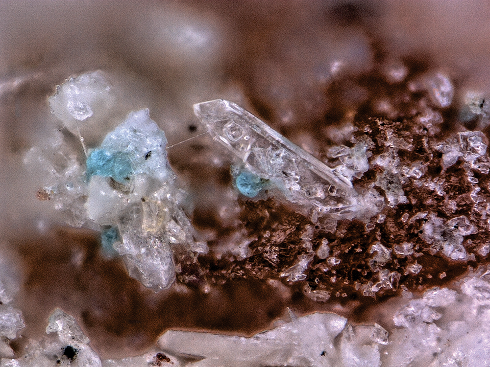
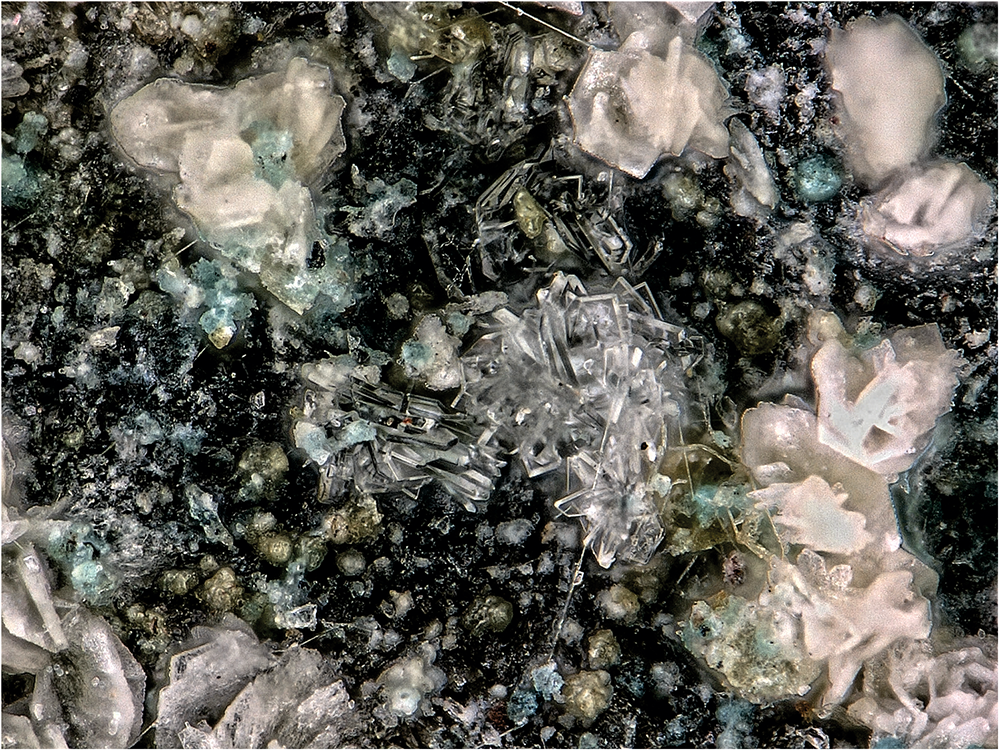
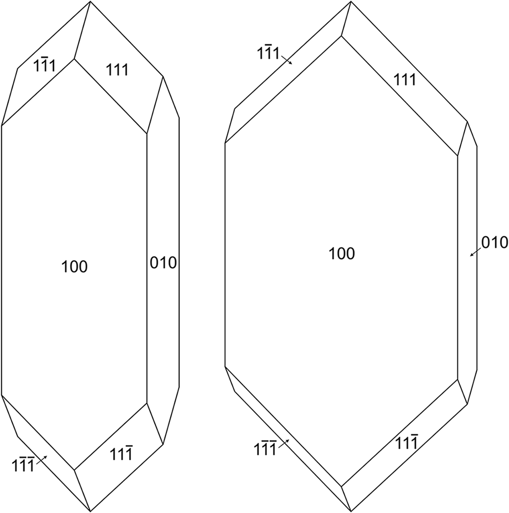
Manganonewberyite crystals are colourless prisms and blades, up to ∼0.15 mm long, occurring as individuals and in irregular groupings (Figs 1 and 2). Crystals are elongated parallel to [001], are more or less flattened on {100} and exhibit the forms {100}, {010} and {111} (Fig. 3). The mineral is transparent and has a white streak. The mineral does not fluoresce in long- or short-wave ultraviolet light. The Mohs hardness is ∼3 based on scratch tests. Crystals are brittle with curved fracture. Cleavage is perfect on {010}. The density measured by flotation in a mixture of methylene iodide and toluene is 2.34(2) g·cm–3. The calculated density is 2.337 g·cm–3 for the empirical formula and 2.351 g·cm–3 for the ideal formula, in both cases using the single-crystal cell. At room temperature, the mineral is insoluble in H2O and dissolves easily in dilute HCl.
Manganonewberyite prism with blue sampleite on holotype specimen #65733. The field of view is 0.38 mm across.

Intergrown colourless blades of manganonewberyite, with cream-coloured niahite and blue sampleite on holotype specimen #65733. The field of view is 0.68 mm across.

Crystal drawings of manganonewberyite prism (left) and blade (right), clinographic projections. Drawn using SHAPE version 7.4 (Shape Software, Kingsport, Tennessee, USA).

Optically, manganonewberyite is biaxial (+) with indices of refraction α = 1.541(2), β = 1.547(2) and γ = 1.559(2) measured in white light. The 2V determined from extinction data analysed with EXCALIBRW (Gunter et al., Reference Gunter, Bandli, Bloss, Evans, Su and Weaver2004) is 71.6(3)°; the calculated 2V is 71.0°. No dispersion was observed and the optical orientation is X = a, Y = b and Z = c. The mineral is nonpleochroic. The Gladstone–Dale compatibility index (Mandarino, Reference Mandarino2007) is 0.007 for the empirical formula and 0.011 for the ideal formula, in both cases in the range of superior compatibility.
Raman spectroscopy
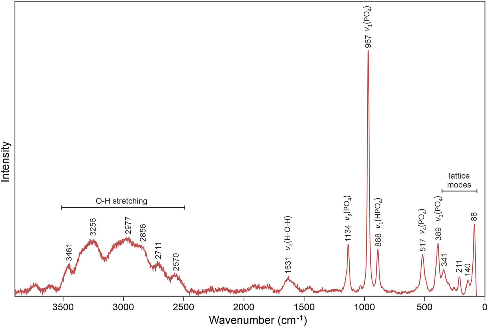
Raman spectroscopy was conducted on a Horiba XploRA PLUS using a 532 nm diode laser, 200 μm slit, 1800 grooves/mm diffraction grating and a 100× (0.9 NA) objective. Because the mineral is very sensitive to the laser, low power (∼2 mW) was used, which provided a very noisy spectrum. The Raman spectrum of manganonewberyite is very similar to that of newberyite (Frost et al., Reference Frost, Palmer and Pogson2011). It is dominated by the stretching and bending vibrations of HPO4 tetrahedra and O–H stretching vibrations. The spectrum from 3900 to 70 cm–1 shown in Fig. 4 includes band assignments based on Frost et al. (Reference Frost, Palmer and Pogson2011).
Baseline-corrected Raman spectrum of manganonewberyite recorded with a 532 nm laser.

Composition
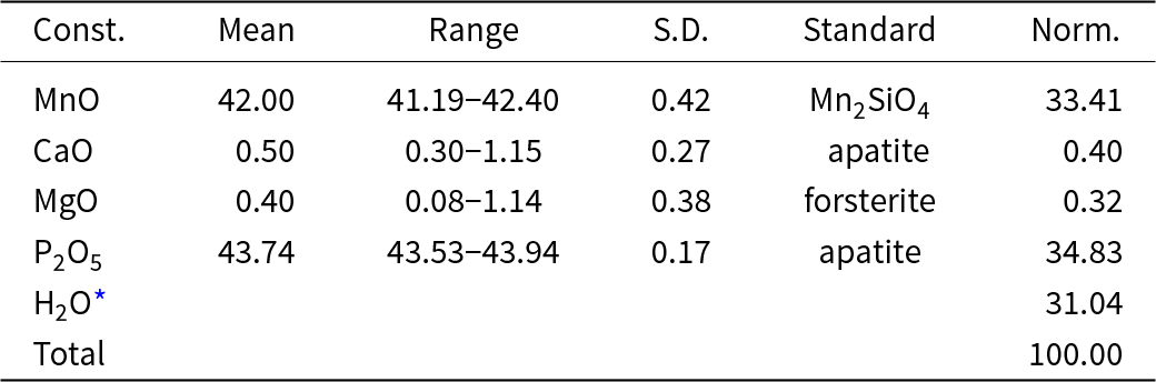
Electron probe microanalyses (EPMA; 7 points) were performed at Caltech on a JXA-iHP200F electron microprobe in WDS mode. Analytical conditions were 15 kV accelerating voltage, 10 nA beam current and 5 μm beam diameter. Insufficient material is available for the determination of H2O, so it is calculated based on the structure. No beam damage was observed during analyses; however, as is common for highly hydrated minerals, much of the H2O in manganonewberyite was lost under vacuum. Analysis under milder conditions using energy dispersive spectroscopy under ambient pressure is unlikely to yield accurate results. The loss of H2O resulted in much higher concentrations for the remaining constituents than are to be expected for the fully hydrated phase; therefore, the other analysed constituents have been normalised to provide a total of 100% when combined with the calculated H2O content. Analytical data are given in Table 1. The empirical formula based on 1 P and 7 O atoms per formula unit (apfu) is (Mn0.960Mg0.016Ca0.015)Σ0.991(H1.02P1.00O4)(H2O)3. The simplified formula is (Mn,Mg,Ca)(PO3OH)(H2O)3 and the ideal formula is Mn(PO3OH)(H2O)3, which requires MnO 34.61, P2O5 34.63, H2O 30.76, total 100 wt.%.
Compositional data in wt.% for manganonewberyite

S.D. – standard deviation
* Based on the structure, i.e., P = 1 and O = 7 apfu
X-ray crystallography and structure refinement
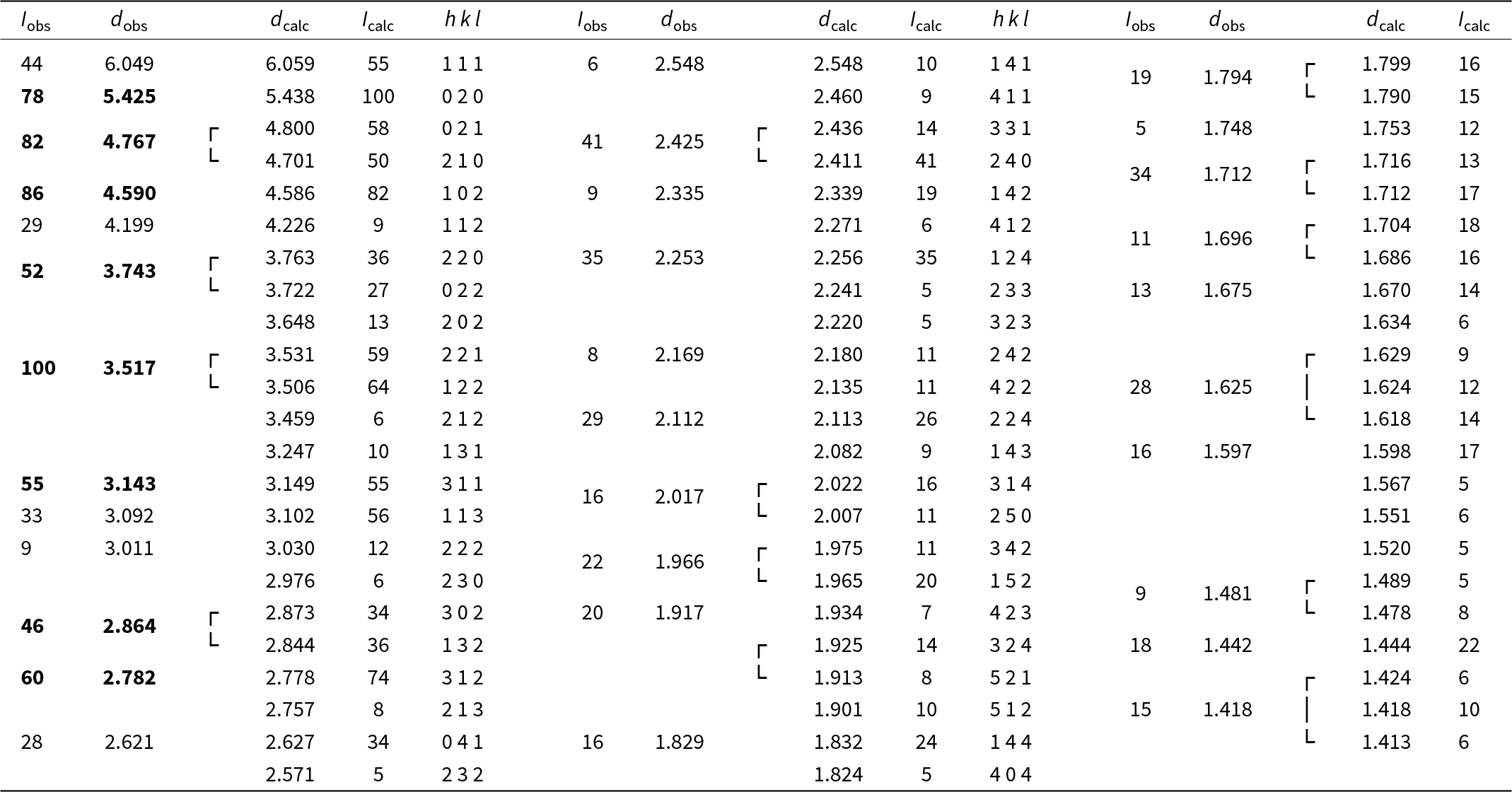
Powder X-ray diffraction data were recorded using a Rigaku R-Axis Rapid II curved imaging plate microdiffractometer with monochromatised MoKα radiation. A Gandolfi-like motion on the φ and ω axes was used to randomise the sample. Observed d-values and intensities were derived by profile fitting using JADE Pro software (Materials Data, Inc.). The powder data are presented in Table 2. The unit-cell parameters refined from the powder data using JADE Pro with whole-pattern fitting (space group Pbca) are a = 10.430(8), b = 10.875(8), c = 10.210(7) Å and V = 1158.1(15) Å3.
Powder X-ray diffraction data (d in Å) for manganonewberyite. Only calculated lines with I > 4.5 are listed*

* The strongest lines are given in bold
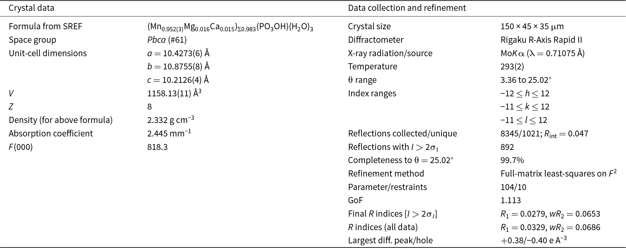
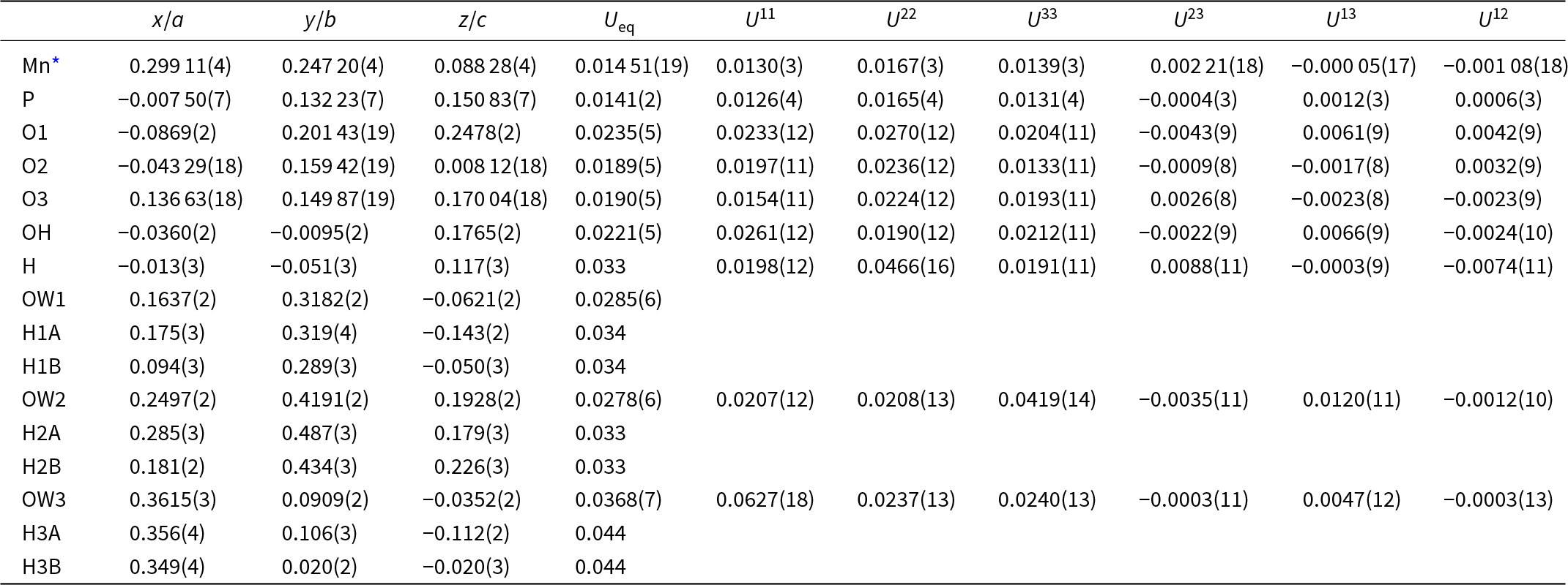
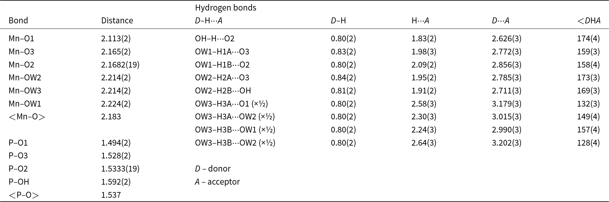
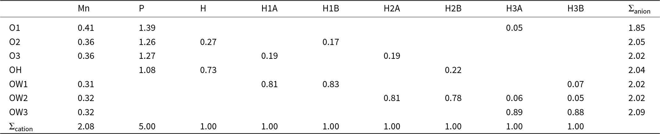
Single-crystal X-ray studies were done on the same diffractometer and radiation noted above. The Rigaku CrystalClear software package was used for processing the structure data, including the application of an empirical absorption correction using the multi-scan method with ABSCOR (Higashi, Reference Higashi2001). The structure was solved using the intrinsic-phasing algorithm of SHELXT (Sheldrick, Reference Sheldrick2015a). SHELXL-2016 (Sheldrick, Reference Sheldrick2015b) was used for the refinement of the structure. After the solution of the structure, atom positions were transformed to correspond to those reported for the structure of synthetic Mn(PO3OH)(H2O)3 (Cudennec et al., Reference Cudennec, Riou and Gerault1989). The occupancy of the Mn site refined to 0.971(3), which is consistent with the site also containing small amounts of Mg and Ca, as suggested by the EPMA. Difference-Fourier syntheses located all H atom positions, which were then refined with soft restraints of 0.82(3) Å on the O–H distances and 1.30(3) Å on the H2O H–H distances. The U eq of the H atom of the (PO3OH) group set to ×1.5 that of its donor O atom. The U eq of each H atom of the H2O groups was set to ×1.2 that of its donor O atom. In the final refinement, the Mn site was modelled with Mg and Ca contents corresponding to the EPMA and with the Mn occupancy refined, yielding (Mn0.952(3)Mg0.016Ca0.015)Σ0.983. Data collection and refinement details are given in Table 3, atom coordinates and displacement parameters in Table 4, selected bond distances in Table 5 and a bond valence analysis in Table 6. The crystallographic information file has been deposited with the Principal Editor of Mineralogical Magazine and is available as Supplementary material (see below).
Data collection and structure refinement details for manganonewberyite

R int = Σ|F o2–F o2(mean)|/Σ[F o2]. GoF = S = {Σ[w(F o2–F c2)2]/(n–p)}1/2. R 1 = Σ||F o|–|F c||/Σ|F o|. wR 2 = {Σ[w(F o2–F c2)2]/Σ[w(F o2)2]}1/2; w = 1/[σ2(F o2)+(aP)2+bP] where a is 0.0308, b is 1.3868 and P is [2F c2+Max(F o2,0)]/3.
Atom coordinates and displacement parameters (Å2) for manganonewberyite

* Occupancy: Mn0.952(3)Mg0.016Ca0.015
Selected interatomic distances (Å) and angles (°) in manganonewberyite

Bond valences (in valence units) for manganonewberyite

The bond valences for the Mn site is based on full occupancy by Mn. Bond-valence parameters are from Gagné and Hawthorne (Reference Gagné and F.C2015). Hydrogen-bond strengths are based on O–O distances according to the relation of Ferraris and Ivaldi (Reference Ferraris and Ivaldi1988).
Discussion
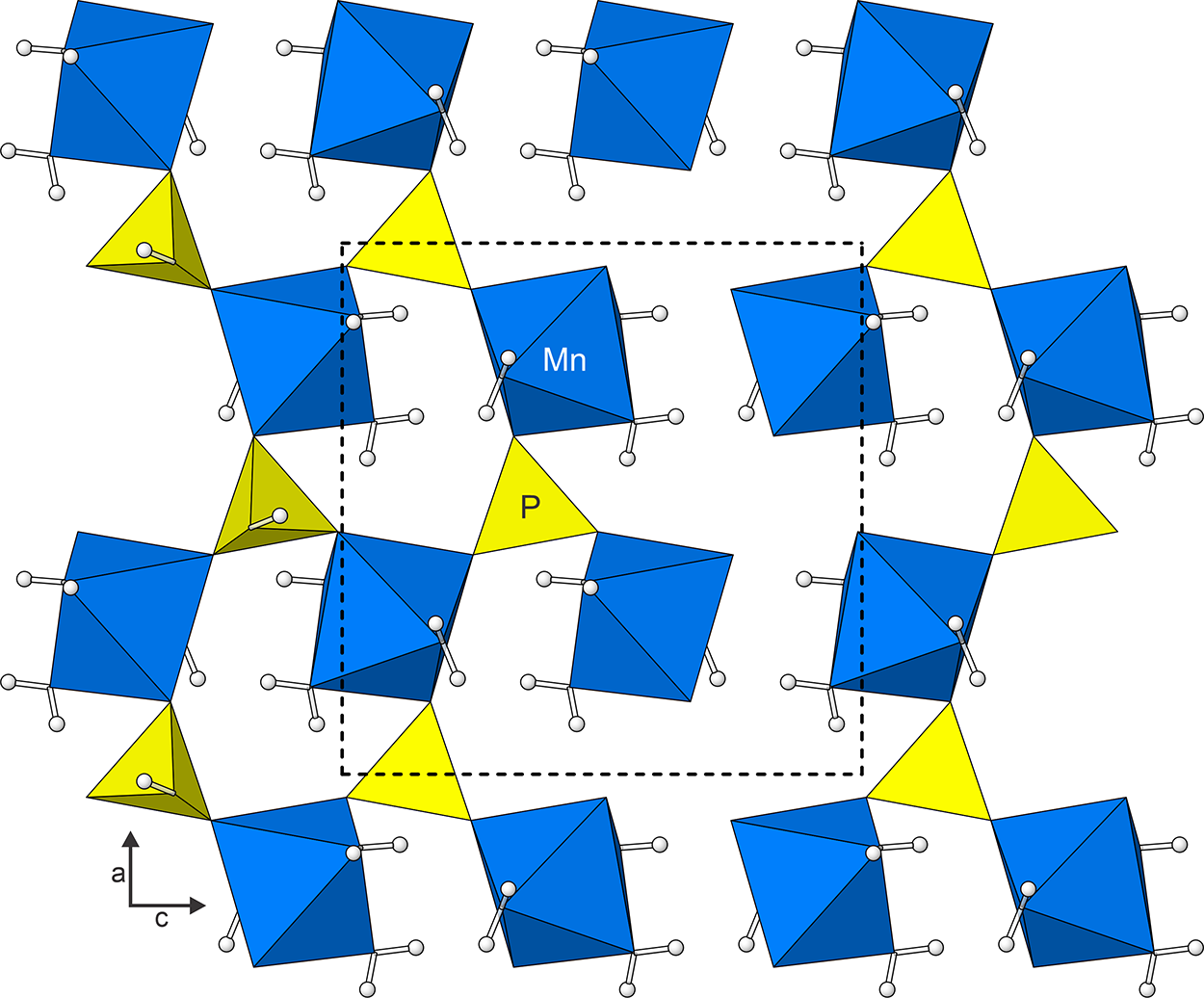
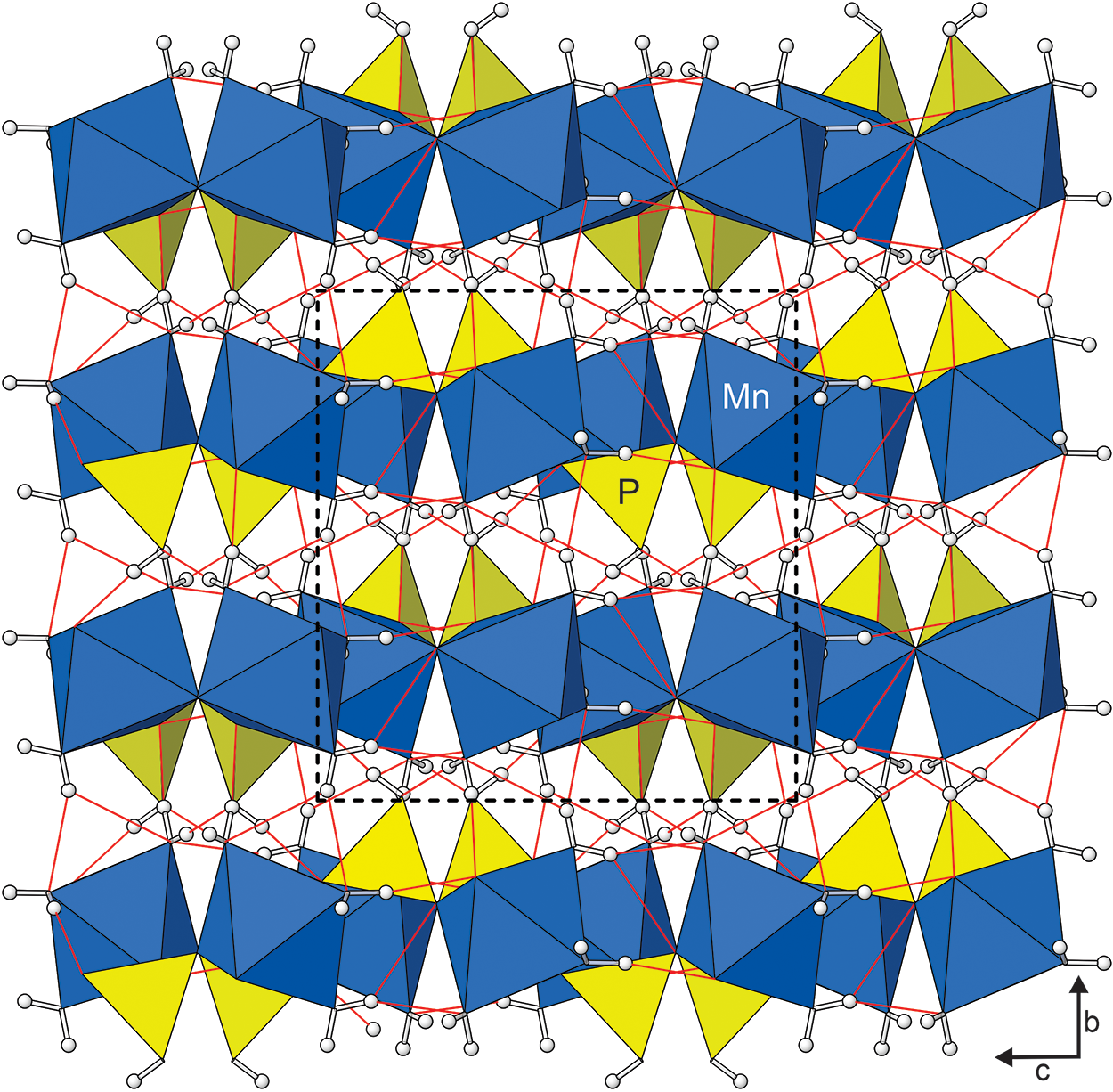
Manganonewberyite is isostructural with newberyite (Abbona et al., Reference Abbona, Boistelle and Haser1979; Bartl et al., Reference Bartl, Catti, Joswig and Ferraris1983) and, as noted above, corresponds to a known synthetic phase (Cudennec et al., Reference Cudennec, Riou and Gerault1989). The MnO3(H2O)3 octahedra and PO3(OH) share their O vertices forming a sheet of alternating octahedra and tetrahedra parallel to {010} (Fig. 5). Successive sheets are linked in the [010] direction only by hydrogen bonds (Fig. 6).
Sheet composed of MnO3(H2O)3 octahedra and PO3(OH) tetrahedra in the structure of manganonewberyite, with H atoms as open spheres. The unit cell outline is shown with dashed lines.

Stacking of polyhedral layers in the structure of manganonewberyite. The hydrogen bonds are shown in red. The unit cell outline is shown with dashed lines. Figs 5 and 6 were created using ATOMS version 6.5 (Shape Software, Kingsport, Tennessee, USA).

The hydrogen positions determined for manganonewberyite are comparable to those reported for the structure of newberyite by both Abbona et al. (Reference Abbona, Boistelle and Haser1979) (based on X-ray diffraction data) and Bartl et al. (Reference Bartl, Catti, Joswig and Ferraris1983) (based on neutron diffraction data). The hydrogen bonds from OH, OW1 and OW2 are straightforward; however, those from OW3 are more complex. (Note that the earlier studies use different atom labelling). Abbona et al. (Reference Abbona, Boistelle and Haser1979) propose a single hydrogen bond from H3A to OW2 and a bifurcated hydrogen bond from H3B to OW1 and OW2. On the other hand, Bartl et al. (Reference Bartl, Catti, Joswig and Ferraris1983) interpret what they refer to as ‘pseudo-bifurcated’ hydrogen bonds from H3A to OW2 and O1 and from H3B to OW1 and OW2, the second bond in each case being longer and weaker. The hydrogen bonds proposed herein are the same as those proposed by Bartl et al. (Reference Bartl, Catti, Joswig and Ferraris1983). Hydrogen-bond contributions are included in the bond-valence analysis in Table 6. Although the bond-valence sum (BVS) for O1 is somewhat low (1.85 valence units), other O sites (O, OH and OW) have very reasonable BVS values, providing further support for the proposed hydrogen-bonding scheme.
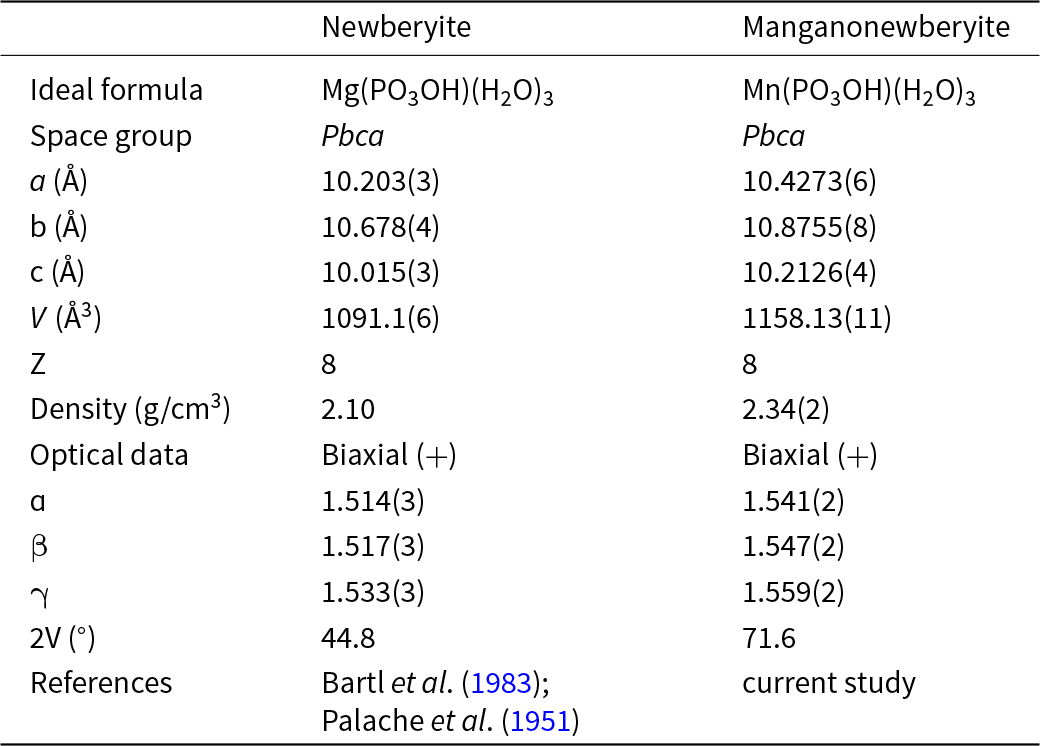
The unit-cell parameters, densities and optical properties of newberyite and manganonewberyite are compared in Table 7. The unit-cell volume of manganonewberyite is ∼6% greater than that of newberyite, a difference that is entirely attributable to the Mn–O vs Mg–O bond lengths. The expansion of the cell in going from newberyite to manganonewberyite is fairly uniform in that the a, b and c cell parameters each increase by ∼2%. In a study of synthetic compounds with the newberyite structure, Antraptseva et al. (Reference Antraptseva, Solod and О.О2021) showed that a complete solid solution exists between Mg(PO3OH)(H2O)3 and Mn(PO3OH)(H2O)3 and that the increase in each of the individual cell lengths is linear. They also reported linear increases in the α and γ indices of refraction. (They did not report data on β). Their values of α and γ for the Mg(PO3OH)(H2O)3 and Mn(PO3OH)(H2O)3 end-members are very close to those for newberyite and manganonewberyite, respectively (Table 7).
Comparison of selected data for newberyite and manganonewberyite

Supplementary material
The supplementary material for this article can be found at https://doi.org/10.1180/mgm.2024.68.
Acknowledgements
Two anonymous reviewers and Structures Editor Peter Leverett are thanked for their constructive comments on the manuscript. A portion of this study was funded by the John Jago Trelawney Endowment to the Mineral Sciences Department of the Natural History Museum of Los Angeles County. The EPMA was carried out at the Caltech GPS Division Analytical Facility, which is supported, in part, by NSF Grant EAR-2117942.
Competing interests
The authors declare none.