Introduction
A non-recurrent inferior laryngeal nerve is an anomaly in which the nerve enters the larynx directly off the cervical vagus nerve, without descending to the thoracic level. This anomaly, which is rarely reported in conventional thyroidectomy, is highly susceptible to damage during surgery.Reference Henry, Audiffret, Denizot and Plan1
Certain imaging signs from computed tomography (CT), which is usually performed before thyroidectomy, may provide possible clues about the existence of a non-recurrent inferior laryngeal nerve, prior to surgery. Thus, it may be possible to avoid iatrogenic injury of a non-recurrent inferior laryngeal nerve.Reference Watanabe, Kawabori, Osanai, Taniguchi and Hosokawa2
The route of the inferior laryngeal nerve may be variable. However, the laryngeal entrance point of the inferior laryngeal nerve, which is a constant anatomical landmark, can be located during thyroidectomy using several thyroid landmarks such as Zuckerkandl's tubercle, Berry's ligament or the inferior constrictor muscle.Reference Yalcin and Ozan3–Reference Pelizzo, Toniato and Gemo5
In recent years, robotic thyroidectomy, with endoscopic magnification, has allowed a more meticulous approach to surgical anatomy.Reference Kang, Jeong, Yun, Sung, Lee and Lee6, Reference Tae, Ji, Cho, Lee, Kim and Kim7 As surgeons do not have tactile sensation during robotic surgery, anatomical landmarks become especially important in identifying a non-recurrent inferior laryngeal nerve.
This paper reports a case of non-recurrent inferior laryngeal nerve identification during robotic thyroidectomy, and describes the relevant radiological and anatomical features of this anomaly.
Case report
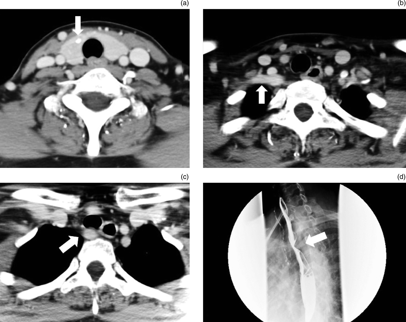
A 38-year-old woman visited our department with suspected papillary microcarcinoma in the right lobe of the thyroid gland, as indicated by thyroid sonography and fine needle aspiration cytology. Pre-operative CT revealed some low density areas with calcified spots in the right thyroid gland (Figure 1a). In addition, a right aberrant subclavian artery was found behind the trachea and oesophagus (Figure 1b & 1c). This was causing extrinsic compression of the oesophagus, as shown on the oesophagogram (Figure 1d).
Axial, contrast-enhanced computed tomography images showing: (a) a suspected papillary carcinoma lesion with calcification (arrow) in the right lobe of the thyroid gland; and the aberrant right subclavian artery (arrows) passing (b) behind the common carotid artery, and (c) behind the trachea and oesophagus. (d) Oesophagogram showing a posterior oesophagus indentation (arrow) caused by the aberrant right subclavian artery.

The patient underwent gasless robotic thyroidectomy via an axillo-breast approach, following the procedure described by Tae et al.Reference Tae, Ji, Cho, Lee, Kim and Kim7 After elevating the skin flap from the right axilla to the anterior central neck area, we created a tunnel in the space between the sternal and clavicular heads of the sternocleidomastoid muscle. The sternal head of the sternocleidomastoid and the sternothyroid muscle were then fixed by an external retractor (Meditech Inframed, Seoul, Korea) to maintain an adequate working space, without using carbon dioxide gas insufflation to expose the thyroid gland.
The robotic arms were docked into position within the surgical field. A dual-channel endoscope was then placed at the centre of the axillary port, and harmonic curved shears, Maryland forceps and ProGrasp™ forceps were used to perform a multidirectional retraction and dissection of the thyroid lobe under three-dimensional (3D) endoscopic magnification (using a da Vinci® standard surgical robotic system).
Initially, upper pole dissection was performed by carefully cutting the branches of the superior thyroid artery on the thyroid capsule using a harmonic scalpel. We then approached the inferior laryngeal nerve, in an orderly manner. The inferior thyroid artery, which was used as a surgical landmark, was held with mild traction as we attempted to locate the inferior laryngeal nerve. In an ordinary recurrent route, the inferior laryngeal nerve may go anteriorly, posteriorly or through the branches of the inferior thyroid artery. However, as we could not detect any inferior laryngeal nerve like tissue, the possibility of a non-recurrent inferior laryngeal nerve was raised, which was in line with the pre-operative CT findings.
Once the inferior thyroid artery had been divided, we retracted the thyroid lobe medially and then dissected it cranially along the thyroid capsule in the tracheo-oesophageal groove.
After approaching Zuckerkandl's tubercle, which can be used as a surgical landmark to locate the laryngeal entrance point of the inferior laryngeal nerve,Reference Yalcin and Ozan3–Reference Pelizzo, Toniato and Gemo5 we found a nerve running perpendicular to Zuckerkandl's tubercle and entering the larynx beside Berry's ligament. Although Berry's ligament also contained a vessel that looked similar to a nerve, using endoscopic magnification, we could easily differentiate the blood-containing vessel lumen from the shiny nerve sheath of the inferior laryngeal nerve (Figure 2).
The thyroid gland (TG) (attached to the trachea) was dissected and retracted medially. A harmonic scalpel (HS) was used to dissect along the tracheo-oesophageal groove and approach Zuckerkandl's tubercle (ZT). Zuckerkandl's tubercle (arrowheads) indicated the location of Berry's ligament, which had a blood-containing vessel (curved arrow). The non-recurrent inferior laryngeal nerve (arrow), with its shiny nerve sheath, branched from the vagus nerve under the right common carotid artery (CCA), and entered the larynx at the lower margin of the inferior constrictor muscle (ICM).
We traced the inferior laryngeal nerve laterally and found that it went under the common carotid artery (Figure 3) and merged with the vagus nerve. The course followed a transverse path that was parallel and superior to the trunk of the transected inferior thyroid artery. The nerve was compatible with a type 2A non-recurrent inferior laryngeal nerve, as defined by Toniato and colleagues.Reference Toniato, Mazzarotto, Piotto, Bernante, Pagetta and Pelizzo8
After right-sided lobectomy and isthmusectomy, the trachea was pushed aside by the harmonic scalpel (HS) to expose the non-recurrent course of the right non-recurrent inferior laryngeal nerve (arrow), which entered the larynx under the inferior constrictor muscle (ICM). CCA = common carotid artery
After dissecting the right thyroid lobe from the trachea and Berry's ligament, we successfully completed the surgery. The pathology revealed a papillary microcarcinoma 0.7 cm in diameter. The patient had no vocal fold paralysis following the surgery.
Discussion
The major complication of thyroidectomy is hoarseness resulting from inferior laryngeal nerve injury and vocal fold paralysis. The identification and preservation of an inferior laryngeal nerve, which commonly recurs at the artery within the thoracic cavity, is fundamental in preventing this complication. However, cases of a non-recurrent inferior laryngeal nerve, wherein the nerve enters the larynx directly from the vagus nerve without recurring, have been observed clinically.
This anomaly was first reported by Stedman in 1823.Reference Stedman9 According to a study by Asgharpour et al., in which 143 cadavers were examined, a non-recurrent inferior laryngeal nerve is very rare, with an incidence of 0.7 per cent.Reference Asgharpour, Maranillo, Sañudo, Pascual-Font, Rodriguez-Niedenführ and Valderrama10 There have been a few reports of non-recurrent inferior laryngeal nerves identified during conventional thyroidectomy. However, to the best of our knowledge, the present study is the first to report a non-recurrent inferior laryngeal nerve identified during robotic thyroidectomy.
One of the main advantages of robotic thyroidectomy, which was developed by Kang et al.,Reference Kang, Jeong, Yun, Sung, Lee and Lee6 is that the surgeon has a 3D field of view and a more accurate sense of perspective. The 3D, high-magnification endoscope can magnify landmark structures, which helps in the preservation of the inferior laryngeal nerve. Toniato et al. recommended the use of palpation for locating the inferior laryngeal nerve during conventional thyroid surgery, if it cannot be immediately identified.Reference Toniato, Mazzarotto, Piotto, Bernante, Pagetta and Pelizzo8 However, it is impossible to palpate the inferior laryngeal nerve with a robotic instrument because of the lack of tactile sensation.Reference Tae, Ji, Cho, Lee, Kim and Kim7 Therefore, it is important to pay particular attention to the embryologic development, image signs and surgical landmarks. These are extremely useful during robotic thyroidectomy for successful preservation of a non-recurrent inferior laryngeal nerve.
A study by Watanabe et al. revealed that an aberrant subclavian artery was always associated with a non-recurrent inferior laryngeal nerve (Figure 4).Reference Watanabe, Kawabori, Osanai, Taniguchi and Hosokawa2 The non-recurrent inferior laryngeal nerve may occur in a variant course of normal recurrent inferior laryngeal nerve development during the embryonic stage. In the embryo, bilateral inferior laryngeal nerves supply the sixth branchial arch. With the descent of the heart, the nerves pass beneath the sixth aortic arch and ascend to the larynx. On the right side, the distal portions of the sixth and the fifth aortic arches disappear, and the recurrent inferior laryngeal nerve moves up to lie beneath the fourth arch, which forms a portion of the subclavian artery. However, sometimes the right fourth aortic arch and the proximal right dorsal aorta are obliterated, and the origin of the subclavian artery becomes anomalous. Thus, the right recurrent inferior laryngeal nerve is free to migrate upward, where it becomes a non-recurrent inferior laryngeal nerve.
Anatomy of the aberrant right subclavian artery (dots) and right non-recurrent inferior laryngeal nerve (arrowhead). The right subclavian artery originates from the left side of the aortic arch (AA), and passes behind the trachea and oesophagus. VN = vagus nerve; CA = carotid artery
Nowadays, CT is a routine examination before thyroid surgery in most institutions. Although the non-recurrent inferior laryngeal nerve itself cannot be seen on the CT scan, it is possible to identify the aberrant position of the right subclavian artery and predict the presence of a non-recurrent inferior laryngeal nerve pre-operatively.Reference Watanabe, Kawabori, Osanai, Taniguchi and Hosokawa2 It is therefore important to be aware of the characteristic course of an aberrant right subclavian artery at the level of the upper mediastinum. It is typically detected on the dorsal side of the membranous wall of the trachea, on the right side of the oesophagus (Figure 1).Reference Watanabe, Kawabori, Osanai, Taniguchi and Hosokawa2
Different anatomical structures have been proposed as useful surgical landmarks for locating the inferior laryngeal nerve. These include the inferior thyroid artery, Zuckerkandl's tubercle, Berry's ligament and the inferior constrictor muscle.Reference Yalcin and Ozan3–Reference Pelizzo, Toniato and Gemo5 Although the relationships between the inferior laryngeal nerve and the aforementioned structures vary, the laryngeal entrance point of the inferior laryngeal nerve is most constant around Berry's ligament and the inferior constrictor muscle.
Yalcin and Ozan emphasised the usefulness of Zuckerkandl's tubercle in locating the entrance point of the inferior laryngeal nerve.Reference Yalcin and Ozan3 Zuckerkandl's tubercle is a lateral or posterior projection from the lateral thyroid lobe, which indicates the point of embryologic fusion of the ultimobranchial body and principal median thyroid process. It therefore forms a constant landmark for the inferior laryngeal nerve during thyroid surgery.Reference Pelizzo, Toniato and Gemo5
• This is the first report of a non-recurrent inferior laryngeal nerve identified during robotic thyroidectomy
• Radiological and surgical anatomical knowledge of this anomaly is important in thyroid surgery
• Pre-operative images of an aberrant subclavian artery can indicate a non-recurrent inferior laryngeal nerve
• Non-recurrent inferior laryngeal nerve is not a contraindication for robotic thyroidectomy
• Use of three-dimensional, high-magnification endoscopic views can aid nerve preservation
In order to locate the non-recurrent inferior laryngeal nerve in the current study, we approached cranially along the thyroid gland, and Zuckerkandl's tubercle was identified. This indicated the entrance point of the non-recurrent inferior laryngeal nerve beside Berry's ligament and the inferior constrictor muscle. Kaisha et al. described a similar anatomical relationship in their cadaver dissection study.Reference Kaisha, Wobenjo and Saidi4
In the present study, the non-recurrent inferior laryngeal nerve could be seen very clearly with the da Vinci endoscope, and it could be traced laterally to the vagus nerve without injury. The images shown in Figures 2 and 3 demonstrate the advantage of having a 3D high-magnification view of the nerve during robotic surgery. The recorded video clips of the surgery also helped us to review the surgical procedure and identify important anatomical landmarks.
In conclusion, a thorough knowledge of the radiological and surgical anatomy of the non-recurrent inferior laryngeal nerve is important for thyroid surgery. Pre-operative CT is helpful for detecting a potential right-sided non-recurrent inferior laryngeal nerve. Although finger palpation and tactile sensation are not possible during robotic thyroidectomy, 3D high-magnification views of important surgical landmarks around the laryngeal entrance point can help thyroid surgeons to safely identify and preserve the non-recurrent inferior laryngeal nerve.
Acknowledgement
The authors would like to thank Ms An-Ting Yu for her assistance in the preparation of the images and figures.