Introduction
Temporal bone computed tomography (CT) is known to deliver relatively high radiation doses due to the need for thin image slices to visualise the structures of the middle and inner ear. Cone beam computed tomography (CBCT), widely used in stomatology and maxillofacial surgery, presents a compelling alternative, offering lower radiation exposure while maintaining satisfactory image quality.
Multiple studies have confirmed the high spatial resolution of temporal bone CBCT and its reduced radiation burden on patients during imaging procedures.Reference Ruivo, Mermuys, Bacher, Kuhweide, Offeciers and Casselman1–Reference Dahmani-Causse, Marx, Deguine, Fraysse, Lepage and Escudé10
However, most comparisons of radiation exposure between CBCT and CT have relied on machine-recorded indices such as the Computed Tomography Dose Index (CTDI), Dose Length Product (DLP) or Dose Area Product (DAP). These indices are not direct patient measurements and have notable limitations.Reference Razafindranaly, Truy, Pialat, Martinon, Bourhis and Boublay2–Reference Helal, Jacob, Elshinnawy, Othman, Al-Dhamari and Paulus4, Reference Nardi, Salerno, Molteni, Occhipinti, Grazzini and Norberti7–Reference Dahmani-Causse, Marx, Deguine, Fraysse, Lepage and Escudé10 For example, CTDI underestimates true exposure as it excludes scatter radiation beyond the field of view (FOV), and DLP and DAP are not directly comparable due to differing units and reliance on conversion factors with inherent uncertainties, particularly for small FOVs.Reference Al-Okshi, Lindh, Salé, Gunnarsson and Rohlin5
Some studies have estimated equivalent or effective doses using cadavers or phantoms, often taking a single measurement per scan and not accounting for variability encountered in clinical practice on living patients.Reference Ruivo, Mermuys, Bacher, Kuhweide, Offeciers and Casselman1, Reference Nardi, Talamonti, Pallotta, Saletti, Calistri and Cordopatri6
Furthermore, imaging parameters varied considerably across studies, reflecting heterogeneity in clinical protocols. In some cases, parameters were standardised specifically for study purposes, limiting real-world applicability.
To our knowledge, no prior research has directly measured radiation doses on live patients undergoing temporal bone CBCT or CT. Despite existing literature, additional evidence supporting CBCT’s dosimetric advantages remains valuable, especially when derived from real-life clinical settings.
The primary aim of this prospective study is to compare radiation exposure across various organs in patients undergoing either temporal bone CT or CBCT. Using direct dosimetric measurements obtained under routine clinical conditions, we seek to quantify the reduction in organ-specific radiation dose achievable with CBCT. Our study also provides average reference dose values for these imaging modalities in clinical practice.
At the ENT department of our University Hospital, intra-operative CBCT is routinely used following cochlear implant surgery to verify electrode positioning before concluding general anesthesia. Therefore, eliminating the need for a separate post-operative CT scan and facilitating out-patient management. This setup presents an opportunity to collect organ-specific dosimetric data during real-time CBCT use.
Conversely, CT remains the standard diagnostic modality for most ear-related indications. Consequently, radiation measurements for CT were obtained from patients undergoing clinically indicated temporal bone CT scans at the same institution, across various clinical scenarios.
Materials and methods
This prospective, single-centre study was conducted to compare radiation exposure between non-injected CT and CBCT for ear imaging. The primary endpoint was the absorbed dose (in milligray [mGy]) to specific organs: ears, eyes, thyroid gland and pelvis. The secondary endpoint was the effective dose, calculated from the DAP or DLP using established conversion coefficients.
Patient selection
Patients included were either undergoing a temporal bone CT at our University Hospital or an intraoperative CBCT for electrode position verification following cochlear implantation. All participants were over 15 years old. Informed non-opposition was obtained after explaining the study objectives and the dosimetric procedure. No additional follow-up was required beyond standard clinical care.
Imaging protocols
CT scans were acquired using a Siemens Somatom Definition Edge scanner with the following parameters: (1) 140 keV, 280 mAs; (2) rotation time: 0.33 sec; (3) thin slices: 0.3 mm thickness; (4) coverage: approx. 20 cm/sec; (5) 128-slice STELLAR detectors; (6) dose optimisation: tin filter and TrueSignal technology; and (7) total: 900 images per acquisition.
CBCT scans were performed with the Xoran xCAT XL IQ™ machine using: (1) 120 keV, 57 mAs; (2) field of view (FOV): 38.7 × 29.9 cm2; (3) voxel matrix: 732 × 732 × 414; and (4) 600 images per acquisition.
Dosimetric measurements
Radiation doses were measured using nanoDot® dosimeters (optically stimulated luminescence technology), which were placed on the skin over the following regions: eyes, ears, thyroid gland and pubis. A control nanoDot® was positioned outside the imaging room to detect background radiation.
Although skin measurements do not directly reflect the internal organ dose, the high penetration capacity of X-rays ensures that skin readings approximate the actual dose absorbed by deeper tissues. Moreover, both CT and CBCT use X-ray technology with similar penetration, allowing for valid comparisons between modalities.11
Dosimetric data were extracted from each nanoDot® and expressed in absorbed dose (mGy). Patients were then classified into either the CT or CBCT group for analysis.
Study design and ethics
The study was conducted per-protocol, with 15 patients per group targeted as the required sample size. All patient data were pseudonymised. A coded list linking identities to anonymised records was stored separately in a secure, password-protected Excel file. Data transfers were conducted using encrypted channels. The study was approved by the local Ethics Committee (Comité de Protection des Personnes). No external funding was received for this research.
Statistical analysis
All statistical analyses were performed using Jamovi software. The significance level (alpha risk) was set at 0.05, and the power was set at 80 per cent.
For comparisons involving categorical variables, Pearson’s chi-squared or Welch’s test was applied, depending on the expected frequency distribution in each group. When comparing categorical and continuous variables, either the Student’s t-test or the Mann–Whitney U test was used, based on sample size, normality of distribution and homogeneity of variances.
Missing data were excluded from all analyses.
Results
The general characteristics of the patients are described in Table 1. A significant age difference was observed between the CT and CBCT groups.
General characteristics of the cohort and comparability of groups

* Median [25%–75%].
Abbreviations: CBCT, cone beam computed tomography; CT, computed tomography.
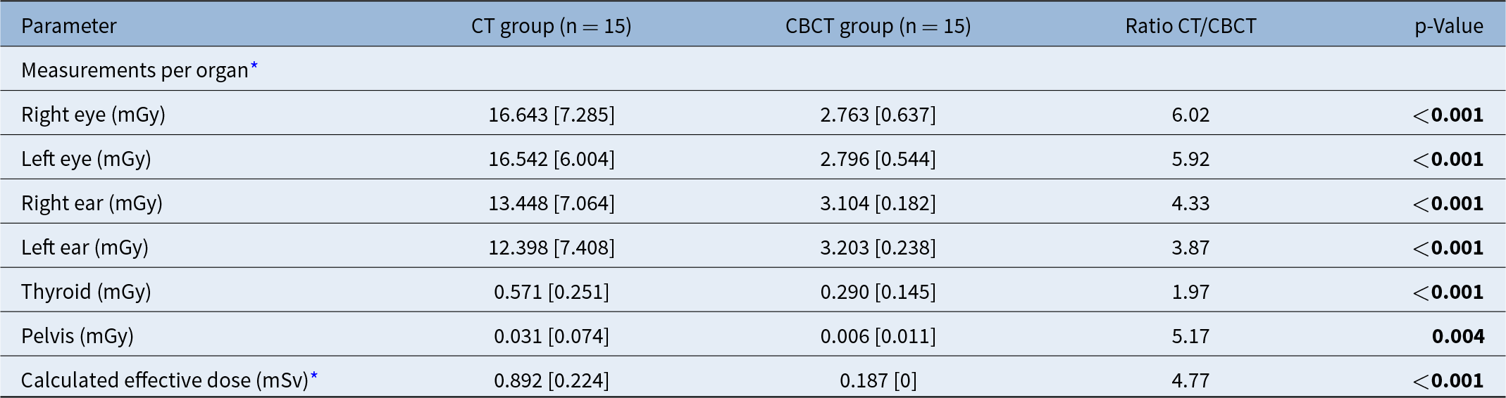
Dosimetric measurements showed a statistically significant reduction in radiation exposure with CBCT compared to CT across all organs studied (p < 0.001 and p = 0.004), as well as in the calculated effective doses (p < 0.001) (Table 2).
Comparison of dosimetry measurements and effective dose between CT and CBCT groups

Note: Values given in boldface are statistically significant.
* Mean [standard deviation].
Control nanoDot® dosimeters consistently recorded zero values, confirming the absence of ambient radiation contamination.
In adult patients, effective doses from CBCT remained constant, as the system delivers a fixed radiation dose per scan, and the age-based weighting coefficient for adults is uniformly equal to 1.
To investigate a potential correlation between patient height and pelvic irradiation, a bivariate analysis was conducted. No significant association was found, either for the full cohort (p = 0.72) or when analysed by imaging modality (CBCT group: p = 0.58; CT group: p = 0.40).
Discussion
The strengths of our study are based on the study of irradiation by organ and on its aspect attached to current practice. Dosimetric measurements were carried out on the patient and under the usual imaging conditions and settings, rather than on global estimates based on indirect data or under conditions codified for the study.
The strengths of this study lie in its focus on organ-specific irradiation and its grounding in real-world clinical practice. Unlike studies based on global estimates or protocols tailored specifically for research purposes, our dosimetric measurements were performed directly on patients under routine imaging conditions. Our results demonstrate a significant reduction in radiation exposure with CBCT compared to CT for temporal bone imaging. Radiation doses were consistently lower for each organ examined. The greatest reduction was observed for the eyes, with CBCT delivering six times less radiation. For the thyroid gland, the difference was smaller but still notable, with a two-fold reduction. The remaining organs, as well as the calculated effective dose, showed reductions of approximately four- to five-fold (Table 2).
These findings support existing literature indicating that CBCT exposes patients to lower radiation doses than CT. However, many previous studies relied on CTDI, DLP or estimated effective doses derived from these indices—methods that have inherent limitations due to indirect calculation and variability in machine parameters.Reference Razafindranaly, Truy, Pialat, Martinon, Bourhis and Boublay2–Reference Al-Okshi, Lindh, Salé, Gunnarsson and Rohlin5, Reference Nardi, Salerno, Molteni, Occhipinti, Grazzini and Norberti7–Reference Dahmani-Causse, Marx, Deguine, Fraysse, Lepage and Escudé10
When comparing effective dose, CTDI and DLP values from one study to another, the values are highly variable, sometimes relying on one or another of these parameters, and depending heavily on the settings of each imaging machine. It is therefore difficult to compare the various results in the literature.
Other research has employed dosimeters embedded in phantoms or cadaver models to compare CBCT and CT exposures.Reference Ruivo, Mermuys, Bacher, Kuhweide, Offeciers and Casselman1, Reference Nardi, Talamonti, Pallotta, Saletti, Calistri and Cordopatri6, Reference Dierckx, Saldarriaga Vargas, Rogge, Lichtherte and Struelens12, Reference Faccioli, Barillari, Guariglia, Zivelonghi, Rizzotti and Cerini13 Although such setups deviate from clinical practice, they offer greater accuracy than indirect estimation methods based on CTDI. These studies have also consistently concluded that CBCT delivers lower radiation doses than CT.
In our cohort, patients in the CBCT group were significantly older, likely because this group primarily included individuals undergoing cochlear implantation, while the CT group comprised patients with a broader range of indications. Nonetheless, age is not expected to influence radiation absorption, so this difference does not affect the validity of the comparisons.
Due to differing clinical indications between groups, image quality could not be directly compared. The CBCT group was evaluated for cochlear implant positioning, while the CT group underwent imaging for varied otologic reasons. Therefore, radiation data could not be correlated with image quality across both modalities.
Struffert et al.Reference Struffert, Hertel, Kyriakou, Krause, Engelhorn and Schick8 evaluated both irradiation and image quality for cochlear implant imaging and found that CBCT offered superior spatial resolution compared to CT, even when using CBCT settings optimised for minimal radiation. These findings suggest that CBCT can achieve excellent imaging results while maintaining low dose levels.Reference Zou, Koivisto, Lähelmä, Aarnisalo, Wolff and Pyykkö14
CBCT’s utility has been particularly highlighted in cochlear implant imaging due to its reduced sensitivity to metal artifacts relative to CT.Reference Razafindranaly, Truy, Pialat, Martinon, Bourhis and Boublay2, Reference Helal, Jacob, Elshinnawy, Othman, Al-Dhamari and Paulus4, Reference Struffert, Hertel, Kyriakou, Krause, Engelhorn and Schick8 This advantage extends to imaging in patients with ossicular prostheses or middle ear implants.
Furthermore, phantom and cadaveric studies—studying temporal bones without pathology or implants— support the superior spatial resolution of CBCT over CT.Reference Ruivo, Mermuys, Bacher, Kuhweide, Offeciers and Casselman1, Reference Nardi, Talamonti, Pallotta, Saletti, Calistri and Cordopatri6, Reference Dahmani-Causse, Marx, Deguine, Fraysse, Lepage and Escudé10, Reference Teymoortash, Hamzei, Murthum, Eivazi, Kureck and Werner15–Reference Erovic, Chan, Daly, Pothier, Yu and Coulson17 As such, CBCT may be a valuable tool for pre-operative assessment to identify surgical challenges.
However, CBCT’s performance is optimal in terms of spatial resolution for structures with high contrast. This observation makes CBCT well-suited for the analysis of the temporal bone and ossicles. But its lower contrast resolution limits its utility in soft tissue evaluation, making it unsuitable for detecting abscesses or conducting oncologic assessments.Reference Faccioli, Barillari, Guariglia, Zivelonghi, Rizzotti and Cerini13, Reference Scarfe and Farman18
Though CBCT has been evaluated for otosclerosis diagnosis, its sensitivity and specificity have been inferior to CT.Reference Liktor, Révész, Csomor, Gerlinger, Sziklai and Karosi19–Reference Révész, Liktor, Liktor, Sziklai, Gerlinger and Karosi21 Future studies should revisit these findings, considering technological advances such as high-resolution CBCT adapted for temporal bone imaging and improved parameter optimisation.
While extensively studied in dental and maxillofacial applications, where it consistently demonstrates higher spatial resolution and lower radiation than CT, the evidence remains low. As in otologic imaging, standardised CBCT settings that optimise the balance between image quality and radiation dose are still under development.Reference Al-Okshi, Lindh, Salé, Gunnarsson and Rohlin5
In conclusion, CBCT of the temporal bone is a promising imaging modality offering significantly reduced radiation, superior spatial resolution and reduced metal artifact compared to CT. Yet, broader adoption in otologic indications beyond cochlear implant evaluation and the minimum radiation doses required to achieve good imaging quality require further validation.
Additionally, practical advantages of CBCT—such as the compact, mobile, cost-effective and user-friendly design of systems like the xCAT XL IQ—enhance its appeal. These devices are well suited for out-patient ENT clinics and could eventually allow direct, radiologist-free imaging access, similar to current practices in dental care. Perhaps the future will see a trend towards this type of practice.
• Numerous studies have demonstrated reduced radiation exposure with cone beam computed tomography (CBCT) for temporal bone imaging
• However, these studies were primarily based on dose estimates (CTDI, DLP, PDS, effective dose) or on phantom models using standardised imaging parameters that do not fully reflect clinical practice
• Our study is the first to use direct organ-specific dose measurements in a cohort of patients undergoing imaging
• Our study demonstrated a significant reduction in radiation exposure with CBCT, ranging from two- to six-fold, depending on the anatomical site assessed
Acknowledgements
This research received no specific funding from public, commercial or non-profit agencies.
Ethical standards
All procedures involving human participants were conducted in accordance with the ethical standards of the institutional and national research committees (Comité de Protection des Personnes) and with the 1975 Helsinki Declaration and its later amendments (2008). Ethical approval was granted by the Comité de Protection des Personnes.
Competing interests
The authors declare no conflicts of interest.