Introduction
According to Grove (Reference Grove1989), the genus Strongyloides Grassi, 1879 includes 52 species of nematode parasites of vertebrates. Domestic mammals, including cats, dogs and farm animals such as cattle, sheep and pigs, are the main hosts for Strongyloides spp. However, few species of Strongyloides have been reported in amphibians, reptiles and birds (Dorris et al., Reference Dorris, Viney and Blaxter2002).
Strongyloides spp. have a complex life cyle which includes a parasitic and a free-living generation. Hosts become infected when free-living infective third-stage larvae (L3) penetrate the skin. These larvae migrate through the host's body and, during this migration, they moult via an L4 stage so that there are adult parasitic female worms present in the gut. The eggs are produced and passed to faeces. In the host faeces the eggs hatch to release first-stage larvae. These L1s have alternative potential developmental fates. In one, they develop via L2–L4 stages into rhabditiform male and female worms, i.e. the free-living adult generation. This type of development is known as indirect, sexual or heterogonic development. The alternative fate of the L1s that hatch from eggs passed in faeces is that they moult via an L2 into infective L3s. This type of development is known as direct, asexual or homogonic development (Viney & Lok, Reference Viney and Lok2007).
In Brazil, Strongyloides ophidiae Pereira, Reference Pereira1929 and Strongyloides cruzi Rodrigues, Reference Rodrigues1968 have been reported to infect reptiles. The former was described in a snake Mastigodryas bifossatus Raddi, 1820 (Pereira, Reference Pereira1929) and the second in a gecko Hemidactylus mabouia Moreau de Jonnès, 1818 (Rodrigues, Reference Rodrigues1968). Pereira (Reference Pereira1929) described S. ophidiae; however, only parasitic females and the eggs were analysed in this species description. Since then, no other S. ophidiae report has been published. Rhabditoid and filarioid larvae, as well as free-living male and female forms of this nematode, were not studied.
Recently, molecular studies have been accomplished for the characterization of Strongyloides spp. (Dorris & Blaxter, Reference Dorris and Blaxter2000; Dorris et al., Reference Dorris, Viney and Blaxter2002; Hasegawa et al., Reference Hasegawa, Hayashida, Ikeda and Sato2009). However, these studies did not include snake Strongyloides species from Brazil. In the present study, we present morphological and molecular data for S. ophidiae, collected from the snake Oxyrhopus guibei Duméril & Bibron Duméril, 1854.
Material and methods
Source of nematode materials
This study was performed with one adult female specimen of O. guibei snake from the municipality of Lençóis Paulista, São Paulo State, Brazil, which was donated to the Center for the Study of Venous and Venomous Animals of São Paulo State University, Botucatu, São Paulo State, Brazil.
Parasitological exams
Parasitological exams revealed the presence of embryonated eggs of the genus Strongyloides. Additionally, snake faeces culture was accomplished in order to obtain L3 larvae, to confirm the presence of Strongyloides. Snake faeces samples were cultured on sterilized filter paper in a Petri dish and maintained in an incubator at 25°C and 80% humidity. Faeces were examined for 7 days, to obtain L1, L2 and L3 stages and also free-living female forms (Hoffmann, Reference Hoffmann1987). Parasitic females were collected from the intestine after necropsy.
Parasitic females, eggs, rhabditoid and filarioid larvae, and free-living female forms were analysed in an image analysis computerized system (Qwin Lite 3.1, Leica Microsystems, Wetzlar, Germany). Arithmetic means of the measurements, followed by maximum and minimum values in parentheses, are presented in the results. The voucher specimens were deposited in the Coleção Helmintológica de Referência at the Departamento de Parasitologia, Instituto de Biociências (CHIBB), at Universidade Estadual Paulista (UNESP), Botucatu city, São Paulo State, Brazil.
DNA extraction and polymerase chain reaction
DNA samples were extracted from a pool of eight S. ophidiae parasitic females and a pool of ten Strongyloides venezuelensis Brumpt, 1934 parasitic females fixed in 70% ethanol, using the Quiamp kit (QIAGEN, GmbH, Hilden, Germany), according to the manufacturer's instructions. Samples were eluted in 100 μl of Tris-EDTA (TE) buffer and 2 μl were used for polymerase chain reaction (PCR) amplification.
An small subunit (SSU) rDNA gene segment was amplified by PCR using the SSUAF primer (5′-AAA GAT TAA GCC ATG CAT G -3′) and SSU22R primer (5′-GCC TGCTGC CTT CCT TGG A-3′) according to Dorris et al. (Reference Dorris, Viney and Blaxter2002). Amplification was carried out in a 10 μl reaction, containing 10 mm Tris-HCl, 1.5 mm MgCl2, 50 mm KCl, pH 8.3, 100 μm each dNTP, 0.2 μm of each primer and 0.5 U of Taq DNA polymerase (GE Healthcare, Bucks, UK).
PCR reactions were performed in a My cycler Thermal Cycler Bio-Rad® (T Gradient; Bio-Rad, Hercules, California, USA) and were conducted for 35 cycles (30 s at 95°C, 30 s at 58.1°C and 30 s at 72°C) preceded by 95°C for 5 min and followed by 7 min at 72°C. PCR products were visualized by ethidium bromide (0.5 μl/ml) staining after electrophoresis in a 2% agarose gel using a horizontal cube Hoefer HE 99 (GE Healthcare) in a solution of TAE 1X (Tris base 0.4 m; acetic acid 0.20 m and EDTA 0.5 m solution, pH 8.0).
DNA sequencing
PCR primers and dNTPs were removed before sequencing: 5 μl of PCR products were incubated with 1 μl of ExoSapIT (GE Healthcare) for 45 min at 37°C, followed by 20 min at 80°C for enzyme inactivation. Amplified fragments were sequenced in both forward and reverse directions using the ABI Prism dGTP BigDye Terminator Ready Reaction kit (Applied Biosystems, Foster City, California, USA), according to the manufacturer's instructions. For each sequencing reaction, approximately 10 ng of purified DNA were combined with 3.2 pmol of primer (sense and/or reverse) used in the amplification reaction. Nucleic acid sequence analysis was performed on an automated Applied Biosystems 377 DNA sequencer.
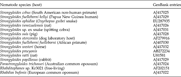
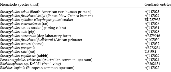
Nucleic acid sequences analysis was performed using the MERGER software package (http://bioweb.pastuer.fr/seqanal/alignment/intro-uk.html) used to produce the consensus sequence for each DNA sample. One sequence of S. ophidiae obtained from Brazilian snakes, 12 different species of Strongyloides and three related species were used in the analysis. Sequences used to construct the phylogenetic tree are shown in table 4. Sequences were aligned with the CLUSTAL X software, version 1.81 (Thompson et al., Reference Thompson, Gibson, Plewniak, Jeanmougin and Higgins1997). Distance method (neighbour-joining/NJ) was used in construction of the phylogenetic tree (Saitou & Nei, Reference Saitou and Nei1987) using a gamma distance (Kimura-2-parameter model) with gamma parameter a = 1. The bootstrap test with 1000 replications was applied to estimate the confidence of branching patterns of the neighbour-joining tree (Felsenstein, 1985). Nodes with bootstrap values lower than 50% are not shown because they were not considered to be supported by the test. The isolates used to construct the phylogenetic tree are shown in table 1. To obtain a better alignment, both pairwise and multiple alignment parameters were changed from the default set. Divergences were estimated by the pairwise alignment using MEGA software (Molecular Evolutionary Genetics Analysis) (Kumar et al., 2001). The distance method (neighbour-joining/NJ) was used in the construction of the phylogenetic tree (Saitou & Nei, Reference Saitou and Nei1987). The bootstrap test was applied to estimate the confidence of branching patterns of the neighbour-joining tree (Felsenstein, 1985).
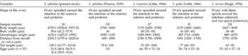
Morphometrical data of parasitic females of different Strongyloides species.

NI, no information.
Results
Morphological characterization of Strongyloides ophidiae
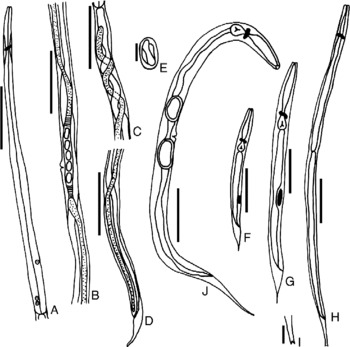
Parasitic females presented a slender body ending in a conical tail. The anterior extremity was truncated and presented stoma; however, details of this structure could not be observed due to poor preservation. The oesophagus was long and filariform, and a nerve ring was observed at the anterior portion of this structure. The reproductive system was didelphic and amphidelphic and the vulva was found located on the mid-ventral surface of the body. The anterior ovary spiralled twice around the intestine and the posterior ovary formed a partial spiral and extended almost to the anal opening. The uterus was short and contained a single row of eggs (2–5) (fig. 1A–D, table 1).
Strongyloides ophidiae morphology. (A) Detail of the oesophagus; (B) detail of the vulva and eggs; (C) detail of the ovary anteriorly spiralled around the intestine; (D) detail of the tail – note the ovary partially spiralled around the intestine; (E) egg; (F–J) free-living forms: (F) L1 rhabditoid larvae; (G) L2 rhabditoid larvae; (H) L3 filarioid larvae; (I) detail of the tip and truncated tail of L3 filarioid larvae; (J) free-living female. Scale bars: 200 μm (A, B, C, D), 30 μm (E), 100 μm (F, G, H, J), 10 μm (I).

Eggs (n = 57) found in the uterus were elliptical, embryonated, with a thin shell and measured 51.8 (48.4–54.5) μm long and 32.4 (30.5–33.6) μm wide. The eggs were slightly smaller than the eggs observed in the faeces (n = 9), which measured 75.8 (39.93–85.62) μm long and 43.9 (37.14–47.95) μm wide (fig. 1E).
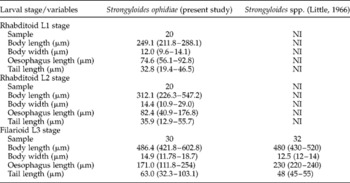
First-stage larvae (L1, n = 20) presented a rhabditiform oesophagus, with a body, isthmus and bulb and an inconspicuous nerve ring involving the isthmus area. A narrowly tapered tail was also observed (fig. 1F, table 2).
Morphometrical data of larvae of different Strongyloides species.

Strongyloides ophidiae (Pereira, Reference Pereira1929), Strongyloides gulae (Little, Reference Little1966), Strongyloides mirzai (Singh, Reference Singh1954) and Strongyloides serpentis (Little, Reference Little1966) have no morphometrical data of larval stages; NI, data not informed.
The second-stage larvae (L2, n = 20) were morphologically similar to the first-stage rhabditoid larvae; however, the body was longer and thinner. The oesophagus was still rhabditoid; however, it was more extended and with a less evident division between the bulb and isthmus. The buccal cavity was less evident than in the L1, the genital primordium was well visible, and this larval stage also presented a thin tail (fig. 1G, table 2).
The infective third-stage larvae (L3, n = 30) were elongated, thinner than the second-stage rhabditoid larvae, and presented a long and filariform oesophagus. This larval stage also presented a thin tail, with a truncated and notched tip (fig. 1H–I, table 2).
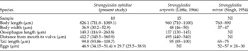
Free-living females (n = 10) had a small, wide body and a thin cuticle with fine transverse striations. The rhabditoid oesophagus presented a short constriction between the anterior portion and the bulb. The nerve ring was located at the posterior end of the oesophageal isthmus. The reproductive system was didelphic and amphidelphic, with the vulva located in the middle of the body and a very short vagina. The body did not present any constriction behind the vulva, which had prominent lips. One single row of eggs was found in the uterus (fig. 1J, table 3). Free-living males were not found.
Morphometrical data of free-living females of different Strongyloides species.

Strongyloides ophidiae (Pereira, Reference Pereira1929) and S. gulae (Little, Reference Little1966) have no morphometrical data of free-living forms; NI, no information.
Molecular characterization of Strongyloides ophidiae
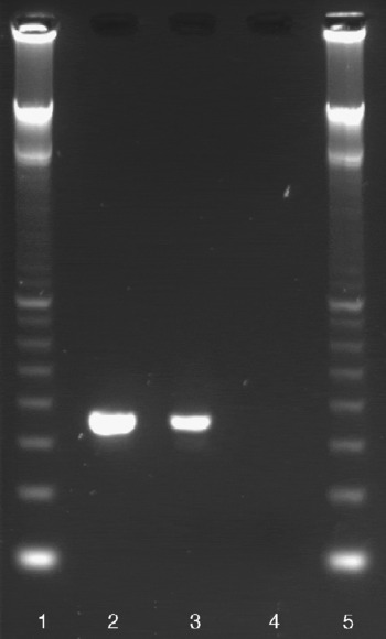
A 350 bp amplification band was obtained by PCR, using DNA samples of S. venezuelensis and S. ophidiae. Lanes 2 and 3 of fig. 2 show a 350 bp amplification band from each DNA sample of S. venezuelensis and S. ophidiae, respectively. The S. ophidiae nucleotide sequence (GenBank accession number EU287935), isolated from O. guibei, presented 98% similarity with Strongyloides procyonis Little, Reference Little1966 (GenBank accession number AB272234.1 and AB205054.1) and 97% similarity with Strongyloides cebus Darling, 1911, Strongyloides stercoralis Bavey, 1876, Strongyloides sp. ex snake and Strongyloides fuelleborni (Von Linstow, 1905) (GenBank accession numbers AJ417025, AF279916.2, AJ417031.1 and AJ407030.1, respectively).
Agarose gel electrophoresis (2%) showing PCR amplification of the SSU rDNA gene with SSUA and SSU22R primer set. Lanes: 1, 100 bp marker; 2, 350 bp amplification band from S. venezuelensis DNA samples; 3, 350 bp amplification band from S. ophidiae DNA samples; 4, reaction containing only reagents (without DNA); 5, 100 bp marker.

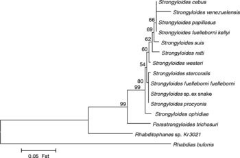
Eleven species of Strongyloides were added from a representative wide host range, including a snake, bovid, rodents, primates and all three recognized parasites of humans (table 4). The Strongyloides SSU sequences were all very similar, with many branch lengths inferred to be very short. The tree constructed using Rhabdias bufonis as the outgroup showed that S. ophidiae was allocated to the same branch of Strongyloides species as S. cebus and S. papillosus and presented a 97–98% similarity relationship with these species (fig. 3).
Nematode species used for phylogenetic analysis plus their corresponding GenBank entries.
Phylogenetic relationships of Strongyloides isolates detected in Brazilian snakes. Molecular phylogenetic analysis of the genus Strongyloides and close relatives based on small subunit ribosomal gene (SSU rDNA) sequences. Rhabdias bufonis was chosen as the outgroup. Numbers at the side of nodes indicate bootstrap support. Sequences used to construct the phylogenetic tree are listed in table 4.

Discussion
There are few reports regarding the occurrence of Strongyloides spp. infecting snakes and, until now, only four species, S. ophidiae (Pereira, Reference Pereira1929), S. mirzai Singh, Reference Singh1954 (Singh, Reference Singh1954), S. serpentis Little, Reference Little1966 (Little, Reference Little1966) and S. gulae Little, Reference Little1966 (Little, Reference Little1966), have been described. The Strongyloides sample studied here was consistent with belonging to the species S. ophidiae but the original description of the species is insufficient to determine this with certainty because only the parasitic females were described and few morphological data were presented by Pereira (Reference Pereira1929). No other studies have been accomplished with Strongyloides spp. from Brazilian snakes. The present study contributes to the morphological characterization of S. ophidiae by providing a description of free-living forms, as well as molecular aspects of this nematode. The parasitic females recovered from O. guibei had ovaries that spiralled around the intestine in the anterior and posterior regions, as reported for S. serpentis by Little (Reference Little1966). According to this author, S. serpentis presents an ovary that spirals twice, anteriorly, and an occasional partial spiral in the posterior region. This characteristic also resembled the parasitic females of S. ophidiae, described by Pereira (Reference Pereira1929). In contrast, the parasitic female of S. mirzai (Singh, Reference Singh1954) differs from the females of S. ophidiae (Pereira, Reference Pereira1929), S. serpentis (Little, Reference Little1966) and S. gulae (Little, Reference Little1966), since it has an ovary that spirals three times around the intestine anteriorly and one posterior spiral.
With regard to parasitic female morphometry, S. ophidiae possessed a larger body length and width, oesophagus length and tail than presented in S. serpentis and S. gulae. The only resemblance among these nematodes was the distance between the vulva and posterior extremity. The eggs in the uterus of S. ophidiae, S. serpentis and S. gulae were of similar lengths. On the other hand, the egg widths of S. serpentis and S. gulae were smaller than that of S. ophidiae. These morphological and morphometric data clearly demonstrate the identity of S. ophidiae.
According to Little (Reference Little1966), eggs and development stages found in faeces are an important factor for species identification. Oxyrhopus guibei faeces were collected directly from the host and immediately analysed. Since all analysed eggs were cleaved, we can conclude that S. ophidiae eggs in faeces contained first-stage larvae. Eggs measured in the present study were slightly larger than those described by Pereira (Reference Pereira1929), but were very similar to those of S. ophidiae obtained from Crotalus durissus terrificus Lautenti, 1768 and Philodryas olfersii (Lichtenstein, 1823) snakes (Santos et al., in preparation).
The first-stage larvae presented a rhabditoid oesophagus, and body length and width that were very similar to the descriptions for Strongyloides spp. from other hosts (Vieira et al., Reference Vieira, Lima and Bessa2006). However, this is the first report of Strongyloides first-stage larvae in snakes. Little (Reference Little1966) and Vieira et al. (Reference Vieira, Lima and Bessa2006) emphasized the importance of the genital primordium characterization in this larval stage; however, this was observed in only a few larvae in the present study.
The second-stage rhabditoid larvae, as well as the first-stage larvae, did not present distinct morphology and morphometry, in comparison to other Strongyloides species; however, these larvae have not been previously reported in snakes either. Morphology analysis of Strongyloides ophidiae third-stage larvae showed that the body length and width, oesophagus length and tail length were larger than in the snake Strongyloides species studied by Little (Reference Little1966). However, these differences in morphology and morphometry do not contribute to the identification of S. ophidiae for two reasons: there are no morphological studies regarding the larval stage in this species, and secondly, according to Vieira et al. (Reference Vieira, Lima and Bessa2006), the Strongyloides spp. L3 larvae can present differences in body length that are not large enough for species identification.
In the free-living female adult stage, the uterus morphology and egg disposition (in a single row) resembled those of S. serpentis and S. gulae (Little, Reference Little1966). Therefore, the free-living female form of Strongyloides also provides little information for use in distinguishing between species. Morphological and morphometric data, mainly of the free-living forms, obtained in the present study, contribute to a better characterization of S. ophidiae, since just the parasitic female was described earlier (Pereira, Reference Pereira1929).
Molecular data on the Strongyloides species have been provided by few authors (Dorris et al., Reference Dorris, Viney and Blaxter2002; Sato et al., Reference Sato, Sauzuki, Osanai, Kamyia and Furuoka2006; Eberhardt et al., Reference Eberhardt, Mayer, Bonfoh and Streit2008; Hasegawa et al., Reference Hasegawa, Hayashida, Ikeda and Sato2009) and only Dorris et al. (Reference Dorris, Viney and Blaxter2002) presented data about Strongyloides of snakes. The amplified 350 bp band, presented in this study, is similar to that found in Strongyloides ex snakes by Dorris et al. (Reference Dorris, Viney and Blaxter2002); however, DNA sequencing analysis showed 97% identity among S. ophidiae and S. cebus, S. stercoralis, Strongyloides sp. ex snake and S. fuelleborni. In the neighbour-joining phylogenetic tree, S. procyonis, S. stercoralis, Strongyloides sp. ex snake and S. fuelleborni were allocated to the same branch. On the other hand, S. ophidiae was allocated to a separate branch, reinforcing the genetic differences among them. When we compared the sequence obtained for S. ophidiae with all other isolates, this presented four different characters represented by three transitions (C ↔ T) at the positions 103, 104, 147 and one transversion at position 145.
Strongyloides ophidiae presented a greater genetic identity (98%) with S. procyonis from Procyon lotor Linnaeus, 1758 (raccoon) (Sato et al., Reference Sato, Sauzuki, Osanai, Kamyia and Furuoka2006). In spite of the genetic proximity between these species, S. procyonis differs from S. ophidiae in that it presents straight ovaries (Little, Reference Little1966). The genomic region analysed in this study is well conserved in the Strongyloides genus, explaining the close identity between S. ophidiae and S. procyonis.
This study presents the first molecular analysis of parasitic and free-living stages of S. ophidiae, in addition to morphological and morphometric data, providing a significant contribution to the characterization of this species.
Acknowledgements
This study was supported by Fundação de Amparo à Pesquisa do Estado de São Paulo (FAPESP – 06/50968-8) and Coordenadoria de Aperfeiçoamento de Pessoal de Nível Superior (CAPES). We are also grateful to Nicola Conran for the English review of the manuscript.