Introduction
Several reports have described the inner-ear dysfunction associated with systemic autoimmune diseases such as rheumatoid arthritis, Wegener's granulomatosis, polyarteritis nodosa, Sjögren's syndrome, systemic lupus erythematosus and progressive systemic sclerosis.Reference Mathews and Kumar1 Chronic inflammatory bowel disease may exhibit extra-intestinal complications, such as joint symptoms and skin lesions; autoimmune sensorineural hearing loss was first reported in 1979.Reference McCabe2 Subsequently, sensorineural hearing loss was recognised as one of the extra-intestinal manifestations of ulcerative colitis.Reference Moris, Milla, Ribacoba and Gonzalez3
Three-dimensional, T2-weighted magnetic resonance imaging (MRI) is an excellent imaging modality with which to visualise the soft tissue structures of the inner ear.Reference Melhem, Shakir, Bakthavachalam, MacDonald, Gira and Caruthers4 However, no previous report has described the inner-ear findings seen on three-dimensional MRI in ulcerative colitis patients presenting with sensorineural hearing loss. To the best of our knowledge, this paper represents the first published report describing the MRI findings of inner-ear obliteration in patients with sensorineural hearing loss associated with ulcerative colitis.
Case report one
A 45-year-old woman presented with an eight-year history of ulcerative colitis. Her ulcerative colitis was successfully managed with multiple plasma exchanges in conjunction with steroid therapy, without requiring surgery. The patient was being followed but was not taking any medication for ulcerative colitis. She was referred to our otolaryngology department with a three-year history of bilateral, progressive sensorineural hearing loss, tinnitus and vertigo. The sensorineural hearing loss had progressed despite steroid administration.
The otoscopic examination was normal. Pure tone audiometry showed a complete sensorineural hearing loss at all frequencies in both ears. Nystagmus was not detected.
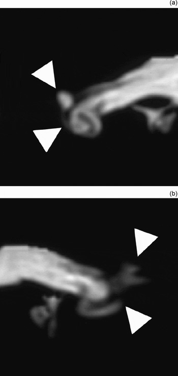
Computed tomography (CT) images of the temporal bone were normal on both sides, with no inner-ear anomalies or abnormal middle-ear findings detected. Intra-cranial MRI was unremarkable. However, three-dimensional, T2-weighted MRI of the inner ear showed bilateral inner-ear obliteration (Figure 1a and b).
Three-dimensional, T2-weighted magnetic resonance images of case one, demonstrating semicircular canal and cochlear obliteration (arrow heads) on the right side (a) and left side (b). (22.5-cm field of view; 288 × 288 matrix; repetition time, 7000 msec; echo time, 250 msec; 90/180 flip angle.)

Since the cochlea on the right side was not totally obliterated, a cochlear implantation was performed on that side. A Nucleus N24 straight electrode (Cochlear Co, Lane Cove, NSW, Australia) was used. Although minor resistance was noted during the insertion of the electrode, the full length of the electrode was successfully implanted. The patient's hearing ability was re-evaluated, and her mean hearing threshold with the cochlear implant was 40 dB, measured by sound field audiometry.
Post-operatively, in her daily life, the patient has no difficulty speaking to others on a one-on-one basis, although she experienced some difficulty with new voices or with many people in conversation. She was also able to talk on the telephone, but only when very familiar with the person to whom she was speaking.
Case report two
A 63-year-old man had an eight-year history of right tinnitus and hearing loss, and a seven-year history of left tinnitus and hearing loss. There was no history of vertigo or dizziness. The patient had a past history of ulcerative colitis, lupus nephritis and systemic lupus erythematosus. Treatment for ulcerative colitis had commenced with prednisone; azathioprine, salazosulphapyridine and mesalazine had also been given, as well as several courses of leukocytapheresis.
The otoscopic examination was normal. Pure tone audiometry showed hearing thresholds of 46.25 dB in the left ear (average thresholds at 0.5, 1, 2 and 4 kHz) and thresholds beyond the scale in the right ear. Nystagmus was not observed.
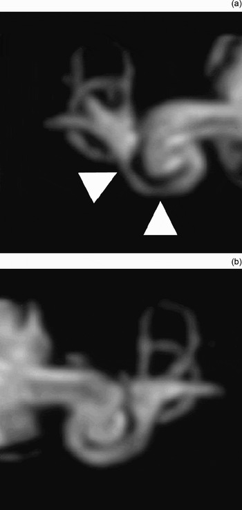
Three-dimensional, T2-weighted MRI of the inner ear showed bilateral inner-ear strictures (Figure 2a and b).
Three-dimensional, T2-weighted magnetic resonance images of case two, demonstrating a stricture of the cochlea (arrow heads) on the right side (a) but not on the left side (b). (22.5-cm field of view; 288 × 288 matrix; repetition time, 7000 msec; echo time, 250 msec; 90/180 flip angle.)

The patient's prednisone was continued, but his hearing level did not improve. Since the patient's ulcerative colitis did not respond to medical therapy, a total colectomy was performed.
Discussion
Sensorineural hearing loss is recognised as one of the extra-intestinal manifestations of ulcerative colitis; an autoimmune mechanism is considered to be one of the causes of inner-ear dysfunction.Reference Mathews and Kumar1 A variety of experimental models have been proposed to explain the pathogenesis of autoimmune inner-ear dysfunction. A specific antibody that binds to a 68-kDa inner-ear antigen has been suggested as one of the important factors contributing to autoimmune sensorineural hearing loss.Reference Billings, Keithley and Harris5 In an experimental guinea pig model, a sterile labyrinthitis following a local immune response caused by either local or systemic administration of antigen resulted in physiological dysfunction, loss of sensory cells, fibrosis and osteoneogenesis in the cochlea.Reference Ma, Billings, Harris and Keithley6 Histopathological study of the temporal bone of a patient with autoimmune sensorineural hearing loss associated with ulcerative colitis showed: (1) missing organs of Corti; (2) a decreased number of spiral ganglion cells; (3) endolymphatic hydrops in the cochlea, saccule and utricle; and (4) fibrosis and osteoneogenesis of the cochlea, semicircular canal and endolymphatic sac.Reference Hoistad, Schachern and Paparella7 Inner-ear fibrosis and osteoneogenesis were observed both in experimental animal models and in a human temporal bone study.
Zavod et al. showed that, in patients with autoimmune sensorineural hearing loss, there was no correlation between the presence of antibodies to inner-ear antigen and the presence of cochlear enhancement on T1-weighted MRI. They concluded that MRI was not helpful in diagnosing or managing autoimmune sensorineural hearing loss.Reference Zavod, Sataloff and Rao8
However, the clinical utility of high-resolution MRI studies of the inner ear has been well established for three-dimensional, gradient-echo sequences,Reference Stillman, Remley, Loes, Hu and Latchaw9, Reference Casselman, Kuhweide, Deimling, Ampe, Dehaene and Meeus10 two-dimensional sequences,Reference Phelps11 and three-dimensional, fast spin-echo sequences.Reference Naganawa, Itoh, Fukatsu, Ishigaki, Nakashima and Kassai12 Compared with two-dimensional studies, three-dimensional studies are useful for assessing inner-ear involvement;Reference Czerny, Rand, Gstoettner, Woelfl, Imhof and Trattnig13 T2-weighted, fast spin-echo MRI is valuable for assessing the extent of inner-ear damage.Reference Krombach, Martiny, Di Martino, Westhofen, Stanzel and Haage14, Reference Lane, Ward, Witte, Bernstein and Driscoll15
We found that, on three-dimensional MRI of both our cases, the inner-ear signals of the membranous labyrinth disappeared. These findings would appear to indicate obliteration of the bony labyrinth and to reflect the inner-ear fibrosis or osteoneogenesis that has been reported from Hoistad and colleagues' human temporal bone study.Reference Hoistad, Schachern and Paparella7
• This paper describes the inner-ear obliteration seen on three-dimensional magnetic resonance imaging (MRI) in patients with sensorineural hearing loss associated with ulcerative colitis
• Cochlear fibrosis or osteoneogenesis may follow autoimmune labyrinthitis
• Three-dimensional inner-ear MRI may be considered in patients with autoimmune sensorineural hearing loss
The diagnosis of autoimmune sensorineural hearing loss can be difficult; no serological, immunological or functional tests have been established to enable a definitive diagnosis.Reference Ruckenstein16 There has been an emphasis on developing Western blotting to detect antibodies bound to the 68-kDa inner-ear antigen or heat shock protein 70; however, the sensitivity and specificity of this test were insufficient to enable it to be used to diagnose autoimmune sensorineural hearing loss.Reference Ruckenstein16 Thus, the diagnosis of autoimmune hearing impairment is generally based on a combination of evidence, including the presence of known autoimmune disease, results of broad tests of autoimmunity and a positive response to steroids. Eventually, the inner-ear involvement revealed by MRI in patients with systemic autoimmune disease may be considered to be evidence of autoimmune inner-ear disease.
Steroids and cyclophosphamide have been used to manage autoimmune sensorineural hearing loss. A prospective, randomised, clinical study found that corticosteroids were useful.Reference Harris, Weisman, Derebery, Espeland, Gantz and Gulya17 Three mechanisms to explain the effect of steroid therapy in autoimmune sensorineural hearing loss have been proposed: immune suppression, anti-inflammatory action and increased sodium transport.Reference Mathews and Kumar1 However, several reported sensorineural hearing loss cases, including ours, showed a poor response to steroid therapy.Reference Moris, Milla, Ribacoba and Gonzalez3, Reference Dowd and Rees18, Reference Kumar, Walsh, Wilson and Carlin19 Cochlear fibrosis or osteoneogenesis may ensue following autoimmune labyrinthitis. In our patients, we found that the inner ears with MRI evidence of obliteration (i.e. both ears in case one and the right ear in case two) had severe hearing loss. This obliteration of the inner ear, as seen using three-dimensional MRI, suggests that the sensorineural hearing loss of patients with systemic autoimmune disease has a poor prognosis. Three-dimensional inner-ear MRI may be considered in patients with suspected autoimmune sensorineural hearing loss.
Acknowledgement
This work was supported in part by grants from the Ministry of Education, Culture, Sports, Science and Technology, Japan.